
Liponeurofibroma
2022-07-06 08:54:10YaKunShaoandJianMinChang
國際皮膚性病學(xué)雜志 2022年2期
Ya-Kun Shao and Jian-Min Chang*
Department of Dermatology, Beijing Hospital, National Center of Gerontology, Institute of Geriatric Medicine, Chinese Academy of Medical Sciences, Beijing 100730, China.
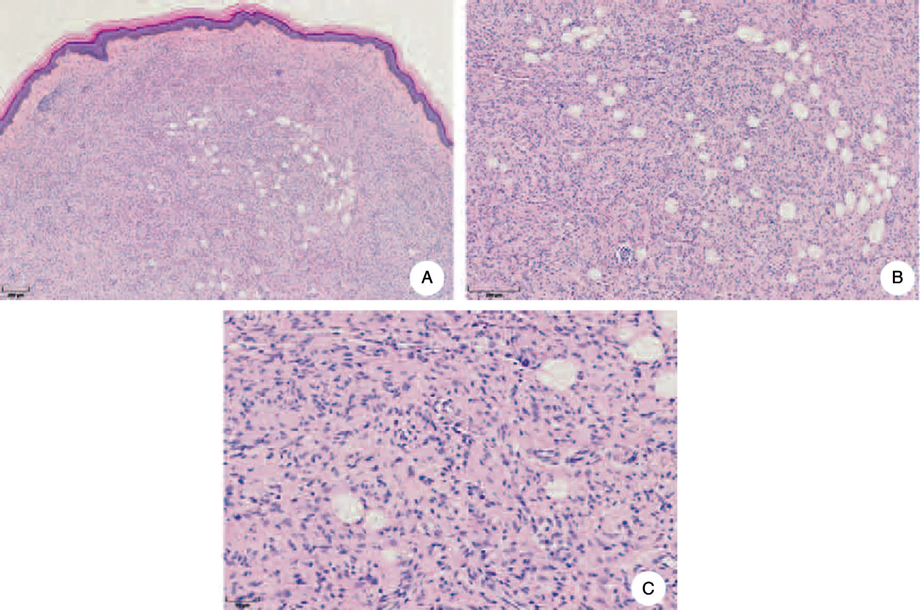
Figure 1. Histopathological examination of cutaneous liponeurofibroma.(A)A well-circumscribed and nonencapsulated tumor mass located in the dermis.The center of the tumor contains scattered fatty tissue(HE staining,40×).(B)Round fat cells with vacuolated cytoplasm and thin nuclei are larger than the surrounding cells. Some adipocytes have intraluminal flap-like structures (HE staining, 100 ×). (C) Fatty change replaces some of the S-shaped and spindle-shaped tumor cells (HE staining, 200 ×).
Histopathology
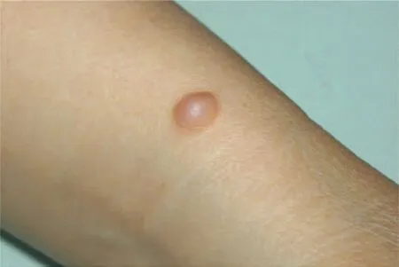
Figure 2. Clinical appearance of cutaneous liponeurofibroma. A dome-shaped, well-circumscribed, and tender cutaneous nodule protruding over the peripheral skin surface.The nodule is light red in color and measures approximately 6cm×4cm, without pedicles.
A liponeurofibroma is a dermal neurofibroma (NF) with an infiltration of abnormal fat cells. Under HE staining,the liponeurofibroma lesion is a well-circumscribed and nonencapsulated tumor mass located in the dermis.Spindle-shaped tumor cells with wavy cytoplasm and elongated nuclei are scattered in the tumor mass. Fatty change is clearly seen inside the tumor, replacing the Sshapedcells.Roundfat cellswithvacuolatedcytoplasmand thin nuclei are larger than the surrounding cells and may gather and fuse into clusters, while some adipocytes have intraluminal flap-like structures (Fig. 1). Compared with deep subcutaneous fat cells,tumor adipocytes are smaller,fewer in number,and vary in size.Liponeurofibromas are classified as focal infiltrating(5.6%)and diffuse infiltrating remaining 10% of lesions are found in persons with an autosomal dominant genetically inherited disease called neurofibromatosis, sometimes accompanied by milkcoffee spots, acoustic neurilemomas, and intellectual abnormalities.1Skin lesions can grow along nerve trunks.Initially, a solitary NF lesion usually occurs as a hard pimple and then gradually enlarges to a nodule with a smooth surface. The lesion varies from mung bean-sized to egg-sized, with varying degrees ofpigmentation. Some skin lesions have pedicles protruding over the peripheral skin surface.The skin lesions have a rubbery texture and remain soft, without causing pain or pruritus. Previous studies have reported some differences between liponeurofibroma and classic NF. Liponeurofibroma generally occurs in older patients than classic NF,while NF is more common in females and frequently develops in the head and neck area.1(1.3%,regularly interspersed).1The incidence of such fatty changes range from 6.9%to 24.6%.2-3
Clinical features
Dermal NF is a benign peripheral nerve sheath tumor that presents as a solitary tumor in 90%of cases(Fig.2).The

- 國際皮膚性病學(xué)雜志的其它文章
- Radiofrequency in Facial Rejuvenation
- Successful Treatment of Severe Pityriasis Rubra Pilaris with Cyclosporine A in An Infant
- Lichen Planus Pigmentosus Inversus: Two Case Reports
- Toe Absence Related to Verrucous Carcinoma
- Dermatoscopy in the Diagnosis of Vulvar Basal Cell Carcinoma: A Case Report
- Skin Organoid Research Progress and Potential Applications