
Toe Absence Related to Verrucous Carcinoma
2022-07-06 08:53:28XiaoLiZhengFeiYiYiQunJiangQianQiuWangGuanZhaoLiangYuePingYinGuoYiZhang
國(guó)際皮膚性病學(xué)雜志 2022年2期
Xiao-Li Zheng, Fei Yi, Yi-Qun Jiang, Qian-Qiu Wang, Guan-Zhao Liang, Yue-Ping Yin,Guo-Yi Zhang*
1Department of STD Clinical Management and Intervention, National Center for Sexually Transmitted Disease Control, Hospital for Skin Diseases (Institute of Dermatology), Chinese Academy of Medical Sciences and Peking Union Medical College, Nanjing,Jiangsu 210042, China; 2Department of Dermatology, Institute of Dermatology, Peking University Shenzhen Hospital, Shenzhen Peking University-The Hong Kong University of Science and Technology Medical Center, Shenzhen 518036, China; 3Department of Pathology, Chinese Center for Disease Control and Prevention, Hospital for Skin Diseases (Institute of Dermatology), Chinese Academy of Medical Sciences and Peking Union Medical College, Nanjing, Jiangsu 210042, China; 4Department of Mycology,Chinese Center for Disease Control and Prevention, Hospital for Skin Diseases (Institute of Dermatology), Chinese Academy of Medical Sciences and Peking Union Medical College, Nanjing, Jiangsu 210042, China; 5STD Reference Laboratory, Chinese Center for Disease Control and Prevention, Hospital for Skin Diseases (Institute of Dermatology), Chinese Academy of Medical Sciences and Peking Union Medical College, Nanjing, Jiangsu 210042, China; 6Outpatient Department, Hospital for Skin Diseases (Institute of Dermatology), Chinese Academy of Medical Sciences and Peking Union Medical College, Nanjing, Jiangsu 210042, China.
Abstract
Keywords: verrucous carcinoma, squamous cell carcinoma, surgical excision, case report
Introduction
Verrucous carcinoma, a rare variant of squamous cell carcinoma,was described a low-grade malignancy.1There are three forms of verrucous carcinoma.2The first type originates in the oropharynx and has been referred to as oral florid papillomatosis. The second type is a giant condyloma acuminata,also termed a Buschke-Lwensteintumor, and is located in the anogenital area. Epithelial tumors are the third form. The most common epithelial site of verrucous carcinoma is the foot,2and it may involve tendons and muscles. Because it generally grows exogenously,bone erosion is rare.Therefore,bone absence due to verrucous carcinoma is exceedingly rare. Here, we reported a rare case of verrucous carcinoma with bone erosion, to improve the knowledgement in this area.
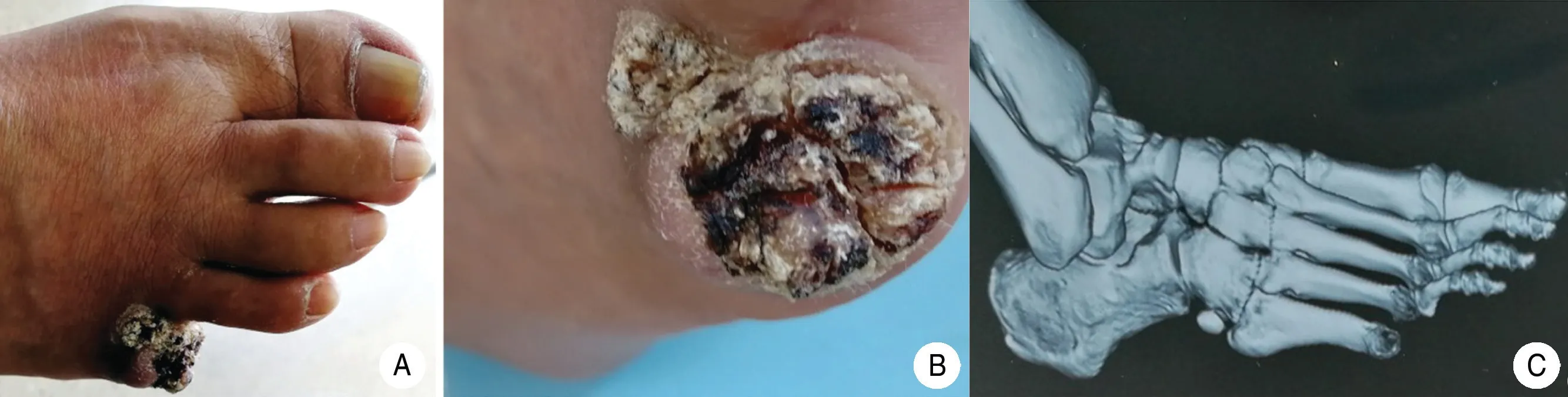
Figure 1. Clinical and three-dimensional computed tomography characteristics of the patient with toe absence caused by verrucous carcinoma.(A)The little toe of the right foot is absent.(B)A white and leather-colored warty tumor with black dirt was observed.(C)Threedimensional computed tomography showed that the fifth toe of the right foot was absent.
Case report
A 64-year-old Chinese male farmer presented with a 6-year history of a slowly growing exogenous hyperkeratotic verrucous lesion on the right fifth toe, which had been absent for 1year (Fig. 1A and 1B). Six years previously, he had undergone nail extraction from this toe. Multiple wound cultures and special stains for fungi and mycobacteria were negative. He had no palpable lymphadenopathy. Three-dimensional computed tomography showed that the fifth toe of the right foot was absent, and no obvious abnormalities were seen in the remaining bone of the right foot (Fig. 1C). On histopathological examination of a skin biopsy specimen,the lesion appeared verrucous with parakeratosis and bleeding in the stratum corneum. Coarse keratohyalin granules in the stratum granulosum and the upper part of the stratum spinosum led us to suspect human papillomavirus infection. Remarkable keratinocyte hyperplasia with consequent infiltration into the superficial dermis was also noted.The peripheral cells were relatively deeply stained and exhibited minimal cytologic atypia. The superficial dermis contained lymphocyte infiltration, and no granulomatous inflammation was observed (Fig. 2A and 2B).The lesion was surgically excised,and the patient was followed up for more than 6months. He had an excellent outcome without recurrence.
Discussion
The typical lesions of verrucous carcinoma initially appear as viral warts.Other less typical manifestations have also been reported, such as bone invasion mimicking a pseudoepitheliomatous reaction resulting in acute osteomyelitis.3Verrucous carcinoma is often misdiagnosed as viral warts, keratoacanthoma, basal cellcarcinoma, cutaneous tuberculosis, and deep mycosis.Early diagnosis of verrucous carcinoma is important because it can invade underlying structures such astendons, muscles, and even bones. The histopathological features of the superficial part of verrucous carcinoma are similar to those of viral warts, especially on imaging examinations; both are exophytic and verrucous, show endophytic growth, and are characterized by acanthosis and papillary hyperplasia.Well-differentiated squamous epithelial cells can be observed in the superficial portions with epidermal hyperkeratosis and parakeratosis, and cytologic atypia is minimal. The border of verrucous carcinoma under microscopy is smooth and pushing. Hematoxylin-eosin staining shows minimal nuclear heterogeneity and nuclear division.
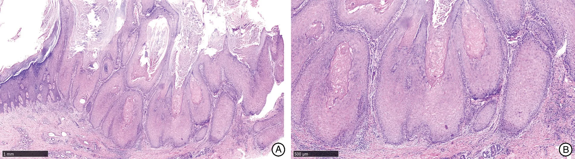
Figure 2. Histopathological characteristics of the patient with toe absence caused by verrucous carcinoma. (H&E staning,×20) (A)Histopathological examination showed that the lesion was exogenous and verrucous and exhibited moderate keratinocyte hyperplasia(H&E staning,×20). (B) The atypia was minimal at higher magnification (H&E staning,×40).
When encountering a man with a wart-like plaque on the foot that responds poorly to conventional treatments,clinicians should maintain a high degree of clinical vigilance for a slowly growing mass and should maintain a low threshold for biopsy because verrucous carcinoma may invade the bones beneath the tumor.The treatment of verrucous carcinoma mainly involves surgery. Extensive resection is usually required to avoid recurrence.4

- 國(guó)際皮膚性病學(xué)雜志的其它文章
- Radiofrequency in Facial Rejuvenation
- Liponeurofibroma
- Successful Treatment of Severe Pityriasis Rubra Pilaris with Cyclosporine A in An Infant
- Lichen Planus Pigmentosus Inversus: Two Case Reports
- Dermatoscopy in the Diagnosis of Vulvar Basal Cell Carcinoma: A Case Report
- Skin Organoid Research Progress and Potential Applications