Protective effect of Citrus reticulata peel extract against potassium dichromateinduced reproductive toxicity in rats
Samir AE Bashandy, Omar A-H Ahmed-Farid, Enayat A. Omara, Sayed A. El-Toumy, Josline Y. Salib
1Pharmacology Department, National Research Centre, 33 El-Bohouth St., Dokki, P.O. 12622, Giza, Cairo, Egypt
2Physiology Department, National Organization for Drug Control and Research, Cairo, Egypt
3Pathology Department, National Research Centre, Cairo, Egypt
4Chemistry of Tannins Department, National Research Centre, Cairo, Egypt
Keywords:Citrus reticulata peel extract Potassium dichromate Sperm Testicular function Oxidative stress Anti-inflammatory
ABSTRACT Objective: To study the protective effect of Citrus reticulata peel extract against potassium dichromate-induced reproductive impairment in male Wistar rats.Methods: A total of 35 male Wistar rats were equally divided into 5 groups. The control group orally received an equivalent volume of saline (1 mL); group 2 was given potassium dichromate at 10 mg/kg/body weight (b.w.) orally for eight weeks; group 3 and 4 orally received Citrus reticulata peel extract at 200 and 400 mg/kg/b.w. respectively, followed by treatment with potassium dichromate. Group 5 was administered with 400 mg/kg/b.w. Citrus reticulata peel extract only. The parameters including oxidative stress (malondialdehyde, antioxidant enzymes), sex hormones (testosterone, luteinizing hormone, follicle-stimulating hormone)and inflammation indexes (tumor necrosis factor-alpha, inter leukin 6) were measured. Sperm characteristics (sperm morphology, motility and count) were also determined.Results: The decrease of male sex hormones concentration in plasma (testosterone, luteinizing hormone), testicular antioxidant parameters (reduced glutathione, catalase, superoxide dismutase, carnitine), sperm motility and sperm count induced by potassium dichromate was mitigated by Citrus reticulata peel extract. Moreover, the peel extract alleviated the increase of sperm abnormalities, follicle-stimulating hormone, testicular malondialdehyde, tumor necrosis factor-alpha and nitric oxide levels caused by potassium dichromate. The sections of rats treated with the potassium dichromate were characterized by some histopathological changes as disorganization of spermatogonial cell layers and deformation of Leydig cells.A positive immune-reaction for p53 was observed in testicular cells of the rats treated with potassium dichromate. The extract of Citrus reticulata peel improved the testicular histology and decreased p53 positive immune-reaction in potassium dichromate administered rats.Conclusions: Citrus reticulata peel extract can alleviate the reproductive toxicity triggered by potassium dichromate via its antioxidant and anti-inflammatory properties. Citrus reticulata peel extract may act as a new natural prophylactically agent for the treatment of potassium dichromate-induced testicular damage.
1. Introduction
Chromium is a dangerous ecological pollutant that is common due to its industrial and agricultural uses. Several industries processes such as chrome plating, welding, painting, textile manufacture,manufacture of stainless steel and wood preservation employ chromium[1]. Potassium dichromate has been notified to encourage reproductive toxicity in the male rats as proved by a decrease of sperm motility, sperm count and testosterone level linked with an increase of sperm abnormalities[2]. It was reported that chromium has many mechanisms to exert its toxicity particularly the increase of free radical species production[3].
Citrus fruits are one of the most familiar fruits belong to the family Rutaceae. The Citrus group included Citrus (C.) reticulate(mandarin) and Citrus sinensis (sweet orange)[4]. The efficiency of fruit remains especially peels as natural phenolic compounds was systematically evaluated[5]. The Citrus peel contains a high rich fraction of hesperidin[6]. The peel fruits of citrus species are known to have antioxidant, anti-inflammatory, anticancer, and anti-lipidemic activities[7-10].
Since fruit remains can be used as a source of active compounds,such as antioxidants, the present study was designed to appreciate the protective role of the water extract of C. reticulate peel rich in flavonoids contrast potassium dichromate motivated testicular injury by the evaluation of oxidative stress parameters, sex hormones,inflammatory markers, sperm characteristics and histopathological changes of testis.
2. Materials and methods
2.1. C. reticulate peel extract
The fruits of C. reticulate were gathered from the Cairo market,Egypt between December 2015 and February 2016. The species was confirmed by a plant taxonomist, and a specimen (C_16) was put in the National Research Centre Herbarium. The fresh fruit peel (1 kg) was swirled by the blender, soaked in a sterile distilled water, warmed at 40 ℃ and then filtered by using Whatman filter paper No.1. The extraction process was repeatedly done. The water extract was condensed by rotary evaporator, and then it was placed in lyophilization to give a dry residue (350 g).
2.2. Chemicals
Methanol [high-performance liquid chromatography (HPLC) grade]and perchloric acid were secured from Loba Co, India. Sulphosalsilic acid and P-aminobenzyl glutamate and pyrogallol were purchased from TMMEDIA Co, India. The 1,1,3,3-tetraethoxypropane,glutathione (GSH) and oxidized glutathione (GSSG) from Sigma Aldrich (USA). Potassium dichromate, monobasic potassium phosphate, nitrites and nitrate were obtained from Al Nasr chemicals(Qualybia, Egypt). All the chemicals used were either HPLC or analytical grade.
2.3. Animals and treatment
A total of 35 adult male Wistar rats (aged 12-15 weeks, weighing 150-170 g)[11] were brought from the National Research Centre,Egypt. They were fed with a standard pellet diet, given water ad libitum and kept at adjusted temperature (22±2) ℃ with 12 h light-dark cycle. Animal handling was carried out according to the principles and guidelines of Ethics Committee of the National Research Centre, Egypt (Approval No. P 15170, 3/6/2016) and followed the recommendations of the National Institutes of Health Guide for Care and Use of Laboratory Animals (Publication No. 85-23, revised 1985).
The 35 rats were divided into 5 groups, with 7 rats in each group.Group 1 (the control group) orally received an equivalent volume of saline (1 mL). Group 2 was orally administered with 10 mg/kg body weight (b.w.) of potassium dichromate[12]. Groups 3 and 4 were pretreated with C. reticulate peel extract orally (200 mg, 400 mg/kg/b.w.,respectively)[13] followed by treatment with potassium dichromate after a half hour. Group 5 was only treated with C. reticulate peel extract at a dose level of 400 mg/kg/b.w. orally. All the treatments continued daily for two months[14]. C. reticulate peel extract or potassium dichromate was dissolved in water.
2.4. Samples collection
The blood (3 mL) was gathered after eight weeks in heparinized tubes from the retro-orbital plexus under local anesthesia by diethyl ether. The animals were killed 8 weeks post-treatment by cervical dislocation under ether anesthesia. Plasma was isolated by centrifugation at 3 000 rpm for 15 min. The body weight was recorded every two weeks while weight of sexual organs was carried out at the end of experiment time (8 weeks). The left epididymides were incubated at 37 ℃ in phosphate buffered saline (PBS) for the evaluation of sperm motility, count and abnormality. The left testes were processed for histological study, and right ones were used for evaluation of malondialdehyde (MDA), reduced GSH, GSSG,superoxide dismutase (SOD), catalase (CAT), nitric oxide (NO),tumor necrosis factor-alpha (TNF-α) and carnitine.
2.5. Testicular homogenate
Each right testis was weighed and homogenized in saline (10%w/v). Samples were centrifuged at 4 500 rpm for 15 min and the supernatant was isolated and stored at -80 ℃ until assay.
2.6. Testicular oxidative stress parameters
Testicular MDA, GSH, GSSG and carnitine[15-17] levels were evaluated by HPLC system of Agilent HP 1200 series (USA) that consisted of a quaternary pump, a column oven, Rheodine injector with 20 μL loop, and variable-wavelength ultraviolet detector. The resulting chromatogram identified the concentration from the sample compared to that of the standard purchased from Sigma Aldrich.
The SOD activity was assayed in the homogenate[18] and was expressed as 1 U/g tissue. The enzyme activity was the activity capable for 50% inhibition of pyrogallol auto-oxidation in 1 min.CAT activity (nmol H2O2/min/mg protein) was measured by a spectrophotometric method based on the decomposition of H2O2[19].
2.7. Testicular inflammatory markers
Testicular NO level was evaluated by HPLC system[20]. TNF-α was assayed by enzyme-immunoassay kit manufactured in R&D Systems, USA.
2.8. Histological analysis
The testis was fixed in 10% formol saline for one week. The samples were subjected to routine histology and stained with hematoxylin and eosin.
2.9. Immunohistochemistry of p53
The paraffin-embedded testis was cut into 5-μm sections and mounted on positively charged slides for p53 immunohistochemistry.Sections were deparaffinized in xylene and rehydrated with a descending series of absolute ethanol, followed by water. The sections were incubated with a primary monoclonal antibody for p53(code No. M7001, dilution 1:50, DAKO) at a dilution of 1:200 for 2 h at 25 ℃ in a humidification chamber. The slides were washed three times in phosphate buffered saline (PBS) for 3 min each. Biotinylated polyvalent secondary antibody (Vectastain Elite ABC Kit; Vector Laboratories, Burlingame, CA) was applied to tissue sections and co-incubated for 30 min. To visualize bound antibodies, sections were washed in PBS and covered with 3,3’-diaminobenzidine substrate and incubated for 10 min.Counterstaining was performed by adding an adequate amount of hematoxylin stain to the slide to cover the entire tissue surface.In negative control slides, the same system was applied with the replacement of the monoclonal antibody by diluted normal bovine serum. Slides were examined with the light microscope (×400)(Olympus BX-50 Olympus Corporation, Tokyo, Japan).
2.10. Hormonal assay
Plasma testosterone, luteinizing hormone (LH) and follicle-stimulating hormone (FSH) levels were determined by enzyme immunoassay kits from Diagnostic products Co. (Los Angeles, CA USA).
2.11. Sperm characteristics
One epididymis was chopped to small pieces in warm PBS (3 mL)to obtain sperm suspension that was incubated for 10 min to allow the sperms to free into buffered solution. The sperm motility was determined by counting motile and non-motile spermatozoa in the same field. The count was repeated until the total count of sperm was about 300. The percentage of motile spermatozoa was computed.The suspension was filtered. An aliquot from the suspension was taken in Neubauer’s counting chamber hemocytometer[21] to elucidate the number of spermatozoa (million/1 mL). To assess the number of abnormal sperm, 1 mL of suspension was stained with 1% eosin. The morphology of spermatozoa was evaluated under a light microscope (×400)[22].
2.12. Statistical analysis
Statistical analysis of the data was performed by using SPSS 17.0(SPSS Corp., Armonk, NY, USA) with one-way analysis of variance followed by the least significant difference. The results were expressed as mean ± standard deviation (mean ± SD). A probability value (P) less than 0.01 was considered statistically significant.
3. Results
3.1. Body and sexual organs weight
A significant derease in the body weight (P<0.01) of potassium dichromate group was observed at 4, 6, and 8 weeks post-treatment compared with the control group. The body weight gain was improved in the group receiving C. reticulata peel extract at 200 or 400 mg/kg/b.w. + potassium dichromate. The body weight gain after eight weeks was 93.77%, 17.66%, 49.02% and 62.47%for the control group, potassium dichromate group, 200 mg/kg/b.w. and 400 mg/kg/b.w. C. reticulata peel extract + potassium dichromate groups, respectively (Table 1). The weight of testis, vas deferens, epididymis, prostate and seminal vesicle in potassium dichromate group or group of 200 mg/kg/b.w. C. reticulata peel extract+potassium dichromate decreased significantly (P<0.01)as compared to those in the control group (Table 2). In addition,prostate and seminal vesicle weight in the group of 400 mg/kg/b.w. C. reticulata peel extract+potassium dichromate decreased significantly (P<0.01) as compared with the control group, while other sexual organs weight did not change significantly. The sexual organ weights of rats administered the peel extract (low or high dose) + potassium dichromate were significantly higher than those of potassium dichromate group. The administration of C. reticulata peel extract only to rats (5th group)had no significant influence on all the parameters of this study.

Table 1. Effect of C. reticulata peel extract on body weight (g) of rats treated with potassium dichromate.

Table 2. Relative reproductive organ weights (g) of potassium dichromate-treated rats with or without C. reticulata peel extract 8 weeks post treatment.
3.2. Oxidative stress and inflammation parameters
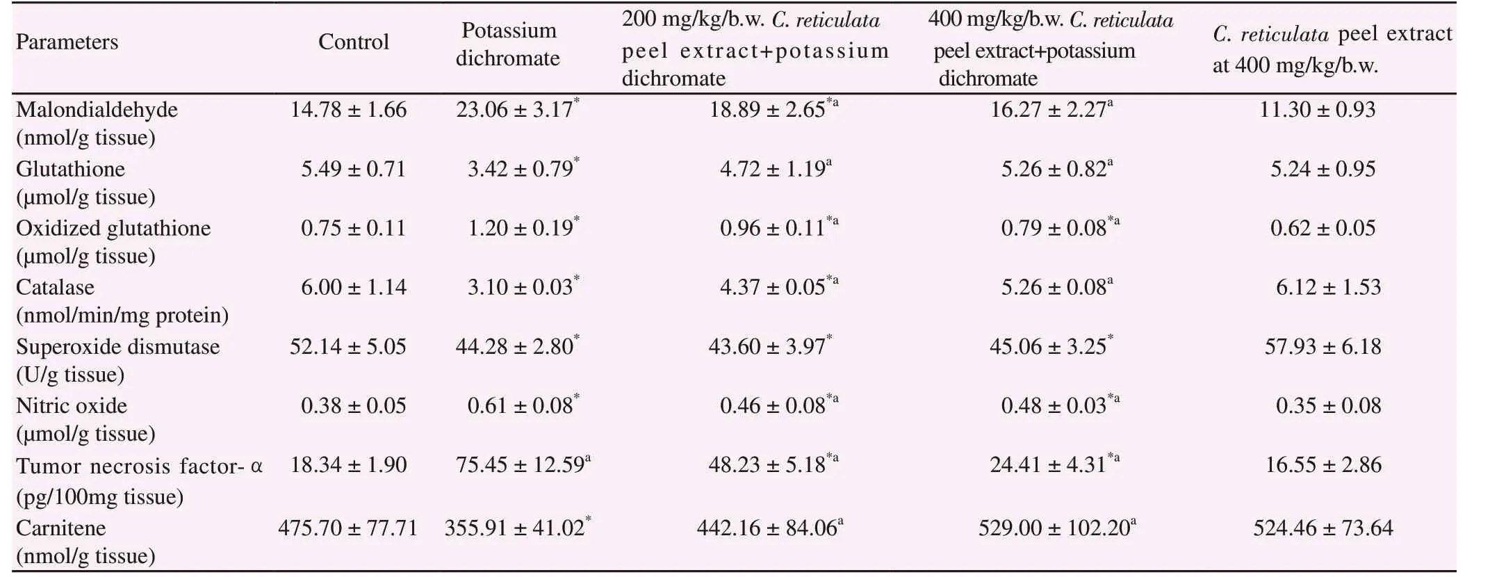
Table 3 confirmed the protective role of C. reticulata peel extract against potassium dichromate which produced testicular oxidative stress and inflammation. The administration of potassium dichromate led to significantly increased testicular MDA, GSSG,NO and TNF-αlevels compared with the control group (P<0.01).On the other hand, significant decreases of testicular GSH, CAT,SOD and carnitine were noticed in the potassium dichromate group.Administration of C. reticulata peel extracts alleviated potassium dichromate-induced toxicity on oxidative stress and inflammation parameters, while administration of C. reticulata peel extract only had no significant influence on these parameters.
3.3. Histological study
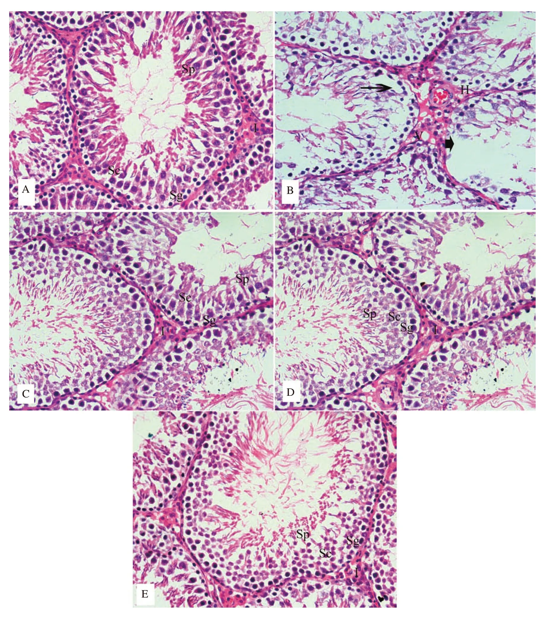
Compared to the control (Figure 1A), testicular tissue damage,marked degenerative changes and necrosis together with disorganization of spermatogonial cell layers were observed in the potassium dichromate section (Figure 1B). Moreover, damaged spermatocytes and spermatids were detected within the tubular lumina of many seminiferous tubules associated with interstitial hemorrhage and vacuolated Leydig cells. The group treated with potassium dichromate + low or high dose of peel manifested partially improvement of spermatogenic cells and mild interstitial hemorrhage(Figure 1 D and E). The testis section of rats given C. reticulata peel extract only appeared normal (Figure 1C).
3.4. Immunohistochemical study
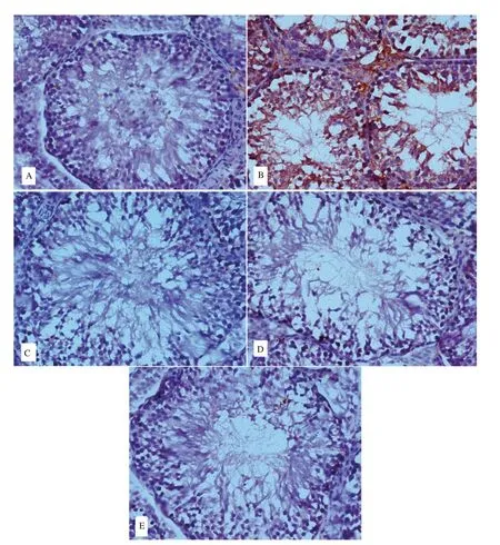
A positive immune-reaction for p53 was detected in spermatogenic cells of the potassium dichromate-treated group (Figure 2B). The rats receiving potassium dichromate + C. reticulata peel extract (low or high dose) showed a decrease of p53 expression (Figure 2 D and E). No immune positive reaction was noticed in the section of rats given C. reticulata peel extract only (Figure 2C).
3.5. Male sex hormones
The rats treated with potassium dichromate displayed significant increased FSH level and significant reduction of testosterone and LH levels (P<0.01). The alteration in hormone levels due to potassium dichromate treatment was significantly alleviated by administration of C. reticulata peel extract. Moreover, the administration of C. reticulata peel extract only showed similar results to the control group (Table 4).
3.6. Sperm characteristics
The sperm motility and count of all treatment groups lowered significantly (P<0.01) in comparison with the control group except potassium dichromate + high dose of C. reticulata peel group which showed no significant difference (Table 5). Although the sperm abnormalities were elevated significantly in the potassium dichromate group and potassium dichromate + low or high dose of C. reticulata peel extract group as compared with the control group,the abnormality rates were significantly lower in extracts-treatmemt groups than in potassium dichromate group. On the other hand,the administration of C. reticulata peel extract only showed similar results to the control group.

Table 3. Testicular oxidative stress and inflammatory parameters of rats treated with C. reticulata peel extract and potassium dichromate.

Table 4. Protective effect of C. reticulata peel extract on potassium dichromate induced alteration of male sex hormones.

Figure 1. Histological study of testicular tissue. A: the control group shows normal spermatogonia (Sg), spermatocytes (Sc), spermatids (Sp), and interstitial cells; B: the potassium dichromate group shows marked disorganization of spermatogenesis, different degenerative changes (arrow head), interstitial haemorrhage (H) and vacuolated of Leydig cells (V); C: Rats treated with C. reticulata peel extract (400 mg/kg/b.w.) show nearly normal structure; D:Rats treated with C. reticulata peel extract at 200 mg/kg/b.w. + potassium dichromate show partially improved spermatogenic cells and mild interstitial haemorrhage (I); E: Rats treated with C. reticulata peel extract at 400 mg/kg/b.w. + potassium dichromate show nearly normal histological characteristic(hematoxylin and eosin; ×400).

Table 5. Protective effect of C. reticulata peel extract on potassium dichromate induced alteration of sperm.

Figure 2. Sections of testis tissue stained with p53 imminohisochemical stain. A: the control group shows no immune reaction expression of p53; B: the potassium dichromate treated group shows remarkable positive immune-reaction for p53; C: Rats treated with C. reticulata peel extract at 400 mg/kg/b.w. show no immune reaction expression of p53; D: C. reticulata peel extract at 200 mg/kg/b.w. + potassium dichromate group shows moderate reduce immune-reaction for p53; E: C. reticulata extract at 400 mg/kg/b.w + potassium dichromate group shows obvious reduce immune-reaction for p53 (Immunohistochemistry P53; ×400).
4. Discussion
In the current study, potassium dichromate showed deleterious effects on the testis and accessory sexual organs. The present findings demonstrated that the administration of potassium dichromate to rats resulted in a significant decline in the weight of the body, testis and accessory sexual organs. The decrease in body weight by potassium dichromate can be referred to a decrease in food intake[23]. The decrease of testosterone level in the potassium dichromate group may lead to a decline in testis and accessory sexual organs since the hormone is responsible for the growth of male reproductive organs[24]. A testicular function is controlled by gonadotropic hormones, FSH and LH[25]. In the study, LH and testosterone levels lowered significantly while FSH level significantly increased in the rats given potassium dichromate. The elevated FSH level in the potassium dichromate group is likely due to a decrease in inhibin production by Sertoli cells[26] that affect negative feedback on the hypothalamic-pituitary axis[27].
In the present study, potassium dichromate treatment motivates testicular oxidative stress as seemed by a rise of testicular MDA,GSSG and a decline of testicular GSH, CAT, SOD and carnitine levels. The mechanism of reproductive deterioration by potassium dichromate might be associated with oxidative stress and disruption of the hypothalamic-pituitary-testicular axis. Elevated oxidative stress impacts the physiological function of the interstitial cells of Leydig, which play a crucial role in the synthesis of testosterone[28].Interstitial hemorrhage and vacuolated Leydig cells were observed in the rats treated with potassium dichromate. The reduction of testosterone concentration in the potassium dichromate group can be attributed to the deformed Leydig cells and a decline of LH level.The estimation of MDA is an evidence of lipid peroxidation and oxidative stress[29]. Elevation of lipid peroxidation may result in a damage to biologically important molecules and tissues[30]. It was concluded that potassium dichromate produced reactive oxygen species and oxidative stress leading to the damage and malfunction of spermatogenic cells[31]. The histological changes noticed in testis sections of the potassium dichromate group as degenerative and necrosis of spermatogonial cell layers may be referred to an increment of testicular MDA level or a descent of the testicular GSH and antioxidant enzymes.
Different flavonoids of citrus fruit peel may play an animated role in repro-protective action, and antioxidant potential[32]. In the study, the C. reticulata peel extract relieved the deleterious effects of potassium dichromate on the weight of sexual organs, sexual hormone level, oxidative stress parameters and testicular pathology.Citrus peel residues contain a large number of bioflavonoids as hesperidin[6]. The antioxidant properties of hesperidin minimized lipid peroxidation-mediated reproductive damage evoked by cisplatin[33] or doxorubicin[34]. The improvement of histoarchitecture in the group treated with C. reticulata peel extract and potassium dichromate can be attributed to a decrease of testicular oxidative stress. GSH is an endogenous antioxidant lessening cell injury originated from free radical reactions[35]. SOD and CAT are effective antioxidant enzymes and protect cells from reactive oxygen species invasion[36]. Carnitine exerted its protective effect via maintaining antioxidant enzymes production[37].
In the present study, potassium dichromate created testicular lesions and alteration of sexual hormones particularly testosterone level,which may lead to a spermatogenic arrest as appeared from reducing epididymal sperm number and increase of sperm abnormalities.Testosterone, secreted from the testis, is reliable for the activation and maintenance of spermatogenesis[38]. It was reported that sperm defects were distributed among welders subjected to chromium compounds[39]. Male germ cells[40] and spermatozoa[41] are more susceptible to oxidative stress than somatic cells because their membranes contain a high percentage of fatty acids. Oxidative stress considered a main helpful agent to infertility lead to sperm damage and deformity. Here, potassium dichromate treatment led to a decrease in sperm motility. Reactive oxygen species generated during chromium reduction may lower spermatozoa motility by affecting the phosphorylation of axonemal proteins essential for spermatozoa movement[42].
The alleviation influence of C. reticulata peel extract on potassium dichromate stimulated sperm abnormalities is most likely to the phenolics-related compounds like flavonoids, leading to maintenance of the normal redox status of cells. Flavonoids are characterized by their antioxidant and metal chelating properties, and they preserve sperm motility and prevent sperm membrane oxidative damage by heavy metals[32]. It appears that C. reticulata peel extract (rich in flavonoids) exerts its protective effect against potassium dichromateevoked deleterious effects on male gonads directly through scavenging free radicals or indirectly via enhancement of antioxidant defense system and decrease testicular TNF-α and NO levels (antiinflammatory mediators). The study results of Leyva-López et al showed the extracts from three Mexican oregano species containing flavonoids had anti-inflammatory activity by lowering reactive oxygen species and NO production in lipopolysaccharide model[43].
P53 may involve in chromium-motivated tissue damage, imbalance of antioxidant defense system and carcinogenesis[44]. P53 plays an important role in the regulation of cell production during normal spermatogenesis either by regulating cell proliferation or more likely by regulating the apoptotic spermatogonia. P53 may represent a risk factor for male infertility[45]. In addition, C. reticulata peel extract reduced a positive immune-reaction of p53 in testicular tissues of rats given potassium dichromate. The protective effects of C.reticulata peel extract appear to be due to its influence on restoring endogenous antioxidants and inhibition of MAD formation.
In conclusion, C. reticulata peel extract lowered the deleterious effects of potassium dichromate on the male reproductive system.C. reticulata peel extract demonstrates its action via reducing inflammatory mediators, inhibiting lipid peroxidation and enhancing antioxidant enzymes. These findings led us to suggest that C.reticulata peel extract may display a new precautionary policy for reducing potassium dichromate-induced male infertility. However,the mechanisms of C. reticulata peel extract in relieving the toxic effects of potassium dichromate on the male reproductive system need further research.
Conflict of interest statement
The authors declare that they have no conflict of interest.
 Asian Pacific Journal of Reproduction2019年6期
- Asian Pacific Journal of Reproduction的其它文章
- Effect of L-carnitine supplementation during in vitro maturation and in vitro culture on oocyte quality and embryonic development rate of bovines
- Stable state of serum inflammatory cytokines during induction of benign prostate hyperplasia in dogs
- Effects of Crassocephalum bauchiense (Hutch) leaf aqueous extract on toxicity indicators and reproductive characteristics in Oryctolagus cuniculus exposed to potassium dichromate
- Prolonged post-thaw culture of embryos does not improve outcomes of frozen human embryo transfer cycles: A prospective randomized study
- Effect of cognitive behavioral therapy on anxiety and depression of infertile women:A meta-analysis
- Human blastomere rotation in early cleavage embryos is not associated with reduced implantation: Evidence from time-lapse videography
