Exosomes derived from microglia overexpressing miR-124-3p alleviate neuronal endoplasmic reticulum stress damage after repetitive mild traumatic brain injury
Yan Wang, Dai Li, Lan Zhang, Zhenyu Yin, Zhaoli Han, Xintong Ge, Meimei Li, Jing Zhao, Shishuang Zhang,Yan Zuo, Xiangyang Xiong, Han Gao, Qiang Liu, Fanglian Chen, Ping Lei,*
Abstract We previously reported that miR-124-3p is markedly upregulated in microglia-derived exosomes following repetitive mild traumatic brain injury.However, its impact on neuronal endoplasmic reticulum stress following repetitive mild traumatic brain injury remains unclear.In this study, we first used an HT22 scratch injury model to mimic traumatic brain injury, then co-cultured the HT22 cells with BV2 microglia expressing high levels of miR-124-3p.We found that exosomes containing high levels of miR-124-3p attenuated apoptosis and endoplasmic reticulum stress.Furthermore, luciferase reporter assay analysis confirmed that miR-124-3p bound specifically to the endoplasmic reticulum stress-related protein IRE1α, while an IRE1α functional salvage experiment confirmed that miR-124-3p targeted IRE1α and reduced its expression, thereby inhibiting endoplasmic reticulum stress in injured neurons.Finally, we delivered microglia-derived exosomes containing miR-124-3p intranasally to a mouse model of repetitive mild traumatic brain injury and found that endoplasmic reticulum stress and apoptosis levels in hippocampal neurons were significantly reduced.These findings suggest that, after repetitive mild traumatic brain injury, miR-124-3 can be transferred from microglia-derived exosomes to injured neurons, where it exerts a neuroprotective effect by inhibiting endoplasmic reticulum stress.Therefore,microglia-derived exosomes containing miR-124-3p may represent a novel therapeutic strategy for repetitive mild traumatic brain injury.
Key Words: apoptosis; C/EBP homologous protein; endoplasmic reticulum stress; exosome; inositol-requiring enzyme 1α; microglia; miR-124-3p; neuron;repetitive mild traumatic brain injury; X-box binding protein 1
Introduction
The rate of traumatic brain injury (TBI) is increasing, and this condition is believed to be responsible for a considerable proportion of global and regional disease burdens (Clark et al., 2022).Because of various measures that have been implemented, such as advance prevention, the construction of specialized neurosurgical intensive care units, the formulation of evidencebased guidelines, and the provision of high-level nursing care, survival rates after TBI have greatly increased and the rate of infection-related mortality due to TBI has greatly decreased over the last few decades (Jiang et al., 2019).Nevertheless, the details of TBI pathophysiology remain unknown, and there is a significant lack of effective therapeutic interventions.Therefore, there is an urgent need for in-depth investigation of TBI pathogenesis, especially regarding the cellular and molecular changes that occur after TBI.
In addition to the primary injury caused by external physical forces, TBI also involves secondary injuries caused by biological processes.The secondary damage that follows TBI is the most significant factor influencing patient outcomes and is considered to be reversible (Aychman et al., 2023).The pathogenesis of the neuronal damage associated with TBI involves multiple processes, including excitotoxicity, inflammation, and more.Neuronal apoptosis occurs as a consequence of various pathophysiological alterations and is the leading cause of long-term neurological dysfunction in patients following TBI (Su et al., 2019; Capizzi et al., 2020).The hippocampus, a region of the brain that is critical for learning and memory, has been demonstrated in experimental and clinical investigations to be especially vulnerable to brain injury (Titus et al., 2013).Damage to hippocampal neurons can continue to develop for days, weeks, or even months after moderate to severe brain injury (Titus et al., 2013), eventually leading to persistent cognitive impairment.In another study, Fluoro-Jade staining of hippocampal sections after brain injury showed acute death of newborn neurons that manifested as nuclear fragmentation, which is characteristic of apoptosis (Gao et al.,2008).The hippocampus and contusional area showed the highest levels of apoptotic neuronal cells between 24 and 48 hours after injury in a study using a controlled cortical impact (CCI) model (Fox et al., 1998).However, the mechanism of TBI-mediated neuronal apoptosis is still poorly understood.
The endoplasmic reticulum (ER) tightly regulates protein production and maturation.ER chaperones and foldases bind to proteins and aid in proper protein folding, preventing protein aggregation under physiological circumstances.Under various stress conditions, dysfunction of the proteostasis network, especially the ER component, leads to accumulation of misfolded proteins in the ER lumen, triggering “ER stress,” which is considered to be a cause of abnormal protein aggregation.In response to ER stress,cells activate a dynamic signaling network known as the unfolded protein response (UPR) (Hetz et al., 2020), which up-regulates the expression of chaperone proteins to aid in protein folding and temporarily prevents protein production.However, when exposed to chronically high levels of ER stress,the UPR can also lead to cellular dysfunction, inflammatory body activation,and apoptosis (Smith et al., 2011).Some studies suggest that long-term functional impairment due to TBI is also associated with the rapid induction of abnormal ER stress levels and ultimately leads neuronal cells to undergo ER stress-induced apoptosis (Hood et al., 2018; Xu et al., 2019).
Lipid vesicles called exosomes vary in diameter from 50 to 150 nm.After being released by their origin cells, they can interact with other cells through specific receptors on the cell surface or be phagocytosed by the destination cells, after which they release their cargos, thereby transferring genetic information and proteins between cells to achieve intercellular communication.Exosomes contain nucleotides (mRNA and microRNA(miRNA)) and proteins (Chopp and Zhang, 2015; Thompson et al., 2016; Liu et al., 2023a, b; Zhang et al., 2023), which makes them an important source of diverse biochemical signaling molecules and allows them to play vital physiological and pathophysiological roles.
A class of post-transcriptional gene expression regulators known as miRNAs is associated with practically all developmental and pathological processes(Brown et al., 2022).In our previous study, we developed a mouse model of repetitive mild traumatic brain injury (rmTBI) to examine the regulatory impact of miRNAs carried by microglial exosomes on neurodegeneration post-rmTBI (Ge et al., 2020).Importantly, the expression level of miR-124-3p in microglial exosomes produced by damaged brain is increased, we added miR-124-3p-riched microglial exosomes to neurons with scratch injury and found that these exosomes can promote amyloid beta breakdown,reduce neurodegeneration and enhance cognitive function through Rela action on ApoE (Ge et al., 2020).In the target gene prediction of miR124-3p, we discovered that besides Rela and ApoE genes related to amyloid beta(Aβ) breakdown, the gene inositol requiring enzyme 1α (IRE1α) related to ER stress was also included and showed a strong correlation.Therefore, our study aims to explore the effects of miR-124-3p-enriched microglial exosomes on the ER stress of neurons and the related cell apoptosis.
In summary, increased levels of extracellular microglial miR-124-3p play a crucial role in the modulation of several pathogenic processes post-rmTBI,possibly including neuronal ER stress.Previously, we reported that miR-124-3p enhances microglial polarization to the anti-inflammatory M2 phenotype and found that microglial exosomes containing miR-124-3p promoted neuronal survival in anin vitroscratch injury model (Huang et al., 2018).In the follow-up mechanism exploration, by predicting the target genes of miR-124-3p, we found that it was strongly correlated with genes related to neuroimmunoregulation, Aβ breakdown, and ER stress.In previous studies,we focused on the effect of miR-124-3p on neuroimmunoregulation and Aβ breakdown (Huang et al., 2018; Ge et al., 2020).However, the exact mechanism by which miR-124-3p regulates ER stress is still unclear.Therefore,in this study, we further explored the role of miR-124-3p-enriched microglia exosomes in the regulation of ER stress in injured neurons after mild traumatic brain injury (mTBI), mainly by detecting the expression of ER stressrelated proteins and interfering with ER stress-related pathways.
Methods
Ethical approval
The Tianjin Medical University Animal Care and Use Committee authorized all experimental procedures (approval No.IRB2022-DWFL-074) on February 22, 2022.All experiments were performed in accordance with the National Institutes of Health’s Guide for the Care and Use of Laboratory Animals (8thed., National Research Council, 2011).All experiments were designed and reported according to the Animal Research: Reporting ofIn VivoExperiments(ARRIVE) guidelines (Percie du Sert et al., 2020).
CCI-induced rmTBI mouse model
To eliminate sex differences as a confounding factor, only adult male C57BL/6J mice (aged 8–10 weeks, weighing 22–25 g, Beijing Vital River Laboratory Animal Technology Co., Ltd., Beijing, China, license No.SCXK (Jing) 2021-0006) were used in this study.The mice were randomly assigned to two groups (n= 25/group): the Sham and rmTBI groups.The rmTBI mouse model was generated as we described previously (Ge et al., 2020).After anesthetization with 4.6% isoflurane (RWD, Shenzhen, Guangdong, China),the mice were affixed to an acrylic cast that we designed, which provides 3.0 mm of space for their heads to accelerate and decelerate at the impact point.Each mouse’s head was shaved, and it was then fitted with a custom helmet constructed from a concave metal disc designed to fit is skulls directly caudal to the bregma to transfer the impact force to the whole brain.The mouse was then placed on the acrylic cast in a prone posture and affixed with surgical tape over the shoulders.The impounder tip of the injury device (Model 6.3 electronically CCI apparatus, American Instrument Co., Ltd., Richmond,VA, USA) was adjusted to its full impact distance, placed in the middle of the helmet surface, and readjusted to deliver the impact.The following settings were used: discharge at 5.0 m/s with a 2.5 mm striking depth.After each impact was delivered, the mice were moved to a well-ventilated cage maintained at 37°C until they recovered consciousness.Four impacts were delivered at 48-hour intervals.The mice in the Sham group underwent the same procedures except for the impact.
Exosomes isolation
To extract exosomes derived from microglia cells in the damaged brain tissue,the mice were anesthetized with 4.6% isoflurane, sacrificed, and subjected to cardiac perfusion with 4°C phosphate buffered saline (PBS) at 3, 7, 14,or 21 days post-last impact (DPI).Next, the bilateral cerebral cortex and hippocampus (Opitz-Araya and Barria, 2011) were removed and placed in 3 mL of Hibernate-A cell culture media (Thermo Fisher Scientific, Waltham,MA, USA) (Ge et al., 2020).The damaged brain tissue was then digested with 20 U/mL papain (Solarbio, Beijing, China) at 37°C for around 15 minutes, and the tissue was fragmented by repeatedly pipetting the solution up and down.To isolate single cells, the tissue fragments were centrifuged for 10 minutes at 2000 ×gand 4°C for and then for 30 minutes at 10,000 ×gand 4°C.The supernatants were then passed through a 0.22-μm filter to remove dead cells and large particles (MilliporeSigma, Billerica, MA, USA).Subsequently,total exosome isolation reagent (Thermo Fisher Scientific) was added to the supernatants at a ratio of 1 mL supernatant to 500 μL reagent, and the samples were stored overnight at 4°C.
The next day, the exosomes were collected by ultracentrifugation at 100,000×gfor 70 minutes at 4°C.After the supernatants were removed, the pellet containing the exosomes was resuspended in 0.35 μL Dulbecco’s PBS (Thermo Fisher Scientific).The samples were then incubated for 1 hour at room temperature with 1.5 μg of rat anti-mouse CD11b double antibody (Thermo Fisher Scientific) in 50 μL of 3% bovine serum albumin.Following this, the samples were incubated for 30 minutes at room temperature with 10 μL of Pierce Streptavidin Plus UltraLink Resin (Thermo Fisher Scientific) in 40 μL of 3% bovine serum albumin to extract the microglial exosomes.The samples were then centrifuged at 800 ×gfor 10 minutes at 4°C, and the supernatants were discarded.The resulting microglia-derived exosomes were temporarily stored at 4°C.
To extract exosomes from microglia culturedin vitro, microglial culture medium was first centrifuged for 10 minutes at 300 ×gat 4°C to eliminate free cells, as we described previously (Guo et al., 2023).The supernatants were then transferred to fresh tubes and centrifuged at 2000 ×gfor 10 minutes at 4°C.The resulting supernatants were passed through a 0.22-μm filter to remove particles bigger than 200 nm and dead cells (MilliporeSigma).The exosomes were then collected by ultracentrifugation at 100,000 ×gfor 70 minutes at 4°C, the supernatants were discarded, and the pellet containing the exosomes was stored at 4°C for 1 day before being used in experiments.
Exosome identity verification
After the exosomes were prepared, as described above, we verified their identity.To extract proteins from the microglial exosomes, the exosomes were resuspended in 100 μL PBS, and 4× loading buffer (Solarbio) was added.The samples were then heated for 10 minutes at 95°C (Eppendorf ThermoMixer F1.5, Hamburg, Germany), and the denatured protein samples were stored at –80°C for later protein detection.Exosome biomarkers (CD9, CD63, CD81,and tumor susceptibility gene 101 (TSG101)) (Abcam, Cambridge, UK) were detected by western blot.In addition, the morphology of the exosomes was assessed using a transmission electron microscope (HT7700, Hitachi, Tokyo,Japan).Twenty microliters of the specimen was applied to a formvar film coated with glow-discharged carbon that was connected to a metal sample grid.The formvar film was then incubated for 5 minutes at room temperature with 50 μL of 1% glutaraldehyde before being washed with 2 mL of distilled water eight times at 2-minute intervals.Following a 30-minute period of drying with filter paper, the grid was treated with an equivalent volume of 10% uranyl acetate for 5 minutes at room temperature, followed by 50 μL of methyl cellulose-uranyl acetate for 10 minutes at 4°C.Then, the excess liquid was removed and a transmission electron microscope was used to observe the samples.Finally, a Nanosight NS300 nanoparticle tracking analysis system(Malvern Panalytical, Malvern, UK) was used to measure and evaluate the diameters of the isolated exosomes.
Exosome quantification
Exosomes obtained by supercentrifugation were lysed with radioimmunoprecipitation assay buffer (Beyotime, Shanghai, China) on ice for 30 minutes to harvest total protein.Then, the protein concentration was measured with a bicinchoninic acid protein quantification kit (Solarbio)according to the manufacturer’s instructions.Depending on the protein content of the exosomes, injured HT22 cells were then treated with 50, 100,or 200 μg/mL of exosomes for 24 hours.
miR-124-3p transfection
To assess the function of miR-124-3p, we transfected cells with an miR-124-3p mimic or inhibitor (Table 1; Gene Pharma, Shanghai, China).In brief,the miR-124-3p mimic or inhibitor was diluted to 20 mM, according to the manufacturer’s instructions (INTERFERin?; Polyplus, Illkirch, France).Cells were seeded into 12-well plates the day prior to transfection with the aim of achieving 30–50% confluency for maximum transfection effectiveness.To enable stable transfection complexes to form between the mimic or inhibitor and the INTERFERin, 0.6 μL miRNA was mixed with 8 μL INTERFERin reagent in200 μL serum-free Dulbecco’s modified Eagle medium/F12 (DMEM/F12) and incubated for around 10 minutes.The transfection mixture was then added to the cell culture plates, at a concentration of 10 nM miRNA per well, and the plates were gently shaken to mix then reincubated for 48 hours.Changes in miR-124-3p levels in the target cells were identified by real-time polymerase chain reaction (RT-PCR) to evaluate transfection efficiency.

Table 1 |Sequences of miR-124-3p mimic/inhibitor
Cell culture
HT22 mouse hippocampus neuronal cells (Cat# CL-0697, RRID: CVCL_0321)purchased from Wuhan Pricella Biotechnology Co., Ltd.were grown in DMEM basic culture medium (Gibco Laboratory, Grand Island, NY, USA) containing 1% penicillin/streptomycin and 10% fetal bovine serum (Thermo Fisher Scientific).BV2 cells (CL-0493, RRID: CVCL_0182) purchased from Wuhan Pricella Biotechnology Co., Ltd.were grown in DMEM/F12 culture media containing 1% penicillin/streptomycin and 10% fetal bovine serum (Thermo Fisher Scientific).The cells were seeded into 6-well plates at a density of 5 ×10⁵cells per cm2and maintained in a 37°C sterilized incubator with a 5% CO2atmosphere.Antibodies to microtubule-associated protein 2 (MAP-2) and ionized calcium-binding adapter molecule 1 (Iba1) (both from Abcam) were used for immunofluorescence staining to verify the purity of HT22 mouse hippocampal neuronal cells and BV2 microglial cells, respectively.Antibody information is shown in Tables 2 and 3.
In vitro experiment design
To investigate ER stress and its impact on neurons after TBI, HT22 neurons were assigned to four groups: untreated cells (Control), normal cells +tauroursodeoxycholic acid (TUDCA) treatment (Control + TUDCA), scratch injury (Injury), and scratch-injury + TUDCA treatment (Injury + TUDCA).TUDCA was used at a concentration of 100 μM.Scratch injury was performed when the cell confluency reached 70%, after which the cells were treated with TUDCA for 24 hours.The same quantity of TUDCA was administered to the normal cells for 24 hours (Additional Figure 1A).
The interaction between HT22 cells and BV2 cells was investigated using a Transwell system with chambers separated by a 0.4-μm filter membrane(Corning Corporation, Corning, NY, USA) that permitted passage of microglial exosomes without intercellular contact (Li et al., 2018).First, BV2 cells were inoculated into the upper chamber of the Transwell system, while HT22 neurons were inoculated into the lower chamber, to establish a coculture system.GW4869 (Selleck, Houston, TX, USA) is a drug that decreases exosome production by inhibiting nSMase2 activity.The cells were randomly assigned to four groups: the normal cell group (Control), the scratch injury group(Injury), the injured cells + miR-124-3p up-regulated microglia coculture group (I + mimic), and the injured cells + miR-124-3p-upregulated microglial exosome + GW4869 group (I + mimic + GW4869).Western blotting was performed to monitor target protein expression after coculturing for 1 day(Additional Figure 1B).
The neurons were assigned to four groups to examine the effect of miR-124-3p derived from microglia exosomes on neuronal ER stress: injured cells +microglial exosome treatment (Injury + EXO), injured cells + miR-124-3poverexpression microglial exosome treatment (Injury + EXO-124), scratch injury group (Injury), and normal cell group (Control).These cells were for 24 hours prior to evaluation (Additional Figure 1C).
In order to analyze the detailed mechanism of IRE1α and ER stress regulation following miR-124-3p transport into neurons, cells were assigned to five groups using a randomized block design: normal control group (Control), scratch injury group (Injury), injured cells + miR-124-3p mimic group (Injury + miR-124-3p mimic), injured cells + negative control group (Injury + NC), and injured cells+ miR-124-3p inhibitor group (Injury + miR-124-3p inhibitor).The cells were cultured for an additional 24 hours after scratch injury (Additional Figure 2A).A functional rescue experiment was performed to determine whether miR-124-3p targets IRE1α and to analyze the role of IRE1α in miR-124-3p–mediated modulation of ER stress in thein vitroTBI model.Injured neurons were assigned to four groups: negative control + lentiviral vector with a si-IRE1α group (NC+ si-IRE1α), negative control + lentiviral vector group (NC + vector), lentiviral miR-124-3p mimic + lentiviral vector with IRE1α overexpression group (miR-124-3p + IRE1α), and lentiviral miR-124-3p mimic + lentiviral vector treatment group (miR-124-3p + vector).Neurons were transfected with lentiviral vector(LV)-IRE1α and LV-si-IRE1α 24 h before injury, and the cells were cultured for an additional 24 hours after scratch injury (Additional Figure 2B).
Establishment of a neuronal scratch injury model
We used the previously described HT22 scratch injury model to investigate ER stress following rmTBIin vitro(Li et al., 2019; Wu et al., 2021).A 10-μL pipettetip was used to make horizontal and vertical scratches on a cell monolayer, with an interval of 4 mm between each line.The detached cells were then removed by rinsing the monolayer three times with neurobasal media.Injured HT22 cells were observed with a microscope 0 and 24 hours after scratch injury.

Table 2 |Primary antibodies used in this study

Table 3 |Secondary antibodies used in this study
Luciferase reporter assay
A luciferase reporter analysis was conducted to investigate whether miR-124-3p directly targets IRE1α mRNA in order to detect miR-124-3p targets related to ER stress.IRE1α luciferase reporter plasmids were constructed by inserting the IRE1α 3′-untranslated region (3′UTR), including all predicted miR-124-3p binding sites, into the pGL3 luciferase reporter vector.In short, the IRE1α fragment was PCR-amplified from mouse genomic DNA and ligated into the pGL3 control vector downstream of the SV40 promoter using the XbaI sites(Promega, Madison, WI, USA).To construct a mutant (MUT) version of the plasmid, the miR-124-3p binding site was removed.
As we reported previously (Li et al., 2019), HT22 neuronal cells were seeded in 96-well plates.Employing Lipofectamine 2000, the miR-124-3p mimic or random oligonucleotides (Gene Pharma) were cotransfected into neurons with the wild-type or mutant pGL3-IRE1α-3′UTR.Following incubation for 2 days, the cells were harvested, and luciferase activity was detected using a Dual-Luciferase Reporter Assay System (Promega).
Lentivirus transfection and RNA interference
A recombinant lentiviral vector overexpressing IRE1α was used to further explore how miR-124-3p modulates ER stress by targeting IRE1α.siRNA lentiviral particles targeting IRE1α (si-IRE1α) or a scrambled siRNA control (sicontrol) (Gene Pharma, Shanghai, China) were transfected into HT22 neurons to knock down IRE1α mRNA expression (Gene Pharma).Two days after transfection, PCR was employed to detect IRE1α mRNA expression levels.
Immunofluorescence staining
HT22 cells were gently washed three times with PBS, fixed in 4%paraformaldehyde for 20 minutes at room temperature, permeablized with 0.3% Triton X-100 at 37°C for 20 minutes, and blocked with 3% bovine serum albumin for 30 minutes.Then, the cells were treated with primary antibodies to Iba1, MAP-2, glucose-regulated protein 78 (GRP78), and NeuN overnight at 4°C.The next day, the cells were rinsed three times with PBS and treated with secondary antibodies for 1 hour at 37°C.Antibody information is shown in Tables 2 and 3.Terminal deoxynucleotidyl transferase-mediated dUTP nick end labeling (TUNEL) reaction mixture from the TUNEL Apoptosis Detection Kit (Roche, Hamburg, Germany) was then added to the HT22 cells, which were incubated for 1 hour, and 4′,6-diamidino-2-phenylindole (Abcam) was used to stain the nuclei.A fluorescence microscope (IX73, Olympus Corporation,Tokyo, Japan) was used to take micrographs of the cells in each group.The number of cells and the fluorescence intensity were determined using ImageJ software (version 1.8.0, National Institutes of Health, Bethesda, MD, USA)(Schneider et al., 2012).
To demonstrate exosome transport into neurons, microglia-derived exosomes were labeled with the dye PKH26 (red, Sigma-Aldrich, St.Louis, MO, USA).In brief, 4 μL of PKH26 dye and exosomes were mixed with diluent C (red, Sigma-Aldrich), followed by incubation in the ultracentrifuge tube for 4 minutes.Then, precooled PBS was used to stop the labeling reaction.The labeled exosomes were ultracentrifuged at 100,000 ×gfor 70 minutes, rinsed with PBS, and then ultracentrifuged at 100,000 ×gonce more.The resulting pellet containing the exosomes was then suspended in PBS and added to the HT22 cell growth medium.The cells were incubated at 37°C for 1 day and then identified by MAP-2 immunofluorescence staining.
Western blot assay
Western blotting was used to detect HT22 cellular proteins 24 hours after scratch injury and brain tissue proteins 14 days after rmTBI injury.An 8%sodium dodecyl sulfate acrylamide gel was used to identify GRP78 (Abcam),IRE1α (CST, Boston, MA, USA), phospho-inositol requiring enzyme 1α (p-IRE1α),spliced X-box binding protein 1 (XBP1s), cleaved caspase12, C/EBP homologous protein (CHOP), cleaved caspase 3, and β-actin (which was used as the internal control).The membranes were incubated with the primary antibody overnight on a shaker at 4°C.On the next day, the membranes were submerged in the appropriate horseradish peroxidase-conjugated secondary antibody at room temperature for 1 hour.Antibody information is shown in Tables 2 and 3.Finally, the membranes were incubated with a chemiluminescence reagent(Millipore Sigma), and a ChemiDoc XRS+Imaging System (Bio-Rad, Hercules,CA, USA) was used for densitometry.The optical density of each band was determined using the Quantity One program (Bio-Rad).
Real-time polymerase chain reaction
TRIzol reagent was used to extract total RNA from cells, exosomes, and tissues(Thermo Fisher Scientific).A NanoDrop One Spectrophotometer (Thermo Fisher Scientific; ND-2000) was used to determine RNA concentration and purity.Next, complementary DNA (cDNA) was generated, and real-time polymerase chain reaction was carried out using a Hairpin-it miR-124-3p/mRNA real-time polymerase chain reaction Quantitation Kit (Gene Pharma).The primer sequences are shown in Table 4.Glyceraldehyde-3-phosphate dehydrogenase (GAPDH) was used as the internal control for IRE1α, and U6 was used as the internal control for miR-124-3p.The real-time polymerase chain reaction conditions were as follows: initial denaturation at for 3 minutes at 95°C, followed by 40 cycles at 95°C for 12 seconds and 62°C for 40 seconds.The RNA concentrations were determined using the 2–ΔΔCtmethod (Livak and Schmittgen, 2001).
5-Ethynyl-2′-deoxyuridine assay
Twenty-four hours after scratch injury, 5-ethynyl-2′-deoxyuridine (EdU)working solution (Beyotime) preheated to 37°C was added to the medium to a final concentration of 10 μM, and the cells were cultured for 2 hours.Next,the culture medium was carefully removed, and the cells were incubated with 1 mL of fixing solution at 37°C for 15 minutes, followed by 0.3% Triton X-100 in PBS at 37°C for 15 minutes.Then the cells were washed, and 200 μL of Click Additive Solution was added to each well according to the BeyoClickTM EdU-594 Cell Proliferation Detection Kit (Beyotime) instructions.Finally, the nuclei were stained, and cell viability was analyzed under a fluorescence microscope(IX73, Olympus Corporation, Tokyo, Japan).

Table 4 |List of the primer sequence for quantitative real-time polymerase chain reaction
Intranasal delivery of exosomes
Cultured microglia cells were first transfected with miR-124-3p mimic using appropriate transfection reagents.Exosomes were then harvested from these miR-124-3p-up-regulated cells and nontransfected microglia and administered to rmTBI mice (lightly anesthetized with isoflurane) intranasally 1 hour following the final injury and then twice more at 24-hour intervals.The total volume administered was adjusted based on the exosome concentration so that each mouse recevied 100 μg of exosomes.The experimental mice were randomly divided into four groups (n= 8/group): rmTBI + untransfected microglial exosomes (rmTBI + EXO), rmTBI + miR-124-3p-up-regulated microglial exosomes (rmTBI + EXO-124), rmTBI + PBS (rmTBI), and sham surgery (Sham).At 14 DPI, the mice were sacrificed by cardiac perfusion with 4°C PBS, and brain samples were harvested for immunofluorescence and western blot analysis.
Statistical analysis
No statistical methods were used to predetermine sample sizes; however,our sample sizes are similar to those reported in a previous publication (Xu et al., 2018).Statistical analyses were performed using GraphPad Prism (version 9.4.1 for Windows, GraphPad Software, San Diego, CA, USA, www.graphpad.com).All data were based on at least three independent experiments.Mice that were unable to eat or drink and mice with skull damage or obvious bleeding in the brain were excluded.The evaluator was blinded to the group assignments.All results were analyzed using Student’st-test (two groups) or one-way analysis of variance followed by the least significant differencepost hocanalysis (more than two groups).The findings are reported as means± standard deviation (SD).WhenP< 0.05, differences were considered statistically significant.The animal weights were similar among the groups.
Results
Inhibiting neuronal ER stress alleviates TBI-induced injury in vitro
To investigate the role of ER stress in TBI pathology, we performed scratch damage tests using cultured HT22 neurons.MAP-2 immunofluorescence staining confirmed pure HT22 neuron cultures that were then used asin vitroTBI models (Additional Figure 3A–C).Western blotting showed that GRP78, p-IRE1α, XBP1s, CHOP, cleaved caspase 12, and cleaved caspase 3 expression levels were higher in neurons following scratch damage than in the Control group (Figure 1A–C).Immunofluorescence staining indicated that GRP78, a well-established ER stress biomarker (Ibrahim et al., 2019), was highly expressed in the scratch injury group (Figure 1D and E).These findings indicate that ER stress is related to TBI and leads to neuronal death.
To determine whether suppressing ER stress can alleviate neuronal injury, we treated injured neurons with TUDCA, a commonly used ER stress inhibitor.Then, the neuronal expression levels of GRP78, p-IRE1α, XBP1s, CHOP,cleaved caspase 12, and cleaved caspase 3 following damage and TUDCA treatment were quantified via western blot analysis.In addition, GRP78 levels were examined via immunofluorescence staining.The results showed that all of the expression levels of all tested proteins were suppressed in the group treated with TUDCA compared with the Injury group (Figure 1A–C).The IF results were consistent with the western blot results (Figure 1D and E).Taken together, these findings clearly indicate that scratch injury significantly induces ER stress in neuronsin vitro.In addition, the induction of ER stress following TBI may lead to neuronal injury, and inhibiting ER stress may reduce apoptosis and have a neuroprotective function.
The miR-124-3p content of microglial exosomes increases after rmTBI
To investigate the role of microglial exosomal miRNAs in pathological alterations following rmTBI, we employed the rmTBI mouse model (Figure 2A)that we developed previously (Ge et al., 2018).Meanwhile microglial exosomes from the combined tissue of the hippocampus and cerebral cortex following rmTBI were collected by ultracentrifugation for miRNA microarray analysis,which showed clear alterations in miR-124-3p content (Additional Figure 4;Ge et al., 2020).In addition, we used real-time polymerase chain reaction to validate miR-124-3p expression changes in microglial exosomes collected from mouse brains after rmTBI.Nanoparticle tracking analysis showed that the peak diameter of the exosomes was 138 ± 4.0 nm (Figure 2B).The identity of the microglial exosomes was verified by staining with exosome biomarkers (CD9,CD63, CD81, and TSG101) (Figure 2C), which showed that the markers were expressed at high levels in the precipitate but low levels in the supernatant.Exosome morphology was assessed by transmission electron microscopy,which showed that they were round particles with double membranes, with a size range of 50–150 nm (Figure 2D).These results indicate that exosomes were the main component of the microglial precipitants.Real-time polymerase chain reaction demonstrated that all rmTBI groups had elevated miR-124-3p levels in their microglial exosomes (Figure 2E).Elevated miR-124-3p content in microglial exosomes has been demonstrated to protect neurons from damage following TBI.Consequently, miR-124-3p was selected as the key miRNA for further investigation.
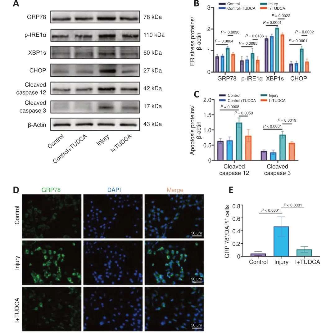
Figure 1 |Neuronal ER stress is significantly induced after TBI in vitro, and inhibition of activated neuronal ER stress protectes against trauma-induced injury.(A–C) Western blot analysis revealed that GRP78, p-IRE1α, XBP1s, CHOP, cleaved caspase 12, and cleaved caspase 3 expression levels were upregulated in cultured HT22 neurons after scratch injury, and that TUDCA treatment attenuated this effect.(D, E)Immunofluorescence staining revealed an increase in the number of GRP78 (green, Alexa Fluor 488)-positive HT22 neurons after scratch injury, which decreased after treatment with TUDCA.Scale bars: 50 μm (original magnification 400×).Data are expressed as mean ± SD (n = 3–5 for western blot, n = 5 for immunofluorescence staining) and were analyzed by one-way analysis of variance followed by the least significant difference post hoc analysis.CHOP: C/EBP homologous protein; ER: endoplasmic reticulum; GRP78:glucose-regulated protein 78; p-IRE1α: phospho-inositol requiring enzyme 1α; TBI:traumatic brain injury; TUDCA: tauroursodeoxycholic acid; XBP1s: spliced X-box binding protein 1.
Microglia-derived exosomes containing miR-124-3p mitigate neuronal damage by inhibiting ER stress
To study the effect of microglia and their microglial exosomal miR-124-3p on neurons following rmTBIin vitro, we cultured BV2 microglial cells and HT22 cells and verified the identity of pure BV2 microglia by Iba1 immunofluorescence staining (Additional Figure 3D).Next, a coculture model was established,and the microglia were transfected with an miR-124-3p mimic.Real-time polymerase chain reaction analysis showed that transfection with the miR-124-3p mimic significantly increased miR-124-3p expression levels in comparison to the Control group (Figure 3A).Then, we used Transwell systems to cultivate two different types of cells in order to mimic interactions between two types of brain cells.Microglia overexpressing miR-124-3p or microglia treated with GW4869,which upregulates miR-124-3p, were co-cultured with damaged neurons (Figure 3B).BCA analysis showed that treatment with GW4869 significantly reduced exosome secretion compared with the Control group (Figure 3C).In addition,treating microglia with miR-124-3p transfection significantly reduced the increase in apoptosis-related protein (CHOP, cleaved caspase 12, and cleaved caspase 3) expression induced by scratch injury, and coculture significantly reduced the increase in GRP78, p-IRE1α, and XBP1s expression in the Injury group.These results suggest that trauma-induced ER stress can be inhibited by coculturing neurons with microglia overexpressing miR-124-3p, which has a protective effect on the damaged neurons.In contrast, GW4869 treatment increased the levels of GRP78, p-IRE1α, XBP1s, CHOP, cleaved caspase 12, and cleaved caspase 3 expression, suggesting that suppressing exosomes reverses ER stress and attenuates the protective influence of coculturing neurons with microglia overexpressing miR-124-3p (Figure 3D–F).Taken together, these findings indicate that miR-124-3p overexpression by microglia inhibited ER stress and reduced cell apoptosis in scratch-injured neurons, possibly by exosome-mediated transfer of miR-124-3p to neurons.
Exosomes derived from microglia overexpressing miR-124-3p play a neuroprotective role by increasing miR-124-3p expression in injured neurons
Exosomes derived from microglia overexpressing miR-124-3p or normal microglia were added to HT22 cells to validate the impact of exosomal miR-124-3p on scratch-injured neurons.Additionally, miR-124-3p levels in HT22 cells and microglial exosomes were detected by real-time polymerase chain reaction (Figure 4A and B).The statistical analysis showed that the miR-124-3p expression level rose after scratch damage and was even higher after exposure to exosomes derived from microglia overexpressing miR-124-3p.However, miR-124-3p expression was not altered significantly in damaged neurons exposed to normal exosomes.Next, exosomes derived from microglia overexpressing miR-124-3p were labeled with PKH26 and introduced to the HT22 neuron culture medium; immunofluorescence staining showed that the HT22 cells phagocytosed the exosomes (Figure 4C).To determine the best concentration of microglial exosomes for treating injured neurons, we tested different concentrations (50–200 μg/mL) and performed western blotting to detect GRP78 concentrations.At a concentration of 200 μg/mL,GRP78 expression in injured neurons decreased significantly, so this concentration was used for the subsequent experiments (Figure 4D and E).Next, we examined the expression of proteins associated with neuronal cell apoptosis after damage and exosome treatment and found that treatment with exosomes derived from miR-124-3p-overexpressing microglia inhibited CHOP and cleaved caspase 12 expression in neurons.Subsequently, we found that the expression levels of the ER stress marker proteins GRP78, p-IRE1α,and XBP1s were also reduced in neurons that exposed to exosomes derived from miR-124-3p-overexpressing microglia compared with those in the Injury group.However, compared with the Injury + EXO-124 group, the expression levels of GRP78, p-IRE1α, XBP1s, CHOP, and cleaved caspase 12 were higher in the Injury + EXO group.This further suggests that the inhibition of ER stress by microglial exosomes is achieved through the exosome-mediated transport of miR-124-3p into neurons (Figure 4F–H).These results suggest that microgliaderived exosomal miR-124-3p suppressed neuronal ER stress in damaged neurons, thereby mitigating neuronal damage.
miR-124-3p targets IRE1α to inhibit ER stress activation in injured neurons
Potential miR-124-3p target genes were explored to investigate the mechanism underlying the impact of miR-124-3p on trauma-induced neuronal ER stress.We found that the IRE1α mRNA 3′UTR, which is associated with the ER stress response, contains miR-124-3p binding sites, suggesting that IRE1α may be a miR-124-3p target.Therefore, we used a luciferase reporter to determine whether miR-124-3p specifically targets the 3′UTR of IRE1α mRNA.The IRE1α 3′UTR mRNA fragment with the potential miR-124-3p binding site or a mutant version with the binding site deleted was specifically cloned into the luciferase reporter vector pGL3 to yield pGL3-IRE1α-3′UTR WT or pGL3-IRE1α-3′UTR MUT, respectively.The plasmids were then cotransfected into damaged neurons with scrambled oligonucleotides or miR-124-3p mimic.The relative luciferase activity of neurons cotransfected with pGL3-IRE1α-3′UTR WT and miR-124-3p was significantly reduced in comparison to the Control group, suggesting that miR-124-3p binds to the 3′UTR region of IRE1α in neurons, thereby suppressing IRE1α expression (Figure 5A and B).
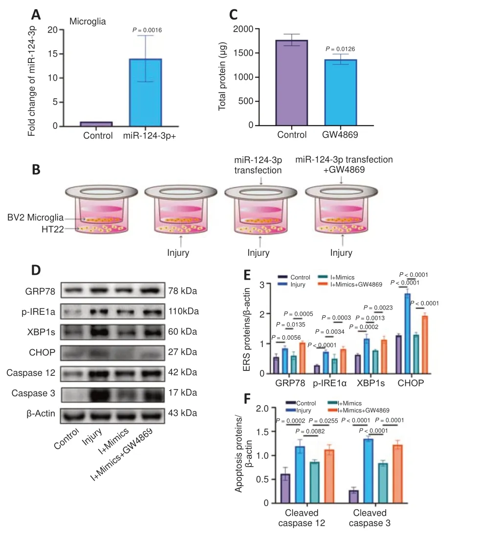
Figure 3 |Exosomes derived from miR-124-3p-overexpressing microglia exert a protective effect on injured neurons by inhibiting neuronal ER stress.(A) Real-time polymerase chain reaction analysis revealed that miR-124-3p expression was significantly upregulated in the cultured microglia after transfection with an miR-124-3p mimic.(B) The neuron and microglia coculture system.Neurons and microglia were cultured together in a Transwell system.(C) Compared with the Control group, the total protein levels were decreased in the GW4869 group.(D–F) Western blot analysis of ER stress-related proteins (GRP78, p-IRE1α, XBP1s, CHOP) revealed that ER stress was induced in cultured neurons after scratch injury; cleaved caspase 12 and cleaved caspase 3 expression levels were upregulated after scratch injury.Neuronal ER stress and apoptosis were inhibited by exosomes derived from the cocultured miR-124-3poverexpressing microglia.This inhibitory effect was alleviated by GW4869 treatment.Data are expressed as mean ± SD (n = 3 for real-time polymerase chain reaction, n = 4–5 for western blot) and were analyzed by Student’s t-test (A, C) or one-way analysis of variance followed by the least significant difference post hoc analysis (E, F).CHOP: C/EBP homologous protein; ER: endoplasmic reticulum; GRP78: glucose-regulated protein 78;p-IRE1α: phospho-inositol requiring enzyme 1α; XBP1s: spliced X-box binding protein 1.
To more clearly validate the impact of miR-124-3p on IRE1α expression in damaged neurons, injured neurons were transfected with an miR-124-3p mimic, a negative control, or an miR-124-3p inhibitor.After 1 day of transfection, real-time polymerase chain reaction analysis showed that the level of miR-124-3p expression was significantly altered in the various transfected groups (Figure 5C and D).In HT22 cells transfected with the miR-124-3p mimic, miR-124-3p expression was clearly greater than in the NC group, but it was lower in HT22 cells transfected with the miR-124-3p inhibitor.In addition, IRE1α expression was significantly lowered by miR-124-3p overexpression, but significantly increased by miR-124-3p inhibition(Figure 5E and F).Next, to evaluate the direct impact of miR-124-3p on ER stress in damaged neurons, we investigated the expression of ER stressrelated proteins (p-IRE1α, GRP78, and XBP1s), as well as the expression of CHOP, cleaved caspase 12, and cleaved caspase 3 in each experimental group.Our findings demonstrated that transfection with an miR-124-3p mimic suppressed ER stress and cell death in damaged neurons, while transfection with an miR-124-3p inhibitor somewhat enhanced both ER stress and cell death (Figure 5G–I).TUNEL staining and EdU detection revealed that the apoptosis rate of the damaged cells increased and their proliferation rate diminished compared with the Control group.However, when the damaged cells overexpressed miR-124-3p, the apoptotic rate was reduced, while the rate of cell proliferation was significantly elevated (Figure 6A–D).These findings are consistent with our earlier results.In conclusion, miR-124-3p may help protect neuronal function by directly targeting IRE1α and inhibiting activation of ER stress in injured neurons.
miR-124-3p overexpression mainly inhibits IRE1α-mediated ER stress in damaged neurons
To further confirm the regulatory effect of miR-124-3p on IRE1α and the role of IRE1α in mediating neuronal injury and ER stress activation,we carried out functional salvage experiments by overexpressing IRE1α in damaged neurons.Before inducing scratch damage, neurons were cotransfected with LV-IRE1α and LV-miR-124-3p or LV-si-IRE1α and an LV-sinegative control.Real-time polymerase chain reaction analysis showed that IRE1α mRNA expression was significantly reduced in the LV-si-IRE1α group but significantly elevated in the LV-IRE1α group compared with the Control group (Figure 7A and B).In addition, western blotting showed that miR-124-3p overexpression suppressed IRE1α expression, while cotransfection with LV-IRE1α restored IRE1α expression.Furthermore, LV-si-IRE1α inhibited IRE1α expression in neurons (Figure 7C and D).Additionally, we found that IRE1α overexpression significantly reduced the inhibitory effect of miR-124-3p overexpression on ER stress activation, and that siRNAmediated suppression of IRE1α mimicked the effect of miR-124-3p on neurons exposed to scratch damage (Figure 7E–H).These results suggest that IRE1α overexpression counteracts the suppression of ER stress by miR-124-3p, and that IRE1α, an miR-124-3p target, plays a significant role in trauma-induced ER stress and may protect damaged neurons by inhibiting its expresson.
I njection of exosomes derived from microglia overexpressing miR-124-3p into rmTBI mice can inhibit ER stress and reduce apoptosis
To elucidate the suppressive effect of exosomes derived from microglia overexpressing miR-124-3p on ER stress after rmTBI, we administered exosomes containing miR-124-3p to the injured brains of rmTBI mice via intranasal delivery.At 14 days post-rmTBI we quantified GRP78, p-IRE1α, and XBP1s expression by western blot and discovered that the expression of all three proteins was decreased by administration of the exosomes.Similarly,compared with rmTBI group, CHOP and cleaved caspase 12 expression levels were significantly decreased in the rmTBI + EXO-124 group (Figure 8A–C).In addition, double immunofluorescence staining for GRP78 and NeuN clearly demonstrated that there were significantly more GRP78-positive cells in the hippocampus of rmTBI mice than in Sham mice, and that administration of EXO-124 mitigated this effect (Figure 8D and E).This result indicates that microglia-derived exosomes rich in miR-124-3p reduced rmTBI-induced ER stress activation and preserved cellular function.Therefore, EXO-124 treatment alleviated neuronal injury after rmTBI.
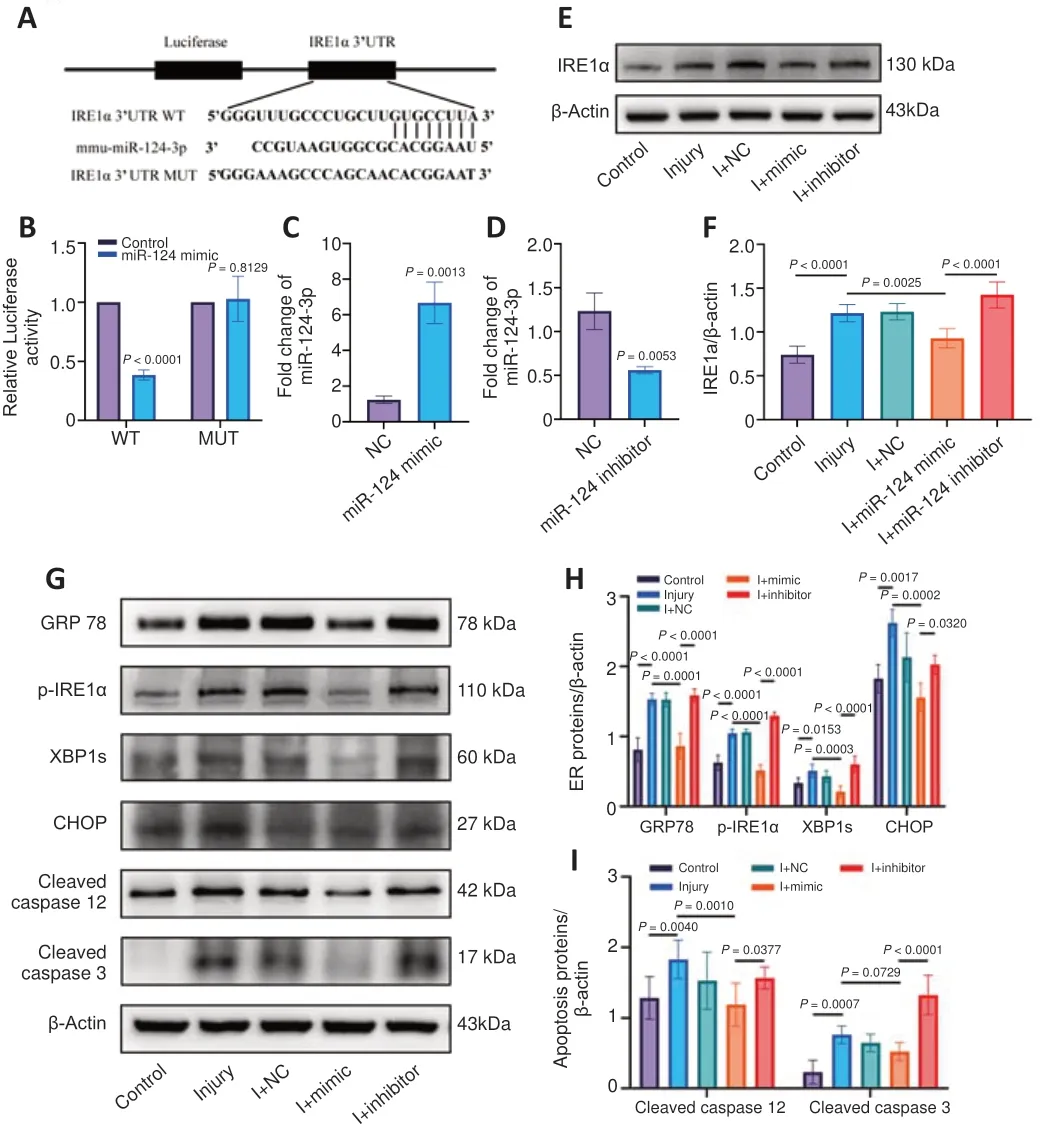
Figure 5 | miR-124-3p targets IRE1α and suppresses activation of ER stress in injured neurons.(A) The WT (IRE1α WT 3′UTR) and MUT (IRE1α MUT 3′UTR) luciferase reporter constructs contained intact and mutated miR-124-3p binding site seed sequences, respectively.(B) The relative luciferase activity of the WT and MUT reporter constructs, which were cotransfected with either the miR-124-3p mimic or a scrambled oligonucleotide.Data are presented as the ratio of luciferase activity in HT22 neurons transfected with the scrambled oligonucleotide to that in neurons transfected with the miR-124-3p mimic.Our findings indicate that miR-124-3p inhibited the luciferase activity of the WT, but not the MUT, reporter construct.Furthermore, miR-124-3p directly targeted IRE1α and downregulated its expression by binding to the 3′UTR.(C, D) Compared with the negative control group, miR-124-3p expression was significantly increased in the mimic group, and miR-124-3p expression was clearly decreased in the inhibitor group.(E, F) Western blot analysis showed that IRE1α expression was upregulated in the cultured HT22 neurons after scratch injury, inhibited in the I + miR-124 mimic group, and increased in the I +miR-124 inhibitor group.(G–I) Western blot analysis of ER stress-related proteins (GRP78,p-IRE1α, XBP1s, CHOP) and apoptosis-related protein revealed that trauma-induced neuronal ER stress and cell apoptosis were inhibited in the I + miR-124 mimic group and promoted in the I + miR-124 inhibitor group.Data are expressed as mean ± SD (n = 3 for relative luciferase activity, n = 4–6 for western blot, n = 3 for real-time polymerase chain reaction) and were analyzed by Student’s t-test (B–D) or one-way analysis of variance followed by the least significant difference post hoc analysis (F, H, I).3′UTR:3′-Untranslated region; CHOP: C/EBP homologous protein; ER: endoplasmic reticulum;GRP78: glucose-regulated protein 78; MUT: mutant-type; p-IRE1α: phospho-inositol requiring enzyme 1α; WT: wild-type; XBP1s: spliced X-box binding protein 1.
Discussion
Recently, a growing number of studies have reported that neuronal apoptosis is closely connected to neurological impairment and mortality after TBI.Here,we showed that continuous ER stress in neurons potentially triggers nerve cell apoptosis and found that IRE1α activation in neurons is a key mechanism mediating abnormal ER stress after TBI.
There is increasing evidence that ER stress-related apoptosis contributes to neuronal death in a variety of neurological disorders, including brain injury.Prior studies have shown that moderate to severe brain trauma can lead to sustained ER stress activation (increased levels of the ER stress marker proteins eIF2α, IRE, GRP78, ATF4, CHOP, etc.) for up to 21 days (Truettner et al., 2007; Begum et al., 2014).Here, we provide convincing evidence that neuronal ER stress is activated after scratch injury.Meanwhile, inhibiting ER stress (by treating cells with TUDCA) can prevent abnormal activation of ER stress after brain injury and subsequent nerve cell apoptosis.One study showed that, in the cerebral cortex and hippocampus, caspase 12 mRNA transcription increases significantly within 3 days after injury and is maintained for at least 5 days following mild to severe craniocerebral injury in rats (Larner et al., 2004).CHOP expression is characteristic of cell apoptosis induced by ER stress (Oyadomari and Mori, 2004).In a CCI mouse model, the CHOP immunoreactive signal in the ipsilateral cerebral hemisphere increased at 6 hours following brain injury and persisted at high levels until the 14thday post-injury (Krajewska et al., 2011).To date, most studies on ER stress and UPR induced by brain injury have focused on the relationship between ER stress and acute neuronal death.In this study, we found that ER stress marker expression and neuronal apoptosis were still increased in mouse brains 14 days after rmTBI, indicating that ER stress is related to persistent neuronal injury induced by rmTBI.
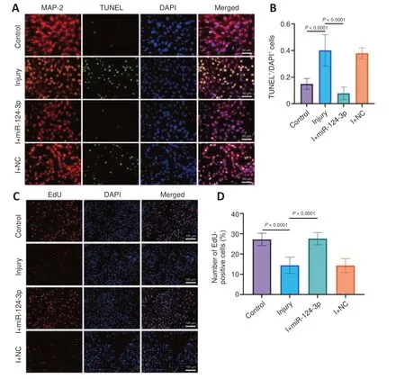
Figure 6 |miR-124-3p reduces apoptosis and promotes proliferation of injured neurons.(A, B) TUNEL (green) and MAP-2 (red, Alexa Fluor 594) double immunofluorescence staining was used to detect apoptosis.Compared with the Control group, the percentage of TUNEL+/DAPI+ cells in the Injury group was markedly increased, and this effect was reversed by treatment with miR-124-3p.Scale bars: 50 μm (original magnification 400×).(C, D) An EdU assay was performed to detect cell proliferation in each group.Compared with the Control group, the percentage of EdU+/DAPI+ cells in the Injury group was markedly decreased, and this effect was reversed by treatment with miR-124-3p.Scale bars: 100 μm (original magnification 200×).Data are expressed as mean ± SD (n = 5–10 for immunofluorescence staining) and were analyzed by one-way analysis of variance followed by the least significant difference post hoc analysis.DAPI: 4′,6-Diamidino-2-phenylindole; EdU: 5-ethynyl-2′-deoxyuridine; MAP-2: microtubule-associated protein 2;TUNEL: terminal deoxynucleotidyl transferase-mediated biotin-deoxyuridine triphosphate nick-end labelling.
The most highly expressed miRNA in brain neuronal cells is miR-124-3p,and research has shown that it may play a role in many disorders of the nervous system (Ge et al., 2020; Jiang et al., 2020; Lu et al., 2020).As the main transmitter of miRNA, exosomes play a crucial role in mediating the interaction between microglia and neurons (Sajja et al., 2016; Gorse and Lafrenaye, 2018; Kempuraj et al., 2018).After TBI, microglial miR-124-3p may be diverted to neurons, where it promotes axon growth, suggesting that microglial exosomes may be taken up by neurons and have a therapeutic effect on TBI (Huang et al., 2018).Thus, in this study we aimed to explore the specific effect and mechanism of EXO-miR-124-3p on neuronal ER stress.Usingin vitroTranswell coculture andin vivoadministration of exogenous exosomes containing high levels of miR-124-3p, we found that exosomederived miR-124-3p can inhibit ER stress in injured neurons.In addition, we found that using GW4869 to suppress exosome release by microglia in a Transwell coculture system inhibited the miR-124-3p-mediated suppression of ER stress, which exacerbated the damage to HT22 cells.This confirms that exosomes are crucial to the interaction between microglia and neurons.
IRE1α, an ER stress receptor, is a major participant in the UPR and a predicted target gene of miR-124-3p.Studies have shown that when dissociated from GRP78, activated IRE1α can induce the production of transcriptionally active XBP1s, which then enters the nucleus, where it induces the transcription of genes encoding proteins associated with protein folding and ER degradation,such as CHOP (Lee et al., 2003; Madhusudhan et al., 2015).The IRE1α endonuclease domain inhibitor MKC3946 has been shown to have a protective impact on a mouse model of AD by inhibiting the production of XBP1s-related ROS, as well as apoptosis (Zhang et al., 2022).In addition,continuous activation of IRE1α in Huntington’s disease can lead to neuronal loss (Hyrskyluoto et al., 2014).In comparison, XBP1s deficiency reduces the loss of motor neurons and the accumulation of mutant SOD1, thus delaying disease and death (Hetz et al., 2009).In the current study, we confirmed that IRE1α is an miR-124-3p target gene via luciferase assay.Using miR-124-3p transfection and siRNA-mediated IRE1α knockdown, we confirmed that miR-124-3p targeting of IRE1α decreases ER stress in injured neurons.Mechanistically, we found that neuronal injury was mediated by miR-124-3p and the downstream p-IRE1α-XBP1s-CHOP signaling pathway.Thus,suppressing abnormal ER stress by targeting IRE1α may be very important approach for reducing neuronal apoptosis after TBI.Interestingly, however,many studies have found that XBP1s has neuroprotective effects.For example, local injection of viruses expressing XBP1s into the substantia nigra can prevent the degeneration caused by neurotoxins produced in Parkinson’s disease (Begum et al., 2014).Similarly, XBP1s expression reduces mutant Huntington protein accumulation in the striatum of HD mice.The apparent inconsistency between these studies and our results may be because different injury models and different experimental specimens were used.
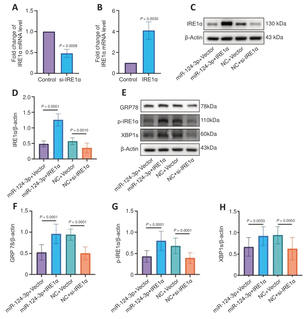
Figure 7 | miR-124-3p overexpression inhibits IRE1α-mediated ER stress in injured neurons.(A, B) Compared with the Control group, IRE1α mRNA expression was significantly increased in the IRE1α group and clearly decreased in the si-IRE1α group.(C, D)Western blot analysis showed that IRE1α overexpression reversed the miR-124-3pinduced inhibition of IRE1α, and that siRNA-mediated IRE1α silencing suppressed IRE1α expression in scratch-injured neurons.(E–H) IRE1α overexpression blocked miR-124-3pinduced inhibition of neuronal ER stress, and siRNA-mediated IRE1α silencing mimicked the effects of miR-124-3p on scratch-injured neurons.Data are expressed as mean ± SD(n = 3 for real-time polymerase chain reaction, n = 11–15 for western blot) and were analyzed by Student’s t-test (A, B) or one-way analysis of variance followed by the least significant difference post hoc analysis (D, F, G, H).ER: Endoplasmic reticulum; GRP78:glucose-regulated protein 78; IRE1α: inositol requiring enzyme 1α; p-IRE1α: phosphoinositol requiring enzyme 1α; siRNA: small interfering RNA; XBP1s: spliced X-box binding protein 1.
There were some limitations to our study.We primarily focused on miR-124-3p overexpression in microglial exosomes in anin vitroTBI model (scratch injury model).The scratch injury model is anin vitromodel of mechanical transection that simulates not only the direct mechanical injury caused by TBI, but also the effects on surrounding cells.It is often used to simulate various pathological changes in the brain after TBIin vitro.The scratch injury model is as simple as it is effective: no special equipment is required, and it is easy to carry out.However, the model still has some shortcomings, in that the mechanical damage parameters cannot be strictly standardized, and the degree of damage cannot be accurately quantified.A further limitation of our study is that we did not perform behavioral analyses of rmTBI mice treated with miR-124-3p-containing exosomes; this will be explored in future studies.In addition, in the future we intended to investigate the impact of EXO-miR-124-3p on basic ER function and cognitive function in mice without rmTBI.Furthermore, a number of studies have recently reported the use of MSC-derived exosomes in the treatment of brain trauma and demonstrated that these exosomes can successfully enhance neurological prognosis by encouraging endogenous angiogenesis and neurogenesis while lowering neuroinflammation.Therefore, future studies should compare the effectiveness of MSC exosomes and microglia exosomes in up-regulating miR-124-3p and ameliorating the effects of rmTBI.
Taken together, our findings show that microglial exosomes containing high levels of miR-124-3p play a crucial role in regulating many pathogenic processes after rmTBI, such as neuronal ER stress.Furthermore, our study demonstrates that miR-124-3p in microglial exosomes can reduce neuronal apoptosis after rmTBI and play a neuroprotective role by being transferred into neurons and targeting the IRE1α-XBP1s-CHOP signaling pathway.The findings from this study may help develop novel treatment modalities based on miRNA-containing microglial exosomes for modulating ER stress following rmTBI.
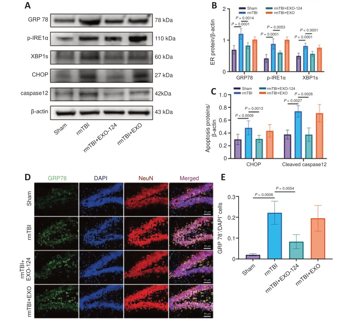
Figure 8 | Intranasal delivery of miR-124-3p-enriched microglial exosomes inhibites ER stress and reduced apoptosis in C57BL/6 mice after rmTBI.(A–C) Western blot analysis showed that GRP78, p-IRE1α, XBP1s, CHOP, and cleaved caspase 12 expression levels in the brain were upregulated after rmTBI, and that miR-124-3p treatment attenuated this effect.(D, E) Representative images of double immunofluorescence staining for GRP78 (green, Alexa Fluor 488) and NeuN (red, Alexa Fluor 594).Compared with the Sham group, GRP78 expression was higher in the rmTBI group.Compared with the rmTBI group, GRP78 expression in the rmTBI+EXO-124 group was lower.Scale bars: 50 μm (original magnification 400×).Data are expressed as mean ±SD (n = 3–7 for western blot, n = 3 for immunofluorescence staining) and were analyzed by one-way analysis of variance followed by the least significant difference post hoc analysis.CHOP: C/EBP homologous protein; DAPI 4′,6-diamidino-2-phenylindole; EXO:exosome; ER: endoplasmic reticulum; GRP78: glucose-regulated protein 78; rmTBI:repetitive mild traumatic brain injury; NeuN: neuron-specific nuclear protein; p-IRE1α:phospho-inositol requiring enzyme 1α; XBP1s: spliced X-box binding protein 1.
Acknowledgments:The authors appreciate Chunsheng Kang, Xiao Liu, Weiyun Cui, and Lei Zhou (Tianjin Neurological Institute) for their technical support.
Author contributions:Study design: PL, DL; methodology: YW, LZ;experimental implementation: YW, LZ, DL, ZY, ZH, XG, ML, JZ, SZ, YZ, XX, HG;technical support: FC; data analysis and interpretion, and figures and tables preparation: YW, LZ, ZY; manuscript draft: YW, LZ, DL; supervion: PL, QL.All authors read and approved the final version of the manuscript.
Conflicts of interest:The authors declare no competing interests.
Data availability statement:All raw miRNA microarray data have been uploaded to GEO (accession number GSE133997).In addition, the datasets used in the project are available from the corresponding author.
Open access statement:This is an open access journal, and articles are distributed under the terms of the Creative Commons AttributionNonCommercial-ShareAlike 4.0 License, which allows others to remix, tweak, and build upon the work non-commercially, as long as appropriate credit is given and the new creations are licensed under the identical terms.
Open peer reviewer:Yi Pang, University of Mississippi, USA.
Additional files:
Additional Figure 1: Flowchart of in vitro experiments for TUDCA treatment,Transwell co-culture and EXO-124 treatment.
Additional Figure 2: Flowchart of in vitro experiments for miR-124-3p transfection and functional rescue experiment.
Additional Figure 3: An in vitro TBI model and identification of cells purity.
Additional Figure 4: miRNA microarray analysis of microglial exosomes isolated from mouse brains at different time points after rmTBI.
Additional file 1: Open peer review report 1.
- 中國神經(jīng)再生研究(英文版)的其它文章
- Does MgSO4 protect the preterm brain? Dissecting its role in the pathophysiology of hypoxic ischemic encephalopathy
- On implications of somatostatin in diabetic retinopathy
- Rebuilding insight into the pathophysiology of Alzheimer’s disease through new blood-brain barrier models
- The functions of exosomes targeting astrocytes and astrocyte-derived exosomes targeting other cell types
- Post-transcriptional mechanisms controlling neurogenesis and direct neuronal reprogramming
- Hypothalamic circuits and aging: keeping the circadian clock updated

