Patellar dislocation following distal femoral replacement after extraarticular knee resection for bone sarcoma:A case report
lNTRODUCTlON
Bone tumors often arise around the knee, especially in the distal femur[1]. Bone sarcomas, such as osteosarcoma, also frequently originate in the distal femur[2]. Standard therapies for bone sarcoma in the distal femur include wide-margin resection and knee reconstruction with tumor endoprosthesis,which is thought to be an effective surgery to control local tumors and restore knee function. However,in cases of tumor invasion extending into the knee joint, extra-articular knee resection is required, which leads to wider resection and functional disorders. Moreover, the incidence of complications, such as aseptic loosening, prosthesis infection, and implant failure, is higher with extra-articular knee resection than with intra-articular knee resection. Initially, a fixed-hinge knee prosthesis was used in these cases;however, aseptic loosening was sometimes noted. Since the introduction of the kinematic rotating hinge prosthesis, the prosthesis survival rate improved considerably. In 2010, Kinkel[3] reported that the five-year implant survival rate was 57%, and the local recurrence rate of primary malignant tumors around the knee was 3%. Eight reports that have ascertained the outcome and complications after extraarticular knee resection and knee reconstruction appear in Table 1[3-10].In summary, following extraarticular knee resection, the endoprosthesis complication rate is higher than that following intraarticular knee resection.

Patellar dislocations are rare complications in these patients. We could find three reports of patellar dislocations after replacement of a tumor endoprosthesis[11-13], but there are no such reports that particularly focus on extra-articular knee resection. Herein, we report a case of patellar dislocation after extra-articular knee resection of a malignant bone tumor in the femur and knee reconstruction with a tumor endoprosthesis without its malalignment. The patient underwent proximal realignment and had an uneventful course.
Night now came on, and there arose a terribly high wind,17 which made them dreadfully afraid. They fancied they heard on every side of them the howling of wolves coming to eat them up. They scarce dared to speak or turn their heads. After this, it rained very hard, which wetted them to the skin; their feet slipped at every step they took, and they fell into the mire18, whence they got up in a very dirty pickle21; their hands were quite benumbed.


CASE PRESENTATlON
Chief complaints
A 36-year-old man with no significant past medical history was admitted to our institution with continuous pain in his left knee for 4 mo.
History of present illness
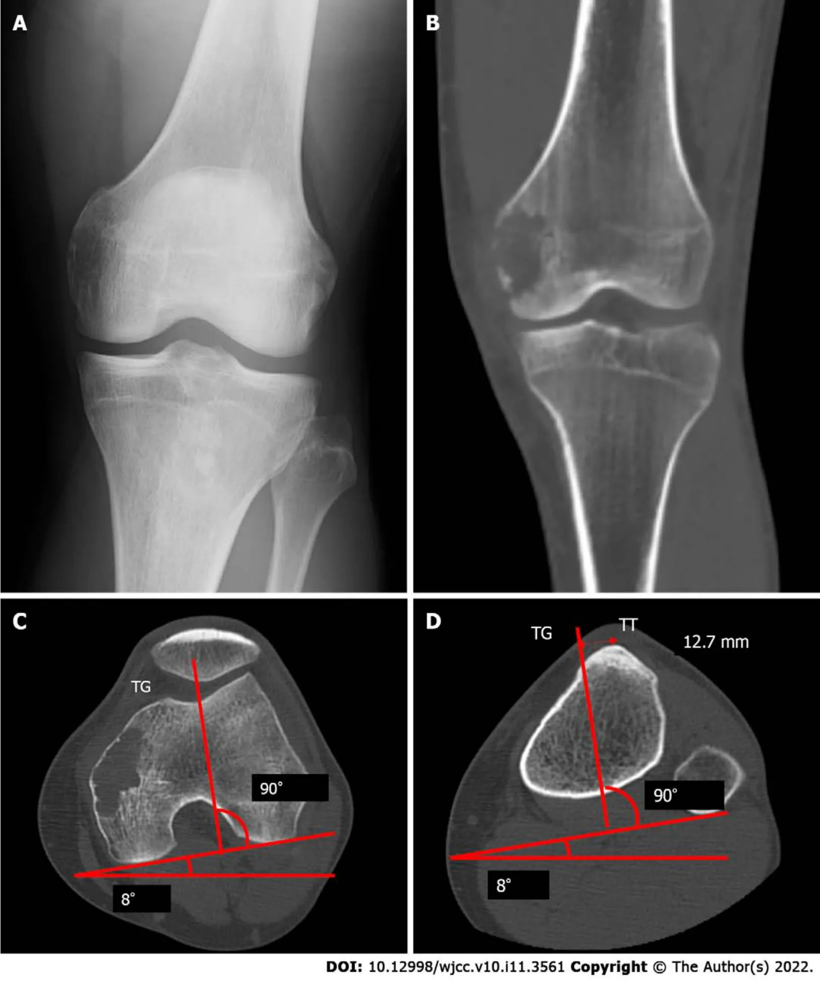
On physical examination, tenderness was observed over the left femoral medial epicondyle without any swelling, redness, or warmth around the joint. Radiological examination and computed tomography(CT) revealed an osteolytic lesion over the femoral medial epicondyle without a sclerotic rim, periosteal reaction, or calcification (Figure 1). Magnetic resonance imaging (MRI) showed a bone tumor. An open biopsy was performed, and reverse transcription-polymerase chain reaction (RT-PCR) and direct sequencing revealed the/fusion gene in the tumor specimens[14]. No other signs of metastasis were found on whole-body CT and positron emission tomography/CT. Extra-articular knee resection and knee reconstruction with a tumor endoprosthesis were performed.
The ROM of the patient was restricted to be between 0° and 60° without pain, swelling, and local heat.
Several studies have reported that patellar dislocations occur after treatment with a tumor endoprosthesis. We could only find three reports on the cause of patellar dislocation and patellar impingement following distal femur replacement in the English literature[11-13]. Among them, only one report included extra-articular tumor resection[12]. Schwab[11] reported three features of distal femoral replacement. First, patellar tendon devascularization may cause scar formation, which leads to patellar baja. Second, if some parts of the extensor musculature are removed, the patellar offset can be increased.This action increases stress on the patellar, which may lead to patellar complications. Finally, knee mobilization can be delayed or insufficient because of fascia deficiency, marginal necrosis, and chemotherapy. The authors also reported that when the Insall-Salvati ratio was > 0.8 and < 1.2, the ROM was usually > 100°. The mean LT/HI ratio of patellar impingement cases was 0.9 and the mean LT/HI ratio without impingement was 1.4 (= 0.07).
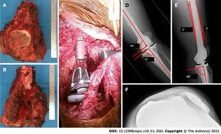
The patient wore a knee brace, which constrained knee extension, from the first postoperative day and began walking with full-body weight-bearing. After walking rehabilitation for six postoperative days, knee instability was noticed. Radiography showed a lateral subluxation of the patellar (Figure 2D and E). However, because the knee subluxation did not progress to dislocation, walking rehabilitation continued. Although a range of motion (ROM) of 30° was allowed during passive motion at 2 wk postoperatively, radiographs revealed no progression of subluxation. However, the axial radiographic view of the patellar (Figure 2F) and CT showed lateral patellar dislocation at 4 wk postoperatively.

History of past illness
The patient had no significant past medical history.
She ran as fast as she could to her little room under the stairs, but because she had stayed too long beyond the half-hour, she could not stop to take off the beautiful dress, but only threw the fur cloak over it, and in her haste she did not make herself quite black with the soot, one finger remaining white
Personal and family history
The patient and his family had no significant past medical history.
Physical examination
The patient was placed in a supine position. A longitudinal incision was made starting distally,medial to the patellar tendon, and progressing proximally towards the lateral aspect. Medially, the distal end of the vastus medialis was resected. Resection was performed at approximately 2 cm from the distal end of the intermuscular septum. Subsequently, the medial head of the gastrocnemius muscle and the semimembranosus muscle attachment was resected. The distal femur was resected at a distance of 12 cm. Patellar osteotomy was performed, preserving the articular capsule of the knee. After dissectingthe patellar tendon, leaving an infrapatellar fat pad over the specimen, the 1-cm proximal tibia was cut above the tibial tuberosity (TT). The resected specimen was removed. The tumor prosthesis was implanted with patellar resurfacing (Figure 2A-C). The vastus medialis and vastus lateralis muscles were attached to the prosthesis. A medial gastrocnemius flap was used for the prosthesis coverage.When subcuticular closure was performed, there was no abnormal patellar position.
Laboratory examinations
But the sly old man, who was terrified for his life that the three rogues might do him some harm, was on his guard, and said to his housekeeper, Nina, take this bladder, which is filled with blood, and hide it under your cloak; then when these thieves come I ll lay all the blame on you, and will pretend to be so angry with you that I will run at you with my knife, and pierce the bladder with it; then you must fall on the ground as if you were dead, and leave the rest to me
Imaging examinations
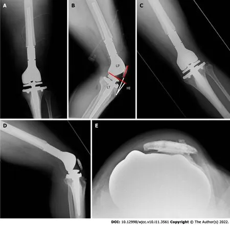
Based on the findings of studies by Kim[15] and Fang[16], the postoperative radiographs showed good results. The angle between the distal portion of the femoral component and the femoral anatomical axis was 96°, and the angle between the proximal portion of the tibial component and the tibial anatomical axis was 91° (Figure 2D). A lateral radiograph also showed good results, with a postoperative femoral sagittal alignment of 3° and a postoperative tibial sagittal alignment of 2°(Figure 2E). The measurement of the patellar position Insall-Salvati ratio was 1.15, and the length of the patellar tendon (LT)/height of the patellar tendon insertion (HI) ratio was 1.27, which implied a good patellar location (Figure 4A and B) according to the report by Schwab[11]. Consequently, we considered that the tumor prosthesis was implanted precisely in terms of the alignment and position.
I alone have kept guard over her, and have gone daily to my own castle to get food and to beat the greedy gold-seekers who forced their way into my dwelling30
After the patient noticed knee instability for six postoperative days, radiography showed a lateral subluxation of the patellar (Figure 2D and E). The axial radiographic view of the patellar (Figure 2F) and CT showed lateral patellar dislocation at 4 wk postoperatively.

FlNAL DlAGNOSlS
The final diagnosis was patellar dislocation following distal femoral replacement after extra-articular knee resection for bone sarcoma.
TREATMENT
The question remains whether extra-articular knee resection could be a cause of patellar dislocation.Compared to intra-articular knee resection, extra-articular knee resection requires a wider margin of resection, including quadriceps muscle resection, which can cause patellar instability. In this case, the reason for patellar dislocation was considered to be an imbalance in the forces controlling patellar tracking. The resection of the distal vastus medialis and the scarring of the distal vastus lateralis could be contributing factors. The resection of the distal vastus medialis reduces the ability to control the patellar medially. It was assumed that scarring of the distal vastus lateralis shortened its length, and the patellar was pulled laterally, although it is unknown how the scar developed.
Laboratory tests revealed:Erythrocytes 5.05 × 10/L (reference range, 4.35 × 10-5.55 × 10/L),hemoglobin 126 g/L (reference range, 137-168 g/L), leukocytes 5.27 × 10/L (reference range, 3.3 × 10-8.6 × 10/L), eosinophils 3.2%, basophils 0.9%, neutrophils 54.6%, lymphocytes 36.6%, monocytes 4.7%,C-reactive protein 0.7 mg/L (reference range, 0-1.4 mg/L), total protein 69.3 g/L (reference range, 66.0-81.0 g/L), aspartate aminotransferase 28.8 U/L (reference range, 13.0-30.0 U/L), alanine aminotransferase 42.2 U/L (reference range, 10.0-42.0 U/L), blood urea nitrogen 23 mg/dL (reference range, 8-20 mg/dL), creatinine 1.12 mg/dL (reference range, 0.65-1.07 mg/dL), creatine phosphokinase 299 U/L(reference range, 59-248 U/L).
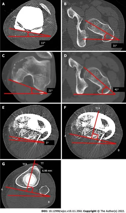
We also measured the rotation of the tibial and femoral components using CT. Before the secondary surgery, the post-primary operative rotational alignment of the femoral component was defined as the angle subtended by the line tangent to the condyles and the axis of the femoral neck. The angle was 20°,which was equal to that of the native alignment (20°) (Figure 3A-D). Moreover, the external axial rotation of the tibial component in relation to the posterior margins of the tibial plateau and the tibial bearing was 5°, which was a good result (Figure 3E).


The secondary surgery consisted of lateral release and proximal realignment and was performed 1 mo postoperatively. Under anesthesia, it was impossible to treat the lateral patellar dislocation of the left knee with a closed reduction and passive ROM of 0°-30°. A skin incision was made which followed the previous skin incision. There was abundant thick scar tissue on the lateral side of the patellar and the vastus lateralis muscle. A lateral release was performed from the tibial component to 20 cm proximally,and scar adhesion between the vastus lateralis muscle and the femur was also released. Although patellar dislocation could be reduced, the ROM was still 30°. We concluded that scar formation of the quadriceps muscle was the reason for the flexion contracture.
Who shall describe their surprise when they at last turned round and beheld99 standing before them a beautiful lady exquisitely100 dressed!With a smile she held out her hand to the Caliph, and asked: Do you not recognise your screech owl? It was she! The Caliph was so enchanted by her grace and beauty, that he declared being turned into a stork had been the best piece of luck which had ever befallen him
Here, indeed, it would be better to be a man than such a poor dumb fish as I am now, said he to himself; if I could only remember the words that the troll says when he changes my shape, then perhaps I could help myself to become a man again
The pie crust technique was applied to the quadriceps muscle along with a longitudinal split of the scar of the vastus lateralis muscle. The removal of scar tissue in the vastus intermedius muscle increased the ROM to 60°. The distal end of the vastus medialis had already been resected and was loosely and indirectly attached to the patellarthe scar tissue. After the vastus medialis obliquus and scar tissues were separated from the patellar, they were advanced over the tendon of the quadriceps muscle and sutured, using the baseball suture technique with polyblend polyethylene suture. Polybutylate-coated braided polyester suture material was also used to reinforce the sutures. We confirmed that 45° flexion of the knee did not tear the sutures or cause patellar dislocation and subsequently closed the surgical wound.
From the first postoperative day, the patient began non-weight-bearing exercises. The ROM exercises were limited to up to 30°. After confirming wound healing at 2 wk postoperatively, full-weight bearing with bilateral crutches and limited ROM exercise up to 60° was allowed. Unlimited ROM exercise was started 3 wk postoperatively. The patient could walk with a lateral crutch and the ROM was 0°-90° at discharge and 4 wk postoperatively.
When Sonali sang on the steps of the state capitol, her voice was unbelievably strong. It was as though she wanted to fill the whole universe with this impassioned prayer so it would reach her daddy. As she sang, I felt it also become a pure prayer to everyone gathered-a prayer that painted a vision. I was delighted when she asked me if she could sing again, this time for Alan s memorial service at Grace Cathedral in San Francisco.
OUTCOME AND FOLLOW-UP
At the time of the outpatient consultation, the patient was able to walk stably without crutches, and the ROM was 0°-100°. The patient could perform his daily activities at 9 mo postoperatively. Radiography revealed no patellar re-dislocation (Figure 4C-E).
DlSCUSSlON
The most common site where bone tumors arise is the distal femur[1,2]. The current standard therapy for malignant bone tumors is wide margin resection and knee reconstruction using a tumor endoprosthesis. Extra-articular knee resection is required if the tumor invades the knee joint. As shown in Table 1, extra-articular knee resection results in various complications. However, extended survival of the salvaged limb can be achieved by taking care of each complication.
At length he made out some sort of track, and though at the beginning it was so rough and slippery that he fell down more than once, it presently became easier, and led him into an avenue of trees which ended in a splendid castle
Akiyama[12] also reported patellar dislocation following distal femoral replacement after extraarticular tumor resection. The authors enumerated the following reasons for patellar dislocation after reconstruction:unconscious femoral rotation, usage of semi-rotating hinge endoprosthesis, insufficient balancing of soft tissues at the time of closure, and scarring of the tightened lateral soft tissues. They also mentioned that it was very difficult to predict good postoperative patellar tracking, even if intraoperative patellar tracking was normal. In their case, the modified Merkow’s realignment procedure of the patellar was implemented with medial plication and lateral release, and no patellar dislocation occurred during the postoperative course.
We used a rotating hinge endoprosthesis, as recommended by Akiyama[12]. In addition, the alignment was favorable according to the abovementioned points and the criteria suggested by Kim[15]. The authors suggested placing the knee components in the position with the overall anatomical knee alignment at an angle of 3.0°-7.5° valgus, femoral component alignment at 2°-8° valgus, femoral sagittal alignment at 0°-3°, tibial coronal alignment at 90°, and tibial sagittal alignment at 0°-7°. In our case, each alignment was 7° valgus, 6° valgus, 3°, 91°, and 2°, respectively (Figure 2D and E).
Usually, to measure the femoral rotational alignment during total knee arthroplasty (TKA), the posterior condylar angle formed by the line tangent to the posterior condyles and the trans-epicondylar axis is used[15]. It was impossible to apply this procedure to our patient because of a distal femur replacement. Therefore, we measured the preoperative TT-TG (12.7 mm), which was within the ideal range (10-15 mm) (Figure 1C and D). We confirmed that the preoperative angle, subtended by the line tangent to the condyles and the axis of the femoral neck, was equal to that of the postoperative angle(Figure 3A-D).
With regard to the tibial rotational alignment, we measured 5° on the axial rotation of the tibial component in relation to the posterior margins of the tibial plateau and the tibial bearing (Figure 3E).The external rotation was within 2°-5°, which could be considered a precise alignment[15]. Furthermore,a more accurate measurement procedure for tibial rotational alignment, as described by Saffi[18],was also applied. The two-dimensional axial CT of the center of the tibial tray to the tip of the tibial tubercle was 6.98 mm. It was outside the cut-off values of < 6 mm and > 10 mm, and we confirmed that the tibial rotational alignment was preferable (Figure 3G). The causes of patellar dislocation reported in earlier cases were not present in our patient, and the implant alignment was favorable. However, the patient still experienced patellar dislocation.
It was impossible to treat the patellar dislocation with closed reduction; therefore, open reduction was performed. Dejour[17] reported that a large distance between the natural TT and the trochlear groove (TG) induce lateral patellar tracking, resulting in patellar instability. These indices can be measured simply using CT, and they are defined as the distance between the middle of the TT to the bottom of the TG. The ideal TT-TG distance is within a range of 10 to 15 mm[17]; TT-TG, in this case,was 12.7 mm (Figure 1C and D).
There are four main treatment procedures for patellar dislocation after TKA:proximal realignment[19], medial patellofemoral ligament (MPFL) reconstruction[20-22], distal realignment including tibial tuberosity osteotomy (TTO)[23], and lateral retinaculum release[24]. When any procedure is performed,Gennip[21], and Goto[25] suggested that alignment of the implant should be assessed first, to determine whether it is within the normal range of alignment. Gennip[21] stated that it is better to consider distal realignment if the TT-TG value is outside the normal range, or if it is not possible to improve patellar mal-tracking with both lateral release and MPFL reconstruction despite a normal TTTG value. Nevertheless, because the present case underwent extra-articular knee resection and distal femoral replacement, there was no point for anchoring a graft on the femoral side, and the anchor point of the thin patellar could be fractured. Therefore, we chose proximal realignments of the quadriceps as the best procedure. Matar[19] reported that patellar proximal realignment for patellar dislocation after TKA is effective.
As to the Gray Wolf, he spent one day, he spent two days, he spent three days in Tsar Afron s Palace, all the while having the shape of the beautiful Tsarevna, while the Tsar made preparations for a splendid bridal. On the fourth day he asked the Tsar s permission to go for a walk on the open steppe.
I myself helped to rescue cattle and things, nothingalive burnt, except a flight of pigeons, which flew into the fire, andthe yard dog, of which I had not thought; one could hear him howlout of the fire, and this howling I still hear when I wish to sleep;and when I have fallen asleep, the great rough dog comes and placeshimself upon me, and howls, presses, and tortures me
As Piedade[26] stated, TTO should not be performed preferentially because of the risk of skin necrosis and fracture of the tibial tubercle. If extra-articular knee resection is performed, further complications, such as skin necrosis and fracture of the tibial tubercle, could also develop. Hence, in cases similar to the present case, TTO should be avoided. Therefore, lateral release and proximal realignment were performed. No patellar dislocation occurred over the 9-mo postoperative follow-up period (Figure 4C-E).
CONCLUSlON
To the best of our knowledge, this is the first report showing that extra-articular knee resection can cause patellar dislocation after distal femoral replacement without malalignment of the prosthesis.Lateral release and proximal realignment are the most effective procedures. As in the present case,when distal femoral replacement with extra-articular knee resection is planned, and the quadriceps muscle is assumed to be resected asymmetrically, proximal realignment may be taken into consideration during the primary surgery if the forces controlling patellar tracking are imbalanced.
The great dog by the yard gate of a nobleman s mansion sitsproudly on the top of his kennel when the sun shines, and barks atevery one that passes; but if it rains, he creeps into his house,and there he is warm and dry
Kubota Y, Tanaka K and Tsumura H conceived the study; Kubota Y and Tanaka K reviewed the literature and contributed to manuscript drafting; Kubota Y, Tanaka K, Iwasaki T and Kawano M contributed data analysis and interpretation; Hirakawa M, Itonaga I and Tsumura H were responsible for the critical revision of the manuscript for important intellectual content; all authors have read and approved the final version of the manuscript.
Informed written consent was obtained from the patients for the publication of this report and any accompanying images.
The authors declare that they have no conflict of interest.
The authors have read the CARE Checklist (2016), and the manuscript was prepared and revised according to the CARE Checklist (2016).
This article is an open-access article that was selected by an in-house editor and fully peer-reviewed by external reviewers. It is distributed in accordance with the Creative Commons Attribution NonCommercial (CC BYNC 4.0) license, which permits others to distribute, remix, adapt, build upon this work non-commercially, and license their derivative works on different terms, provided the original work is properly cited and the use is noncommercial. See:https://creativecommons.org/Licenses/by-nc/4.0/
Japan
Yuta Kubota 0000-0003-1426-148; Kazuhiro Tanaka 0000-0002-5138-8952; Masashi Hirakawa 0000-0002-0045-6214; Tatsuya Iwasaki 0000-0002-9407-3547; Masanori Kawano 0000-0002-1497-8689; Ichiro Itonaga 0000-0003-3071-711X; Hiroshi Tsumura 0000-0002-5384-822X.
Chen YL
A
Chen YL
 World Journal of Clinical Cases2022年11期
- World Journal of Clinical Cases的其它文章
- Pleomorphic adenoma of the left lacrimal gland recurred and transformed into myoepithelial carcinoma after multiple operations:A case report
- Thyrotoxicosis after a massive levothyroxine ingestion:A case report
- Contrast-enhanced ultrasound manifestations of synchronous combined hepatocellular-cholangiocarcinoma and hepatocellular carcinoma:A case report
- Papillary thyroid microcarcinoma with contralateral lymphatic skip metastasis and breast cancer:A case report
- Del(5q) and inv(3) in myelodysplastic syndrome:A rare case report
- Ultrasound-guided local ethanol injection for fertility-preserving cervical pregnancy accompanied by fetal heartbeat:Two case reports
