
Definition and classification of acute-on-chronic liver diseases
2022-06-22 08:49:04YuanYaoZhangZhongJiMeng
World Journal of Clinical Cases 2022年15期
INTRODUCTION
It is estimated that at least 1.5 billion people worldwide suffer from chronic liver diseases (CLDs), and an average of 2 million people die of CLDs each year[1,2]. The latest data from research investigating the global burden of disease released by
in 2020 show that the disability-adjusted life years caused by CLD in 2019 have increased by 33.0% over the past 30 years, accounting for 1.8% of the global burden. These data indicate that CLD imposes an increasing burden on public health[3], likely because most CLD patients are in a stable state for a long time without obvious symptoms or signs in the early stage. In most cases, CLD patients are often unaware of their disease and are exposed to various liver injury factors until the onset of symptoms, such as nausea, vomiting, abdominal distension, jaundice,
, and require hospitalization. At this point, the disease progressed to severe hepatitis, decompensated cirrhosis, and even acute-on-chronic liver failure (ACLF) characterized by high short-term mortality[4,5], placing a serious economic burden on the family and society. These patients are collectively referred to as acute-on-CLD (AoCLD) patients[6]. Given such a large group of patients, it is of great importance for clinicians to quickly identify patients at a high risk of death and make corresponding clinical decisions that can improve the prognosis of patients and save medical resources. To date, the definition of AoCLD is still vague, and a consensus concept of the clinical classification is lacking. Therefore, a definitive definition and classification of AoCLD is urgently needed.
DEFINITION OF AOCLD
In the early 1990s, Kohn
[7], for the first time, proposed the concept of AoCLD and mentioned that“AoCLD may lead to hepatic encephalopathy”. At the end of the 1990s, AoCLD was preliminarily defined as a type of disease with hepatic encephalopathy based on CLD[8,9]. In 2009, the definition of AoCLD was expanded to “acute decompensation occurring on CLD”[10]. With increasing attention to ACLF, some scholars[11] described AoCLD as acute liver injury (ALI) superimposed on CLD and further classified AoCLD into ACLF and non-ACLF. However, other scholars defined AoCLD as ALI on the basis of CLD and patients who did not meet the ACLF criteria[12]. In 2019, Caracuel
[13]proposed another interpretation of the concept of AoCLD, arguing that AoCLD is a clinical syndrome characterized by decompensated cirrhosis, portal hypertension, and visceral hyperdynamic circulation.Recently, in the Chinese ACLF multicentre prospective cohort study launched by the Chinese ACLF Consortium, AoCLD was redefined as an acute exacerbation of liver cirrhosis and non-cirrhotic CLD,including ACLF and non-ACLF (other unstable CLD)[6,14-16]. The evolution of the definition of AoCLD is listed in Table 1.
Considering the evolution of the definition of AoCLD (Table 1), there are two necessary conditions as follows: An underlying disease of CLD and acute exacerbation of the disease in a short period. CLD refers to a cluster of diseases with varying degrees of intrahepatic inflammatory necrosis and/or fibrosis caused by different aetiologies with a history of at least 6 mo. CLDs usually include cirrhosis and noncirrhotic chronic liver diseases[16], including different forms of chronic hepatitis [chronic hepatitis B(CHB) and chronic hepatitis C], alcohol-associated liver disease, metabolic associated fatty liver disease,autoimmune liver disease, genetic metabolic liver disease and chronic drug-induced liver injury. Acute exacerbation is manifested as the new occurrence of acute inflammatory necrosis in the liver under the attack of different inducements (such as hepatitis virus mutation, overlap virus infection, bacterial infection, excessive alcohol intake, drugs or immune damage), causing further aggravation of the original inflammation and/or fibrosis and leading to liver dysfunction, decompensation, or even liver failure[17-19]. The period of the onset of acute aggravation varies in different basic states of CLD. UponALI, acute exacerbation usually presents in patients with chronic hepatitis within 1 wk[20,21], and acute decompensation of liver cirrhosis (LC-AD) usually occurs within 1 mo[6]. Since the definition of ACLF has not been unified in Eastern and Western countries, the time window of the acute exacerbation of ACLF is unclear; however, most studies suggest that ACLF patients display increased mortality at 28 d and that the most adverse outcomes (death or liver transplantation) occur within 3 mo[22-24]. Notably,if ALI occurs in CLD patients without underlying intrahepatic inflammation or fibrosis, AoCLD should not be diagnosed[25].

Thus, AoCLD can be defined as a cluster of diseases in which ALI or acute decompensation occurs in patients with pre-existing CLD, triggered by different precipitants. AoCLD may histologically present with intrahepatic mild to severe inflammatory necrosis and/or advanced fibrosis and clinically manifest as significantly increased alanine aminotransferase (ALT)/aspartate aminotransferase (AST) and total bilirubin (TBil) levels within 1 wk, acute decompensation of liver cirrhosis, or liver failure within 1 mo.
CLINICAL CLASSIFICATIONS OF AOCLD
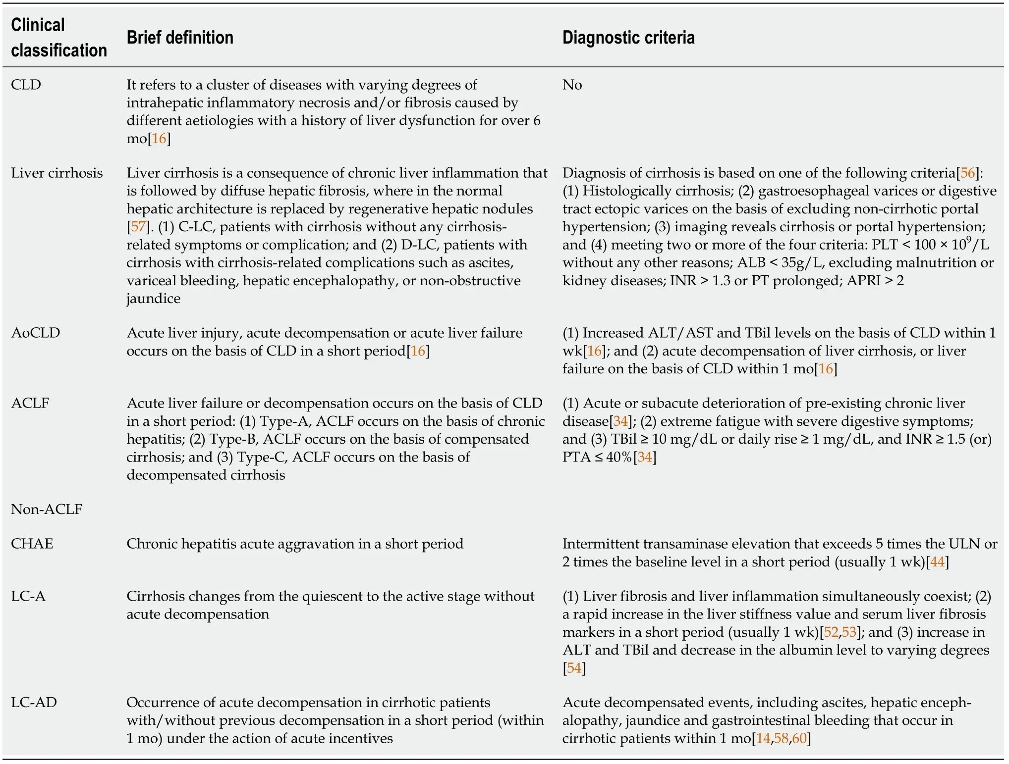
Inflammation and/or fibrosis in patients with chronic hepatitis or compensatory cirrhosis can be alleviated or even reversed (for compensatory cirrhosis) if a proper treatment regimen is applied, such as continuous nucleos(t)ide analogue (NUC) treatment for CHB[26,27]. Once patients with cirrhosis develop acute decompensation, the prognosis is poor, and the median survival time is approximately 5 years[28]. A mild ALI imposed on CLD may not lead to liver dysfunction, and liver injury can recover upon active treatment with minimal effects on the quality of life and longevity. However, once massive or submassive hepatic necrosis occurs, ACLF is triggered with a short-term (28-d) mortality rate of more than 15%[19,29]. The prognosis of AoCLD highly differs depending on both the various types of CLD and the degree of ALI. Therefore, AoCLD can be divided into ACLF and non-ACLF according to the degree of ALI; furthermore, according to the basic state of CLD (non-cirrhotic chronic liver diseases,compensatory cirrhosis, or decompensated cirrhosis), non-ACLF can be further divided into chronic hepatitis with acute exacerbation (CHAE), the active phase of liver cirrhosis (LC-A), and liver cirrhosisacute decompensation (LC-AD) (Figure 1). A brief definition and the diagnostic criteria for each clinical type of AoCLD are shown in Table 2.

ACLF
Between articles, he took breaks by watching the nearby traffic and pedestrians6. The coffee shop was next to a huge parking lot. The lot was for customers of a grocery store, movie rental7 store, pharmacy8, bank, and restaurant. Kevin considered his outdoor seat the perfect place for one of his favorite activities—people watching.
Looking at this, the lawyer thought maybe there s still a chance, but the wife was frowning7 when she answer. This is always the problem, you always think so highly8 of yourself, never thought about how I feel, don t you know that I hate drumsticks?”
When they said that a visitor was asking for her, and then proceeded each one to tell breathlessly a different tale of wonder, in which she could only distinguish the words, oxen, gold, club, giant, lion, she thought they were all out of their minds
NON-ACLF AOCLD
CHAE
In patients with chronic hepatitis, the presence of various precipitants, such as HBV reactivation, leads to acute exacerbation of liver inflammation and/or focal necrosis of hepatocytes, which is manifested as repeated or continuous increases in the serum ALT and/or AST levels, a decrease in albumin or the albumin/globulin ratio, an increase in the TBil level, and even the presence of abnormal coagulation function[17]. The 2015 edition of the Asia-Pacific Liver Association clinical practice guidelines for hepatitis B define CHAE as an intermittent transaminase elevation that exceeds 5 times the ULN or 2 times the baseline level[44]. CHAE clinically manifests as the activation of chronic hepatitis, which can be classified as mild, moderate, and severe according to the degree of inflammation and fibrosis of liver tissue[45].
Notably, due to the particularity of the natural history of HBV infection, HBV-related ALI occurring in a state of chronic HBV infection can be divided into the following two situations: one situation involves a mild transient liver injury that does not activate HBV or require anti-HBV treatment, and the patient is still in a state of “chronic HBV infection” after recovery from ALI[46,47], while in the other situation, the precipitants persist, leading to severe liver damage and even HBV activation, and anti-HBV treatment is needed to control the disease progression. In this case, “chronic HBV infection”transitions into “chronic hepatitis B”[48]. Therefore, ALI occurring under a chronic HBV infection status should not be diagnosed as CHAE.
LC-A
In patients with C-LC, the remaining liver cells can maintain liver functions, such as synthesis and catabolism, even in the presence of portal hypertension[56,57]. D-LC is defined as the occurrence of ascites, hepatic encephalopathy, jaundice, or oesophageal-gastric varices bleeding in patients with C-LC[56,58,59]. Both C-LC and D-LC patients may experience acute decompensation in a short period (within 1 mo) under the action of acute inducement, which is called LC-AD[58].
LC-AD
According to the status of inflammatory activity in liver tissue, liver cirrhosis can be divided into the active and quiescent stages[49,50]. In patients with quiescent cirrhosis, the liver is histologically characterized by pseudolobules and does not show hepatocyte necrosis, lymphocyte infiltration or new fibrogenesis. The presence of acute aggravating factors leads to intrahepatic inflammatory cell infiltration, hepatocyte necrosis and new fibrogenesis, indicating that liver cirrhosis transitioned to the active phase[51]. LC-A is histologically defined as a state of active intrahepatic inflammation and fibrogenesis in patients with cirrhosis and clinically manifests as a sharp increase in serum liver fibrosis markers(laminin, hyaluronic acid, pro-peptide of type III procollagen and collagen IV) or liver stiffness within a short period (usually 1 wk)[52,53] and is accompanied by elevated ALT and TBil levels and decreased albumin[54]. LC-A can occur in both compensatory LC (C-LC) and decompensated LC (D-LC). Due to obvious portal hypertension in patients with D-LC, haemodynamic disorders are very likely to occur,causing hepatic tissue ischaemia and hypoxia, and immunodeficiency renders D-LC patients vulnerable to secondary infections, leading to sepsis and further liver injury; thus, D-LC is rarely in a quiescent state[55]. Therefore, LC-A occurs in patients with C-LC more commonly than in those with D-LC.
LC-AD is mainly manifested by the following two types of pathophysiological changes: portal hypertension and liver dysfunction. The complications of LC-AD may interact with each other, forming a vicious cycle and promoting the progression of LC-AD[59]. Studies have shown that the prognosis of LC-AD patients with previous decompensation is worse than that of LC-AD patients without previous decompensation[28,60], likely because D-LC patients are more prone to intractable ascites and endotoxaemia than C-LC patients. Under the triple attack of immune injury, ischaemia and hypoxia,and endotoxaemia, patients with liver cirrhosis experience massive/submassive necrosis of the liver tissue, resulting in rapid deterioration, and easily develop ACLF[18,61,62].
CONCLUSION
The present review preliminarily summarized the definition, aetiology and inducement, and clinical types of AoCLD (Figure 1). However, the aetiology and precipitants of AoCLD are complex, and the clinical classification and definition of AoCLD are divergent; in particular, the diagnostic criteria for ACLF are controversial. For individual clinical types of AoCLD, the relevant factors, including the assessment of the degree of the disease and the prognosis, need to be characterized, and the mechanism driving the progression of AoCLD needs to be further clarified. Therefore, a multicentre, prospective cohort study is needed to systematically analyse the clinical characteristics and prognostic factors of the individual clinical types of AoCLD, which could provide an evidence-based definition and characterization of the diseases.
FOOTNOTES
On he went, away, away, away, but he sought the snuff-box in vain all up and down the neighbouring countries, and very soon he came to the end of all his money
the National Science and Technology Major Project, No. 2018ZX10723203 and No. 2018ZX10302206; the Foundation for Innovative Research Groups of Hubei Provincial Natural Science Foundation, No. 2018CFA031; the Project of Hubei University of Medicine, No. FDFR201902, No. 2020XGFYZR05, and No. YC2020015; and the Project of Science and Technology Plan of Shiyan, No. 20Y08 and No. 19Y27.
In the diagnosis of ACLF, attention should be given to discriminating ACLF from ALF or subacute LF(SALF) in which liver failure develops within 2 wk or 26 wk, respectively, in patients without preexisting chronic liver injury[35]. Therefore, the difference between ACLF and ALF/SALF mainly lies in the presence or absence of underlying chronic liver injury. Hepatitis B virus (HBV) infection is the main cause of ACLF[36]. The diagnosis of HBV-associated ACLF is sometimes difficult due to the complexity of the natural history of HBV infection. According to the European Association for the Study of the Liver, the natural history of chronic HBV infection is divided into five stages, namely, hepatitis B e antigen (HBeAg)-positive chronic HBV infection, HBeAg-positive CHB, HBeAg-negative chronic HBV infection, HBeAg-negative CHB, and hepatitis B surface antigen (HBsAg)-negative stage[37]. The nomenclature is based on the description of the following two main characteristics of the history of HBV infection: infection and hepatitis. In a state of chronic HBV infection, there is limited or no chronic inflammation or fibrosis in the liver[25]. Studies have shown that the survival rate of chronic HBVinfected patients is comparable to that of non-HBV-infected patients[38]. Therefore, ALF/SALF rather than ACLF should be diagnosed once liver failure occurs in patients with chronic HBV infection or CHB whose intrahepatic inflammation and fibrosis have completely disappeared for more than half a year upon NUC treatment[39,40]. In contrast, ACLF should be diagnosed once liver failure occurs in CHB patients with active intrahepatic inflammation and/or fibrosis. However, since most patients with chronic HBV infection lack liver histological evidence, the status of liver inflammatory activity and fibrosis can be judged only indirectly by referring to the levels of serum ALT/AST and liver stiffness(detected by transient elastography)[41,42]. Thus, for patients with chronic HBV infection who do not show obvious signs and symptoms of active hepatitis but may have different degrees of inflammatory activity and fibrosis histologically, the dynamic monitoring of the ALT/AST levels and liver stiffness could facilitate the assessment of liver inflammatory activity and fibrosis, respectively, which is helpful for distinguishing ALF/SALF from ACLF[41,43].
Zhang YY wrote the manuscript; Meng ZJ contributed to the conception of the study and revised the manuscript.
Then the heart of the man was greatly stirred, and he stretched out his arms to his wife, but she waved her hands and said, We have seen no one yet; it is too soon
The authors declare that they have no conflict of interest.
ACLF refers to a syndrome that occurs in CLD patients under the action of various ALI factors and is characterized by acute jaundice, coagulatory dysfunction, and rapid disease progression with high mortality[12,30]. The differences in the aetiology of CLD and the induction of acute injury between Eastern and Western countries have led to nonuniform diagnostic criteria for ACLF. The definition of ACLF in Asia focuses on liver failure caused by ALI, while the definition in Europe and America pays more attention to systemic multiorgan failure, considering liver failure an unnecessary condition[31]. In 2015, the World Gastrointestinal Organization proposed consensus definitions of ACLF integrated fromthe East and the West and proposed the following three clinical types of ACLF based on the different CLDs: Type A (based on chronic hepatitis), type B (based on compensatory cirrhosis), and type C (based on decompensated cirrhosis)[22,32,33]. The Chinese Medical Association summarized the definition of ACLF in the Guidelines for the Diagnosis and Treatment of Liver Failure as follows: Acute liver failure(ALF) occurs on the basis of CLD (with or without cirrhosis), mainly manifesting as jaundice (serum TBil attaining a level over 10 times the upper limit of normal (ULN) value or a daily increase ≥ 17.1 μmol/L) and a bleeding tendency (prothrombin activity ≤ 40% or international normalized ratio ≥ 1.5),accompanied by failure of one or more extrahepatic organs with significantly increased mortality within 28 d and 3 mo after onset[34]. According to the basic status of CLD, ACLF was also classified into three clinical types consistent with those of the World Gastrointestinal Organization.
This article is an open-access article that was selected by an in-house editor and fully peer-reviewed by external reviewers. It is distributed in accordance with the Creative Commons Attribution NonCommercial (CC BYNC 4.0) license, which permits others to distribute, remix, adapt, build upon this work non-commercially, and license their derivative works on different terms, provided the original work is properly cited and the use is noncommercial. See: https://creativecommons.org/Licenses/by-nc/4.0/
China
Yuan-Yao Zhang 0000-0002-7630-1796; Zhong-Ji Meng 0000-0003-0401-535X.
Chen YL
The queen rode happy home to the hut, and happier still was the man, who had been sitting there in great anxiety, for now he was freed from all the power of the evil spirits
A
Chen YL
1 Li M, Wang ZQ, Zhang L, Zheng H, Liu DW, Zhou MG. Burden of Cirrhosis and Other Chronic Liver Diseases Caused by Specific Etiologies in China, 1990-2016: Findings from the Global Burden of Disease Study 2016. Biomed Environ Sci 2020; 33: 1-10 [PMID: 32029053 DOI: 10.3967/bes2020.001]
2 Moon AM, Singal AG, Tapper EB. Contemporary Epidemiology of Chronic Liver Disease and Cirrhosis. Clin Gastroenterol Hepatol 2020; 18: 2650-2666 [PMID: 31401364 DOI: 10.1016/j.cgh.2019.07.060]
3 GBD 2019 Diseases and Injuries Collaborators. Global burden of 369 diseases and injuries in 204 countries and territories, 1990-2019: a systematic analysis for the Global Burden of Disease Study 2019. Lancet 2020; 396: 1204-1222[PMID: 33069326 DOI: 10.1016/S0140-6736(20)30925-9]
4 Kuo CC, Huang CH, Chang C, Chen PC, Chen BH, Chen WT, Ho YP. Comparing CLIF-C ACLF, CLIF-C ACLF
, and CLIF-C ACLF-D Prognostic Scores in Acute-on-Chronic Liver Failure Patients by a Single-Center ICU Experience. J Pers Med 2021; 11 [PMID: 33572927 DOI: 10.3390/jpm11020079]
5 Lin X, Huang X, Wang L, Feng S, Chen X, Cai W, Huang Z. Prognostic Value of Acute-On-Chronic Liver Failure (ACLF)Score in Critically Ill Patients with Cirrhosis and ACLF. Med Sci Monit 2020; 26: e926574 [PMID: 32978936 DOI:10.12659/MSM.926574]
6 Qiao L, Wang X, Deng G, Huang Y, Chen J, Meng Z, Zheng X, Shi Y, Qian Z, Liu F, Gao Y, Lu X, Liu J, Gu W, Zhang Y, Wang T, Wu D, Dong F, Sun X, Li H. Cohort profile: a multicentre prospective validation cohort of the Chinese Acuteon-Chronic Liver Failure (CATCH-LIFE) study. BMJ Open 2021; 11: e037793 [PMID: 33419900 DOI:10.1136/bmjopen-2020-037793]
7 Kohn CL, Brozenec S, Foster PF. Nutritional support for the patient with pancreatobiliary disease. Crit Care Nurs Clin North Am 1993; 5: 37-45 [PMID: 8448001]
8 Clemmesen JO, Larsen FS, Kondrup J, Hansen BA, Ott P. Cerebral herniation in patients with acute liver failure is correlated with arterial ammonia concentration. Hepatology 1999; 29: 648-653 [PMID: 10051463 DOI:10.1002/hep.510290309]
9 Clemmesen JO, Gerbes AL, Gülberg V, Hansen BA, Larsen FS, Skak C, Tygstrup N, Ott P. Hepatic blood flow and splanchnic oxygen consumption in patients with liver failure. Effect of high-volume plasmapheresis. Hepatology 1999; 29:347-355 [PMID: 9918909 DOI: 10.1002/hep.510290206]
10 Agarwal B, Shaw S, Shankar Hari M, Burroughs AK, Davenport A. Continuous renal replacement therapy (CRRT) in patients with liver disease: is circuit life different? J Hepatol 2009; 51: 504-509 [PMID: 19615775 DOI:10.1016/j.jhep.2009.05.028]
11 Jagadisan B, Srivastava A, Yachha SK, Poddar U. Acute on chronic liver disease in children from the developing world:recognition and prognosis. J Pediatr Gastroenterol Nutr 2012; 54: 77-82 [PMID: 21691224 DOI:10.1097/MPG.0b013e318228d7da]
12 Tasneem AA, Luck NH. Acute-On-Chronic Liver Failure: Causes, Clinical Characteristics and Predictors of Mortality. J Coll Physicians Surg Pak 2017; 27: 8-12 [PMID: 28292360]
13 Caracuel L, Sastre E, Llévenes P, Prieto I, Funes T, Aller Má, Arias J, Balfagón G, Blanco-Rivero J. Acute-on-chronic liver disease enhances phenylephrine-induced endothelial nitric oxide release in rat mesenteric resistance arteries through enhanced PKA, PI3K/AKT and cGMP signalling pathways. Sci Rep 2019; 9: 6993 [PMID: 31061522 DOI:10.1038/s41598-019-43513-y]
14 Zhang Y, Xu BY, Wang XB, Zheng X, Huang Y, Chen J, Meng ZJ, Gao YH, Qian ZP, Liu F, Lu XB, Shi Y, Shang J, Li H, Wang SY, Yin S, Sun SN, Hou YX, Xiong Y, Li BL, Lei Q, Gao N, Ji LJ, Li J, Jie FR, Zhao RH, Liu JP, Lin TF, Chen LY, Tan WT, Zhang Q, Zou CC, Huang ZB, Jiang XH, Luo S, Liu CY, Zhang YY, Li T, Ren HT, Wang SJ, Deng GH,Xiong SE, Liu XX, Wang C, Yuan W, Gu WY, Qiao L, Wang TY, Wu DD, Dong FC, Hua J. Prevalence and Clinical Significance of Portal Vein Thrombosis in Patients With Cirrhosis and Acute Decompensation. Clin Gastroenterol Hepatol 2020; 18: 2564-2572.e1 [PMID: 32109631 DOI: 10.1016/j.cgh.2020.02.037]
15 Long L, Li H, Deng G, Wang X, Lu S, Li B, Meng Z, Gao Y, Qian Z, Liu F, Lu X, Ren H, Shang J, Wang S, Zheng Y,Yan H, Yin S, Tan W, Zhang Q, Zheng X, Chen J, Luo S, Zhao J, Yuan W, Li T, Zheng R, Liu J, Liu X, Gu W, Li S, Mei X, Chen R, Huang Y. Impact of Hepatic Encephalopathy on Clinical Characteristics and Adverse Outcomes in Prospective and Multicenter Cohorts of Patients With Acute-on-Chronic Liver Diseases. Front Med (Lausanne) 2021; 8: 709884[PMID: 34409052 DOI: 10.3389/fmed.2021.709884]
16 Ouyang R, Li H, Xia J, Wang X, Zheng X, Huang Y, Meng Z, Gao Y, Qian Z, Liu F, Lu X, Shi Y, Shang J, Liu J, Deng G,Zheng Y, Yan H, Zhang W, Qiao L, Jiang X, Wang H, Zhong G, Li B, Chen J. Lower platelet counts were associated with 90-day adverse outcomes in acute-on-chronic liver disease patients. Ann Palliat Med 2021; 10: 9342-9353 [PMID:34412498 DOI: 10.21037/apm-21-1019]
17 Crismale JF, Friedman SL. Acute Liver Injury and Decompensated Cirrhosis. Med Clin North Am 2020; 104: 647-662[PMID: 32505258 DOI: 10.1016/j.mcna.2020.02.010]
18 Dienes HP, Drebber U. Pathology of immune-mediated liver injury. Dig Dis 2010; 28: 57-62 [PMID: 20460891 DOI:10.1159/000282065]
19 Thawley V. Acute Liver Injury and Failure. Vet Clin North Am Small Anim Pract 2017; 47: 617-630 [PMID: 28065578 DOI: 10.1016/j.cvsm.2016.11.010]
20 Green TJ, Sivilotti ML, Langmann C, Yarema M, Juurlink D, Burns MJ, Johnson DW. When do the aminotransferases rise after acute acetaminophen overdose?
2010; 48: 787-792 [PMID: 20969501 DOI:10.3109/15563650.2010.523828]
21 Wu HL, Kao JH, Chen TC, Wu WH, Liu CH, Su TH, Yang HC, Chen DS, Chen PJ, Liu CJ. Serum cytokine/chemokine profiles in acute exacerbation of chronic hepatitis B: clinical and mechanistic implications.
2014;29: 1629-1636 [PMID: 24730549 DOI: 10.1111/jgh.12606]
22 Jalan R, Yurdaydin C, Bajaj JS, Acharya SK, Arroyo V, Lin HC, Gines P, Kim WR, Kamath PS; World Gastroenterology Organization Working Party. Toward an improved definition of acute-on-chronic liver failure.
2014; 147:4-10 [PMID: 24853409 DOI: 10.1053/j.gastro.2014.05.005]
23 Chen T, Yang Z, Choudhury AK, Al Mahtab M, Li J, Chen Y, Tan SS, Han T, Hu J, Hamid SS, Huei LG, Ghazinian H,Nan Y, Chawla YK, Yuen MF, Devarbhavi H, Shukla A, Abbas Z, Sahu M, Dokmeci AK, Lesmana LA, Lesmana CRA,Xin S, Duan Z, Guo W, Ma K, Zhang Z, Cheng Q, Jia J, Sharma BC, Sarin SK, Ning Q. Complications constitute a major risk factor for mortality in hepatitis B virus-related acute-on-chronic liver failure patients: a multi-national study from the Asia-Pacific region.
2019; 13: 695-705 [PMID: 31650510 DOI: 10.1007/s12072-019-09992-x]
24 Hernaez R, Liu Y, Kramer JR, Rana A, El-Serag HB, Kanwal F. Model for end-stage liver disease-sodium underestimates 90-day mortality risk in patients with acute-on-chronic liver failure.
2020; 73: 1425-1433 [PMID: 32531416 DOI: 10.1016/j.jhep.2020.06.005]
25 Sugawara K, Nakayama N, Mochida S. Acute liver failure in Japan: definition, classification, and prediction of the outcome.
2012; 47: 849-861 [PMID: 22825549 DOI: 10.1007/s00535-012-0624-x]
26 Bedossa P. Reversibility of hepatitis B virus cirrhosis after therapy: who and why?
2015; 35 Suppl 1: 78-81[PMID: 25529091 DOI: 10.1111/Liv.12710]
27 Saffioti F, Pinzani M. Development and Regression of Cirrhosis.
2016; 34: 374-381 [PMID: 27170391 DOI:10.1159/000444550]
28 Nilsson E, Anderson H, Sargenti K, Lindgren S, Prytz H. Patients with liver cirrhosis show worse survival if decompensation occurs later during course of disease than at diagnosis.
2018; 53: 475-481 [PMID:29513096 DOI: 10.1080/00365521.2018.1447599]
29 Cao Z, Liu Y, Cai M, Xu Y, Xiang X, Zhao G, Cai W, Wang H, Wang W, Xie Q. The Use of NACSELD and EASL-CLIF Classification Systems of ACLF in the Prediction of Prognosis in Hospitalized Patients With Cirrhosis.
2020; 115: 2026-2035 [PMID: 32858565 DOI: 10.14309/ajg.0000000000000771]
30 Sarin SK, Choudhury A, Sharma MK, Maiwall R, Al Mahtab M, Rahman S, Saigal S, Saraf N, Soin AS, Devarbhavi H,Kim DJ, Dhiman RK, Duseja A, Taneja S, Eapen CE, Goel A, Ning Q, Chen T, Ma K, Duan Z, Yu C, Treeprasertsuk S,Hamid SS, Butt AS, Jafri W, Shukla A, Saraswat V, Tan SS, Sood A, Midha V, Goyal O, Ghazinyan H, Arora A, Hu J,Sahu M, Rao PN, Lee GH, Lim SG, Lesmana LA, Lesmana CR, Shah S, Prasad VGM, Payawal DA, Abbas Z, Dokmeci AK, Sollano JD, Carpio G, Shresta A, Lau GK, Fazal Karim M, Shiha G, Gani R, Kalista KF, Yuen MF, Alam S, Khanna R, Sood V, Lal BB, Pamecha V, Jindal A, Rajan V, Arora V, Yokosuka O, Niriella MA, Li H, Qi X, Tanaka A, Mochida S,Chaudhuri DR, Gane E, Win KM, Chen WT, Rela M, Kapoor D, Rastogi A, Kale P, Sharma CB, Bajpai M, Singh V,Premkumar M, Maharashi S, Olithselvan A, Philips CA, Srivastava A, Yachha SK, Wani ZA, Thapa BR, Saraya A,Shalimar, Kumar A, Wadhawan M, Gupta S, Madan K, Sakhuja P, Vij V, Sharma BC, Garg H, Garg V, Kalal C, Anand L,Vyas T, Mathur RP, Kumar G, Jain P, Pasupuleti SSR, Chawla YK, Chowdhury A, Song DS, Yang JM, Yoon EL; APASL ACLF Research Consortium (AARC) for APASL ACLF working Party. Correction to: Acute-on-chronic liver failure:consensus recommendations of the Asian Pacific association for the study of the liver (APASL): an update.
2019; 13: 826-828 [PMID: 31595462 DOI: 10.1007/s12072-019-09980-1]
31 Le?o GS, Lunardi FL, Picon RV, Tovo CV, de Mattos AA, de Mattos ?Z. Acute-on-chronic liver failure: A comparison of three different diagnostic criteria.
2019; 18: 373-378 [PMID: 31053547 DOI: 10.1016/j.aohep.2019.01.001]
32 Tang X, Qi T, Li B, Li H, Huang Z, Zhu Z, Tu M, Gao J, Zhu C, Jiang X, Yu X, Lu G, Xiong M, He Q, Zhou F, Wen W,Chen J, Hou J. Tri-typing of hepatitis B-related acute-on-chronic liver failure defined by the World Gastroenterology Organization.
2021; 36: 208-216 [PMID: 32445263 DOI: 10.1111/jgh.15113]
33 Mu X, Tong J, Xu X, Chen J, Su H, Liu X, Pang F, Zhai X, Wang L, Wang Y, Guan C, Wang F, Hu J. World Gastroenterology Organisation classification and a new type-based prognostic model for hepatitis B virus-related acute-onchronic liver failure.
2021; 45: 101548 [PMID: 33554865 DOI:10.1016/j.clinre.2020.09.009]
34 Zhang Q, Li Y, Han T, Nie C, Cai J, Liu H, Liu Y. Comparison of current diagnostic criteria for acute-on-chronic liver failure.
2015; 10: e0122158 [PMID: 25785855 DOI: 10.1371/journal.pone.0122158]
35 Zhang Y, Guo X, Ma Z, Wang B, Lu H, Qi X. Characteristics and in-hospital outcomes of COVID-19 patients with acute or subacute liver failure.
2021; 53: 1069-1070 [PMID: 34140208 DOI: 10.1016/j.dld.2021.05.027]
36 Gu WY, Xu BY, Zheng X, Chen J, Wang XB, Huang Y, Gao YH, Meng ZJ, Qian ZP, Liu F, Lu XB, Shang J, Li H, Wang SY, Sun X. Acute-on-Chronic Liver Failure in China: Rationale for Developing a Patient Registry and Baseline Characteristics.
2018; 187: 1829-1839 [PMID: 29762630 DOI: 10.1093/aje/kwy083]
37 Hadziyannis SJ. Unrevealing the natural course of the so-called "inactive HBsAg or HBV carrier state".
2007;1: 281-284 [PMID: 19669350 DOI: 10.1007/s12072-007-9004-7]
38 Villa E, Fattovich G, Mauro A, Pasino M. Natural history of chronic HBV infection: special emphasis on the prognostic implications of the inactive carrier state
chronic hepatitis.
2011; 43 Suppl 1: S8-14 [PMID: 21195374 DOI: 10.1016/S1590-8658(10)60686-X]
39 Oketani M, Uto H, Ido A, Tsubouchi H. Management of hepatitis B virus-related acute liver failure.
2014; 7: 19-26 [PMID: 26183504 DOI: 10.1007/s12328-013-0447-1]
40 Mochida S, Nakayama N, Ido A, Takikawa Y, Yokosuka O, Sakaida I, Moriwaki H, Genda T, Takikawa H. Revised criteria for classification of the etiologies of acute liver failure and late-onset hepatic failure in Japan: A report by the Intractable Hepato-biliary Diseases Study Group of Japan in 2015.
2016; 46: 369-371 [PMID: 26615003 DOI:10.1111/hepr.12626]
41 Sonneveld MJ, Brouwer WP, Hansen BE, Chan HL, Piratvisuth T, Jia JD, Zeuzem S, Chien RN, Choi H, de Knegt RJ,Wat C, Pavlovic V, Gaggar A, Xie Q, Buti M, de Man RA, Janssen HLA; SONIC-B Study Group. Very low probability of significant liver inflammation in chronic hepatitis B patients with low ALT levels in the absence of liver fibrosis.
2020; 52: 1399-1406 [PMID: 32886813 DOI: 10.1111/apt.16067]
42 He T, Li J, Ouyang Y, Lv G, Ceng X, Zhang Z, Ding J. FibroScan Detection of Fatty Liver/Liver Fibrosis in 2266 Cases of Chronic Hepatitis B.
2020; 8: 113-119 [PMID: 32832390 DOI: 10.14218/JCTH.2019.00053]
43 Rahayu ES, Mariyatun M, Putri Manurung NE, Hasan PN, Therdtatha P, Mishima R, Komalasari H, Mahfuzah NA,Pamungkaningtyas FH, Yoga WK, Nurfiana DA, Liwan SY, Juffrie M, Nugroho AE, Utami T. Effect of probiotic
Dad-13 powder consumption on the gut microbiota and intestinal health of overweight adults.
2021; 27: 107-128 [PMID: 33505154 DOI: 10.3748/wjg.v27.i1.107].]
44 Sarin SK, Kumar M, Lau GK, Abbas Z, Chan HL, Chen CJ, Chen DS, Chen HL, Chen PJ, Chien RN, Dokmeci AK, Gane E, Hou JL, Jafri W, Jia J, Kim JH, Lai CL, Lee HC, Lim SG, Liu CJ, Locarnini S, Al Mahtab M, Mohamed R, Omata M,Park J, Piratvisuth T, Sharma BC, Sollano J, Wang FS, Wei L, Yuen MF, Zheng SS, Kao JH. Asian-Pacific clinical practice guidelines on the management of hepatitis B: a 2015 update.
2016; 10: 1-98 [PMID: 26563120 DOI:10.1007/s12072-015-9675-4]
45 Zhou XJ, Huang WB. [Evaluation of different grading and staging systems of chronic hepatitis and problems in application].
2008; 37: 636-640 [PMID: 19094592]
46 Salpini R, Battisti A, Colagrossi L, Di Carlo D, Fabeni L, Piermatteo L, Cerva C, Lichtner M, Mastroianni C, Marignani M, Maylin S, Delaugerre C, Morisco F, Coppola N, Marrone A, Angelico M, Sarmati L, Andreoni M, Perno CF,Ceccherini-Silberstein F, Svicher V. A snapshot of virological presentation and outcome of immunosuppression-driven HBV reactivation from real clinical practice: Evidence of a relevant risk of death and evolution from silent to chronic infection.
2019; 26: 846-855 [PMID: 30974483 DOI: 10.1111/jvh.13101]
47 Zhang ZQ, Shi BS, Lu W, Huang D, Wang YB, Feng YL. Quantitative serum HBV markers in predicting phases of natural history of chronic HBV infection.
2021; 296: 114226 [PMID: 34217779 DOI:10.1016/j.jviromet.2021.114226]
48 Huang SC, Yang HC, Kao JH. Hepatitis B reactivation: diagnosis and management.
2020; 14: 565-578 [PMID: 32448008 DOI: 10.1080/17474124.2020.1774364]
49 de Oliveira da Silva B, Ramos LF, Moraes KCM. Molecular interplays in hepatic stellate cells: apoptosis, senescence, and phenotype reversion as cellular connections that modulate liver fibrosis.
2017; 41: 946-959 [PMID: 28498509 DOI: 10.1002/cbin.10790]
50 Liu X, Xu J, Rosenthal S, Zhang LJ, McCubbin R, Meshgin N, Shang L, Koyama Y, Ma HY, Sharma S, Heinz S, Glass CK, Benner C, Brenner DA, Kisseleva T. Identification of Lineage-Specific Transcription Factors That Prevent Activation of Hepatic Stellate Cells and Promote Fibrosis Resolution.
2020; 158: 1728-1744.e14 [PMID: 31982409 DOI: 10.1053/j.gastro.2020.01.027]
51 Fung J, Lai CL, Chan SC, But D, Seto WK, Cheng C, Wong DK, Lo CM, Fan ST, Yuen MF. Correlation of liver stiffness and histological features in healthy persons and in patients with occult hepatitis B, chronic active hepatitis B, or hepatitis B cirrhosis.
2010; 105: 1116-1122 [PMID: 19920809 DOI: 10.1038/ajg.2009.665]
52 Koch A, Horn A, Dückers H, Yagmur E, Sanson E, Bruensing J, Buendgens L, Voigt S, Trautwein C, Tacke F. Increased liver stiffness denotes hepatic dysfunction and mortality risk in critically ill non-cirrhotic patients at a medical ICU.
2011; 15: R266 [PMID: 22082207 DOI: 10.1186/cc10543]
53 Zhu C, Qi X, Li H, Peng Y, Dai J, Chen J, Xia C, Hou Y, Zhang W, Guo X. Correlation of serum liver fibrosis markers with severity of liver dysfunction in liver cirrhosis: a retrospective cross-sectional study.
2015; 8: 5989-5998 [PMID: 26131195]
54 Fujita K, Masaki T. Serum Biomarkers of Liver Fibrosis Staging in the Era of the Concept "Compensated Advanced Chronic Liver Disease".
2021; 10 [PMID: 34362121 DOI: 10.3390/jcm10153340]
55 Bothou C, Rüschenbaum S, Kubesch A, Quenstedt L, Schwarzkopf K, Welsch C, Zeuzem S, Welzel TM, Lange CM.Anemia and Systemic Inflammation Rather than Arterial Circulatory Dysfunction Predict Decompensation of Liver Cirrhosis.
2020; 9 [PMID: 32357568 DOI: 10.3390/jcm9051263]
56 Xu XY, Ding HG, Li WG, Xu JH, Han Y, Jia JD, Wei L, Duan ZP, Ling-Hu EQ, Zhuang H. Chinese guidelines on the management of liver cirrhosis (abbreviated version).
2020; 26: 7088-7103 [PMID: 33362370 DOI:10.3748/wjg.v26.i45.7088]
57 Ginès P, Krag A, Abraldes JG, Solà E, Fabrellas N, Kamath PS. Liver cirrhosis.
2021; 398: 1359-1376 [PMID:34543610 DOI: 10.1016/S0140-6736(21)01374-X]
58 Pfortmueller CA, Wiemann C, Funk GC, Leichtle AB, Fiedler GM, Exadaktylos AK, Lindner G. Hypoglycemia is associated with increased mortality in patients with acute decompensated liver cirrhosis.
2014; 29: 316.e7-316.12 [PMID: 24332992 DOI: 10.1016/j.jcrc.2013.11.002]
59 Bernardi M, Caraceni P. Novel perspectives in the management of decompensated cirrhosis.
2018; 15: 753-764 [PMID: 30026556 DOI: 10.1038/s41575-018-0045-2]
60 D'Amico G, Morabito A, D'Amico M, Pasta L, Malizia G, Rebora P, Valsecchi MG. New concepts on the clinical course and stratification of compensated and decompensated cirrhosis.
2018; 12: 34-43 [PMID: 28681347 DOI:10.1007/s12072-017-9808-z]
61 Yue S, Zhou H, Wang X, Busuttil RW, Kupiec-Weglinski JW, Zhai Y. Prolonged Ischemia Triggers Necrotic Depletion of Tissue-Resident Macrophages To Facilitate Inflammatory Immune Activation in Liver Ischemia Reperfusion Injury.
2017; 198: 3588-3595 [PMID: 28289160 DOI: 10.4049/jimmunol.1601428]
62 Van den Broecke A, Van Coile L, Decruyenaere A, Colpaert K, Benoit D, Van Vlierberghe H, Decruyenaere J.Epidemiology, causes, evolution and outcome in a single-center cohort of 1116 critically ill patients with hypoxic hepatitis.
2018; 8: 15 [PMID: 29383510 DOI: 10.1186/s13613-018-0356-z]
 World Journal of Clinical Cases2022年15期
- World Journal of Clinical Cases的其它文章
- Diet and intestinal bacterial overgrowth: Is there evidence?
- Spontaneous liver rupture following SARS-CoV-2 infection in late pregnancy: A case report
- Metastasis of liver cancer to the thyroid after surgery: A case report
- Solitary primary pulmonary synovial sarcoma: A case report
- Knot impingement after arthroscopic rotator cuff repair mimicking infection: A case report
- Clear aligner treatment for a four-year-old patient with anterior crossbite and facial asymmetry: A case report