
Endoscopic ultrasonography in the evaluation of condition and prognosis of ulcerative colitis
2022-06-22 08:49:10RuiFangJinYiManChenRenPinChenHuaJunYe
World Journal of Clinical Cases 2022年15期
INTRODUCTION
Ulcerative colitis (UC) is a chronic, nonspecific, clinically heterogeneous colitis characterized by recurrent attacks and nonspecific clinical symptoms, which have a great impact on patients' physical and mental health and quality of life. In recent years, UC has gradually become a worldwide social and public health problem that must be addressed[1,2].
The diagnosis of UC is relatively difficult. At present, the disease is usually diagnosed through pathological biopsy, enteroscopy, clinical symptoms, and physical findings; however, it is difficult to accurately evaluate the severity of the disease, and there are some limitations in clinical applications[3,4]. In recent years, with the continuous improvement of medical technology, the application of endoscopic ultrasonography (EUS) in the diagnosis and treatment of UC has attracted widespread attention. It has the advantages of both endoscopy and ultrasonography, can effectively evaluate the changes in intestinal wall thickness and hierarchical structure, and allows monitoring for changes in disease activity and severity[5,6].
In this study, we investigated the value of EUS in the evaluation of the status and prognosis of UC.
When at length he asked the father if he would give him one of his daughters to wife, the two eldest jumped up, ran into their bedrooms to put on splendid dresses, for each of them fancied she was the chosen one
MATERIALS AND METHODS
Baseline data
This study was approved by the ethics committee of our hospital. Patients who underwent endoscopic ultrasound to investigate UC at our hospital between March 2019 and December 2020 were eligible for enrollment. Inclusion criteria were as follows: (1) Diagnostic criteria for UC[7] were met; (2) patients or their families provided written informed consent to participate in the study; (3) patient adherence to,and completion of, the investigation; and (4) age > 18 years. Exclusion criteria were as follows: (1)malignant tumors of the digestive system; (2) history of psychosis; (3) history of allergy; (4) kidney,liver, and other organ dysfunction; (5) pregnancy or lactation; (6) contraindications to endoscopy; (7)administration of relevant treatment before inclusion in the study; (8) infectious enteritis (including
,
,
, acute gastroenteritis, and amoebic bowel disease); (9) Beh?et’s disease and Crohn’s disease; and (10) radiation enteritis and ischemic colitis.
Never had Sir Red been so eaten up with envy as he was now, and he racked his brain day and night to find something quite impossible to demand from the brothers
EUS
A Fujinon SP701 micro ultrasonic probe (Fujinon, Omiya, Japan), with frequency of 20 MHz and rotary scanning mode, and a Fujifilm SU-9000 ultrasonic probe (Fujifilm, Minato City, Japan) with frequencies of 7.5, 10, and 12 MHz were used. Before the examination, the patients were examined, and those with contraindications were excluded from analysis. Four hours before the examination, patients were instructed to consume three packets of polyethylene glycol electrolyte powder, diluted in 3 L of warm water, within two hours. After verifying passage of clear water, endoscopy was performed with the patient in the left lying position. Lubricant was applied to the endoscope, and the investigator expanded the patient’s anus moderately. Air and degassed water in the intestinal cavity were aspirated, prior to the endoscope being withdrawn.
Afterwards I noticed Lucas licking the Neosporin off of his sore. My first thought was, Well, that was a complete waste of time and blood! Then I got an idea. I ll put some Neosporin on his pill. He seems to like the taste of Neosporin. And you know what? It did not work either! So much for shouting with glee(,) and becoming rich of my idea for a Neosporin Flavored Cat Treat .
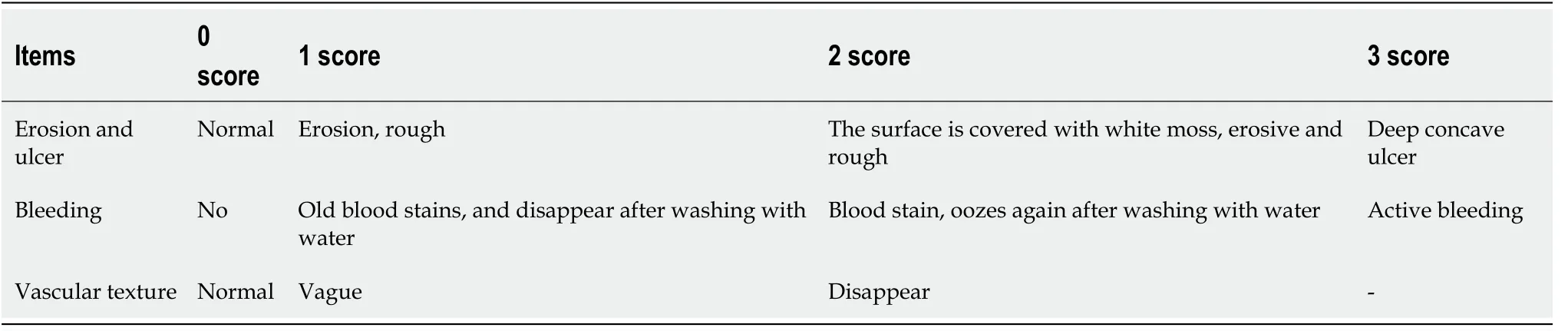
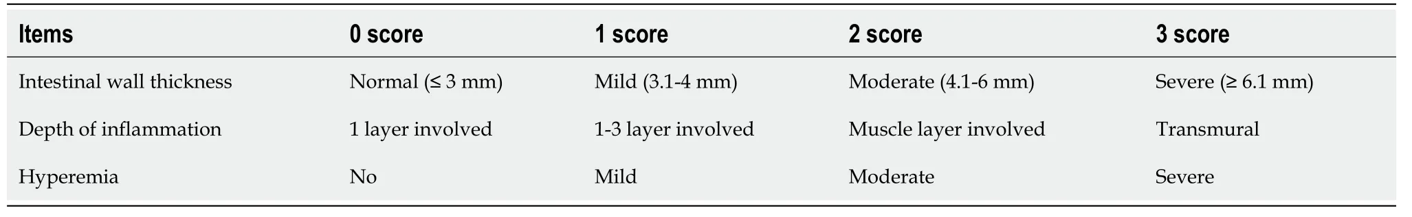
All images were evaluated by physicians with extensive experience in reading endoscopic ultrasound images in our hospital. The UC endoscopic index of severity (UCEIS) and EUS-UC scores were then calculated. The scoring criteria are shown in Table 1 and Table 2 for UCEIS scoring and EUS-UC scoring,respectively.
Evaluation of disease severity
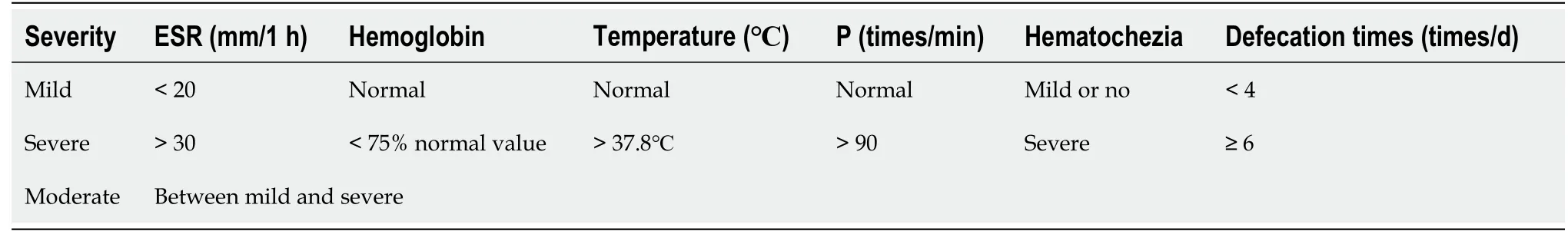
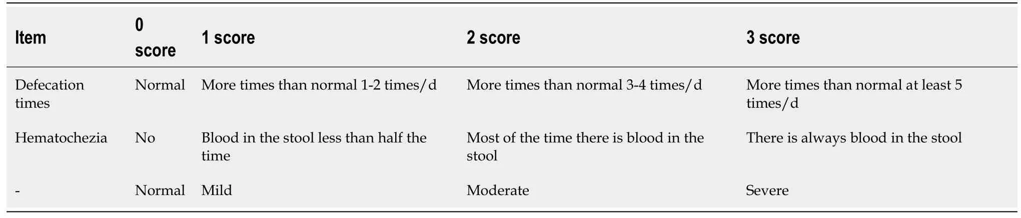
Disease severity was evaluated using the Mayo scale and the modified Truelove and Witts scores. The evaluation criteria are shown in Table 3 and Table 4 for the modified Truelove and Witts and the Mayo evaluation criteria, respectively.
With these and like words the wolf comforted the Prince, and warned him specially17 not to touch the wall or let the horse touch it as he led it out, or he would fail in the same way as he had done with the bird
Treatment method
The UCEIS and EUS-UC scores were higher in moderate cases than in mild cases. The UCEIS and EUSUC scores were higher still in severe cases than in moderate cases, and the differences were statistically significant. The UCEIS and EUS-UC scores in moderate cases were higher than in mild cases. The UCEIS and EUS-UC scores in severe cases were higher than in moderate cases, and the differences were statistically significant. The UCEIS and EUS-UC scores were significantly and positively correlated with disease severity. The UCEIS and EUS-UC scores after 2 mo of treatment and after 6 mo of treatment were lower than the UCEIS and EUS-UC scores before treatment.
Observation indexes
This study investigated the value of EUS in the evaluation of the severity and prognosis of ulcerative colitis.
Statistical analysis
SPSS 22.0 software (IBM Corp., Armonk, NY, USA) was used for data analysis. Measurement data were analyzed using the
-test and are expressed as mean ± SD. Enumerated data were analyzed using the
test and are expressed as
(%). The Spearman method was used for correlation analysis. A
value <0.05 indicated a statistically significant difference.
ONCE there was a gentleman1 who married, for his second wife, the proudest and most haughty1 woman that was ever seen. She had, by a former husband, two daughters of her own humor, who were, indeed, exactly like her in all things. He had likewise, by another wife, a young daughter, but of unparalleled goodness and sweetness of temper, which she took from her mother,2 who was the best creature in the world.
RESULTS
Of the 79 patients with UC included in the analysis, 46 were men and 33 were women. The average age was 49.96 years (range 21-78 years). The lesions were located in the left colon in 17 cases, the rectum in 17 cases, the sigmoid colon in 23 cases, and were extensive in 22 cases.
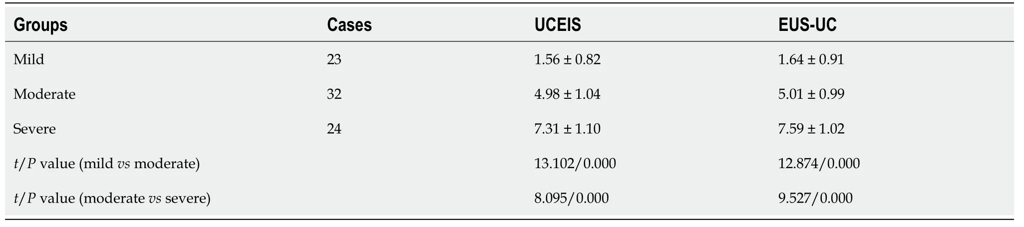
UCEIS and EUS-UC scores of patients with different Mayo scores
According to the Mayo disease activity index, there were 23, 32, and 24 cases of mild, moderate and severe UC, respectively. The UCEIS and EUS-UC scores were higher in moderate cases (4.98 ± 1.04 and 5.01 ± 0.99, respectively) than in mild cases (1.56 ± 0.82 and 1.64 ± 0.91, respectively). Furthermore, the UCEIS and EUS-UC scores were higher still in severe cases (7.31 ± 1.10 and 7.59 ± 1.02, respectively)than in moderate cases, and the differences were statistically significant (
< 0.05) (Table 5).
At last the noise grew so loud that he lost patience, and he stooped to pick up a stone to hurl10 into the midst of the clamour, when suddenly his arm seemed to stiffen11, and the next moment he was a stone himself!That day his sister, who thought her brother s steps were long in returning, took out the knife and found the blade was red as blood
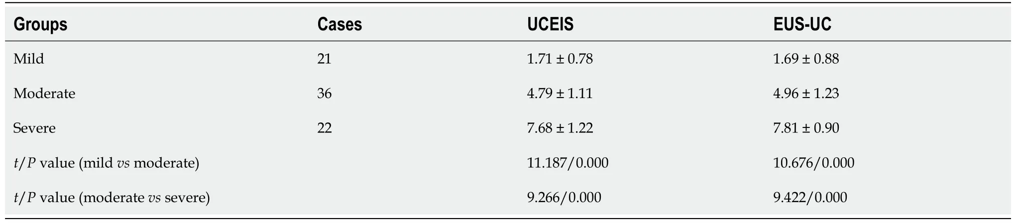
UCEIS and EUS-UC scores of patients with different modified Truelove and Witts scores
Among the 79 cases in this study, 21, 36 and 22 patients were classified as having mild, moderate, andsevere UC according to the modified Truelove and Witts scores. The UCEIS and EUS-UC scores in moderate cases (4.79 ± 1.11 and 4.96 ± 1.23, respectively) were higher than in mild cases (1.71 ± 0.78 and 1.69 ± 0.88, respectively). Furthermore, the UCEIS and EUS-UC scores in severe cases (7.68 ± 1.22 and 7.81 ± 0.90, respectively) were higher than in moderate cases, and the differences were statistically significant (
< 0.05) (Table 6).
O Tsar s Majesty50 the old woman answered, I have with me a marvelous piece of linen stuff, so wondrously51 woven that I will show it to none but thee.
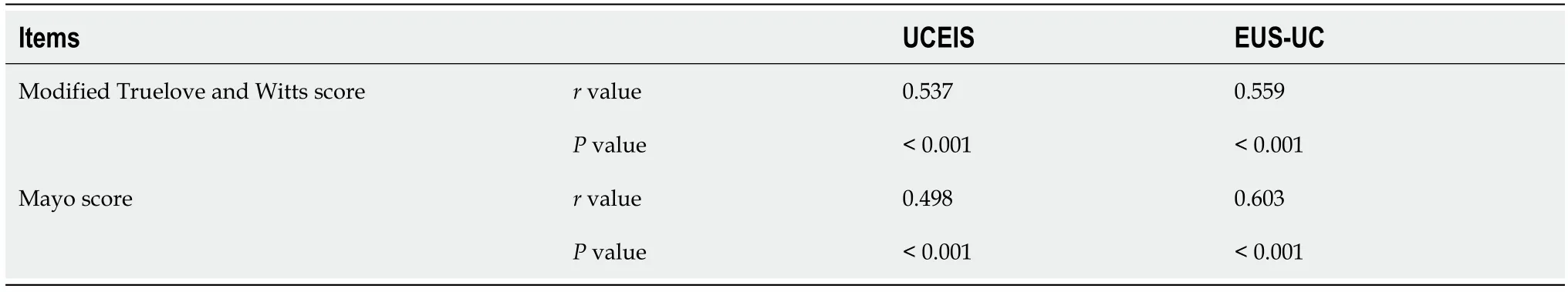
Correlation between UCEIS and EUS-UC scores and disease severity
According to our analysis, the UCEIS and EUS-UC scores were significantly and positively correlated with disease severity (modified Truelove and Witts scores, Mayo score) (
< 0.05) (Table 7).
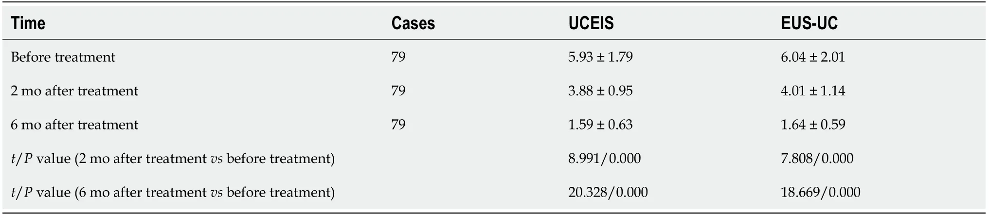
UCEIS and EUS-UC scores before and after treatment
The UCEIS and EUS-UC scores after 2 mo of treatment (3.88 ± 0.95 and 4.01 ± 1.14, respectively) and after 6 mo of treatment (1.59 ± 0.63 and 1.64 ± 0.59, respectively) were lower than the UCEIS and EUSUC scores before treatment (5.93 ± 1.79 and 6.04 ± 2.01, respectively) (
< 0.05) (Table 8).
First, endoscopy was performed to determine the degree and scope of the lesion; then, endoscopic ultrasound was performed at and around the lesions. Depending on the specific condition of the lesion,each was scanned using one of the two following methods. (1) Degassed water filling method: This method can be implemented in many cases. First, excess air is removed, and 200–300 mL of degassed water is injected into the intestinal cavity through the endoscopic working channel to ensure that the focus and ultrasonic probe are submerged in water; and (2) Direct contact method: The ultrasonic probe at the tip of the endoscope is placed in direct contact with the intestinal mucosa. During the examination, the body position was changed according to the specific investigational needs to ensure that the degassed water effectively filled the space between the probe and the focus. The distance between the probe and the focus was set at 1 cm to ensure optimal ultrasonic images could be acquired at all levels of the intestinal wall.
DISCUSSION
The clinical diagnosis and treatment efficacy of UC are mainly established through histopathological examination, barium enema, and endoscopy. However, histopathological studies and routine endoscopy can only evaluate surface lesions of the intestinal mucosa. Therefore, it is difficult to accurately determine the degree of damage to the intestinal mucosa and submucosa, and the structural changes in the intestinal wall, which can affect the accuracy of the evaluation of the severity of UC[8-10]. Therefore, accurate diagnosis and evaluation of UC is still a research hotspot.
EUS can effectively clarify the disease status of patients with ulcerative colitis, stratify the disease severity, and accurately evaluate the treatment response, to provide an objective basis for the clinical formulation or further adjustment of a patient’s treatment plan.
My fascination3 with the ocean escalated4 as the family spent the summer on the eastern end of Long Island, on the shore of the Atlantic Ocean. I was an early riser, and by age ten I was permitted to go down to the beach in the morning to collect shells on my own. Every day I would dress quickly, grab my bucket and head for the beach. I would climb the sand dunes5 that hid the ocean from view and sit quietly at the top and watch the waves roll into shore as I ate my breakfast roll.
When detecting UC, EUS can accurately determine the location of enteritis. The imaging manifestations include abnormally increased thickness of the intestinal wall, abnormal hierarchical structure of the intestinal wall, and irregular or unclear boundaries between various layers of the intestinal wall.Some patients with UC have submucosal vasodilation and extraintestinal lymph node enlargement[15,16]. Other scholars have pointed out that through ultrasonic endoscopy, we can clearly evaluate the condition of the anal canal, terminal ileum, rectum, ileocecal valve, colon, and sigmoid colon with the help of enteroscopy, and we can perform cytological and pathological examination of potentially diseased regions[17,18].
Patients with UC have different degrees of bleeding, mucosal erosion, ulceration, congestion, inflammatory infiltration, and intestinal wall thickening. In addition, the modified Truelove and Witts score and Mayo score are important scales for clinical evaluation of the severity of UC. In this study, these scales and EUS were used to diagnose and evaluate patients with UC. The results showed significant differences in UCEIS and EUS-UC scores in patients with UC, in line with the different disease severity scores according to the modified Truelove and Witts and Mayo scores. Our findings suggest that EUS can accurately detect lesions in patients with UC and can provide an objective reference for the evaluation of disease severity, which is of great guiding significance for diagnosis and treatment. In addition, some studies have pointed out that even if the clinical symptoms of patients with UC are completely relieved and abnormal endoscopic findings are resolved, persistent histological inflammation may still occur in the body, affecting the disease prognosis. EUS can provide more information about UC lesions, especially in the submucosa, muscle layer, serosa, and even outside the serosa.Therefore, EUS may be of great significance in evaluating the severity and prognosis of UC[19,20].
To verify the above findings, the patients included in this study received treatment for UC and were followed up after 2 and 6 mo of treatment. The results showed that the ultrasonic endoscopy findings(UCEIS and EUS-UC scores) of the patients after treatment were improved compared with those before treatment. This confirms that ultrasonic endoscopy also has value in assessing the treatment effect and prognosis of UC. Additionally, the technique can guide clinical prevention and intervention in subsequent exacerbations, so as to improve the treatment effect and prognosis of the disease as a whole.This study confirmed that EUS has significant clinical value in the early treatment of UC, in improving patient prognosis, formulating an effective treatment plan in a timely manner, and, to a certain extent,can assist in the provision of an individualized treatment plan.
However, this study was limited by a small sample size, and some small differences cannot be effectively reflected. In our future research, we will strive to expand the sample size and explore the application value of EUS in individualized treatment.
CONCLUSION
EUS is an auxiliary examination method widely used in clinical practice in recent years. It combines the advantages of digestive endoscopy and ultrasonography, can provide clear images of the gastrointestinal wall and its internal structure, and accurately locate, measure, and evaluate submucosal edema and wall thickening[11,12]. Ultrasound endoscopy involves the installation of an ultrasound probe on the front of the endoscope, which can transmit colonic mucosal images to the computer processing center and present the relevant content on the monitor. Through the display screen, the device can accurately and clearly illustrate the subtle changes in the large intestinal mucosa, including submucosal lesions, diverticula, hemangiomas, polyps, pigmentation, bleeding, ulcers, erosions, and inflammation. The image is more realistic and clearer, and biopsy forceps can be inserted through the endoscope channel to obtain tissues for pathological examination from ultrasound regions of interest, soas to evaluate the nature of lesions[13,14]. Relevant data showed that EUS can directly locate diseased intestinal segments, and the examination results are less affected by subjective and objective factors,thus ensuring the accuracy of diagnosis and evaluation.
ARTICLE HIGHLIGHTS
Research background
Ulcerative colitis (UC) is usually diagnosed through histopathology, enteroscopy, clinical symptoms,and physical findings; however, it is difficult to accurately evaluate disease severity.
Research motivation
In this study, the UC endoscopic index of severity (UCEIS) and endoscopic ultrasonography (EUS)-UC scores of patients were assessed in relation to disease severity, and the correlations between UCEIS and EUS-UC scores and disease severity was also analyzed.
Research objectives
The UCEIS and EUS-UC scores of patients with different disease severities were counted and analyzed.The correlations between UCEIS and EUS-UC scores and disease severity were analyzed. The UCEIS and EUS-UC scores before and after treatment were statistically analyzed.
Research methods
Patients who underwent endoscopic ultrasound to investigate UC at our hospital were eligible for enrollment. A Fujinon SP701 micro ultrasonic probe, with frequency of 20 MHz and rotary scanning mode, and a Fujifilm SU-9000 ultrasonic probe with frequencies of 7.5, 10, and 12 MHz were used.Disease severity was evaluated using the Mayo scale and the modified Truelove and Witts scores.Treatments included oral mesalazine with or without corticosteroids, and intravenous infusion of infliximab. Colonoscopy was repeated 2 and 6 mo after the initiation of treatment.
Research results
According to the consensus on the diagnosis and treatment of inflammatory bowel disease in 2018,targeted interventions were administered according to the patient’s condition. Treatments included oral mesalazine with or without corticosteroids, and intravenous infusion of infliximab. Colonoscopy was repeated 2 and 6 mo after the initiation of treatment.
Research conclusions
EUS can effectively clarify the disease status of patients with ulcerative colitis, stratify the disease severity, and accurately evaluate the treatment response, to provide an objective basis for the clinical formulation or further adjustment of a patient’s treatment plan.
Research perspectives
We will study the application value of this scheme in individualized therapy.
FOOTNOTES
Jin RF and Ye HJ designed this retrospective study; Jin RF wrote the manuscript; Jin RF, Chen YM, Chen PP and Ye HJ sorted the data; all author read and confirmed the revised manuscript.
The shop was air-conditioned at a slightly chilly3 temperature. About eight small round tables were inside. Outside were four bigger tables. Each had four white metal chairs around it, and several big umbrellas provided4 shade. Even on hot afternoons, there was usually a pleasant breeze5.
Wenzhou Science and Technology Bureau, No. Y2020296.
The study was approved by the Ethics Committee of The First Affiliated Hospital of Wenzhou Medical University.
Patients were not required to give informed consent to the study because the analysis used anonymous clinical data that were obtained after each patient agreed to treatment by written consent.
The authors have nothing to disclose.
No additional data are available.
This article is an open-access article that was selected by an in-house editor and fully peer-reviewed by external reviewers. It is distributed in accordance with the Creative Commons Attribution NonCommercial (CC BYNC 4.0) license, which permits others to distribute, remix, adapt, build upon this work non-commercially, and license their derivative works on different terms, provided the original work is properly cited and the use is noncommercial. See: https://creativecommons.org/Licenses/by-nc/4.0/
China
One day his queen presented him with a baby daughter as beautiful as the dawn, and the king himself was so happy and delighted that, for a whole week, he forgot to hunt, and spent the time in public and private rejoicing
Rui-Fang Jin 0000-0001-6904-179X; Yi-Man Chen 0000-0002-1126-9393; Ren-Pin Chen 0000-0002-3211-4702; Hua-Jun Ye 0000-0003-4476-5382.
That doesn t mean it has been easy. I went though, some bad experiences, like being picked last for baseball games (it was hard for me to catch the ball) or the way people looked at me funny when I was out in public. But mostly, people accepted me and helped me. In elementary school, the other kids would help me open my locker3, pick up books or do some of the other tasks that were hard for me.
Wang JL
A
Wang JL
1 Walsh AJ, Bryant RV, Travis SP. Current best practice for disease activity assessment in IBD.
2016; 13: 567-579 [PMID: 27580684 DOI: 10.1038/nrgastro.2016.128]
2 Kinoshita K, Katsurada T, Nishida M, Omotehara S, Onishi R, Mabe K, Onodera A, Sato M, Eto K, Suya M, Maemoto A,Hasegawa T, Yamamoto J, Mitsumori D, Yoshii S, Ono K, Sakamoto N. Usefulness of transabdominal ultrasonography for assessing ulcerative colitis: a prospective, multicenter study.
2019; 54: 521-529 [PMID: 30519747 DOI:10.1007/s00535-018-01534-w]
3 Yan B, Feagan B, Teriaky A, Mosli M, Mohamed R, Williams G, Yeung E, Yong E, Haig A, Sey M, Stitt L, Zou GY,Jairath V. Reliability of EUS indices to detect inflammation in ulcerative colitis.
2017; 86: 1079-1087[PMID: 28760533 DOI: 10.1016/j.gie.2017.07.035]
4 Sugiura K, Kato S, Ishibashi A, Aoyama T, Kani K, Yakabi K. [Comparison of transabdominal ultrasound with quantitative power Doppler and colonoscopic findings for the evaluation of colonic inflammation in active ulcerative colitis].
2020; 117: 695-705 [PMID: 32779587 DOI: 10.11405/nisshoshi.117.695]
5 Allocca M, Fiorino G, Bonovas S, Furfaro F, Gilardi D, Argollo M, Magnoni P, Peyrin-Biroulet L, Danese S. Accuracy of Humanitas Ultrasound Criteria in Assessing Disease Activity and Severity in Ulcerative Colitis: A Prospective Study.
2018; 12: 1385-1391 [PMID: 30085066 DOI: 10.1093/ecco-jcc/jjy107]
6 Bermejo F, Aguas M, Chaparro M, Domènech E, Echarri A, García-Planella E, Guerra I, Gisbert JP, López-Sanromán A;en representación de GETECCU. Recommendations of the Spanish Working Group on Crohn's Disease and Ulcerative Colitis (GETECCU) on the use of thiopurines in inflammatory bowel disease.
2018; 41: 205-221[PMID: 29357999 DOI: 10.1016/j.gastrohep.2017.11.007]
7 Rubin DT, Ananthakrishnan AN, Siegel CA, Sauer BG, Long MD. ACG Clinical Guideline: Ulcerative Colitis in Adults.
2019; 114: 384-413 [PMID: 30840605 DOI: 10.14309/ajg.0000000000000152]
8 Bots S, Nylund K, L?wenberg M, Gecse K, Gilja OH, D'Haens G. Ultrasound for Assessing Disease Activity in IBD Patients: A Systematic Review of Activity Scores.
2018; 12: 920-929 [PMID: 29684200 DOI:10.1093/ecco-jcc/jjy048]
9 Kucharzik T, Koletzko S, Kannengiesser K, Dignass A. Ulcerative Colitis-Diagnostic and Therapeutic Algorithms.
2020; 117: 564-574 [PMID: 33148393 DOI: 10.3238/arztebl.2020.0564]
10 Fodor I, Serban O, Serban DE, Farcau D, Fufezan O, Asavoaie C, Man SC, Dumitrascu DL. The value of abdominal ultrasonography compared to colonoscopy and faecal calprotectin in following up paediatric patients with ulcerative colitis.
2021; 23: 153-160 [PMID: 33626119 DOI: 10.11152/mu-3005]
11 Mohammed Vashist N, Samaan M, Mosli MH, Parker CE, MacDonald JK, Nelson SA, Zou GY, Feagan BG, Khanna R,Jairath V. Endoscopic scoring indices for evaluation of disease activity in ulcerative colitis.
2018; 1: CD011450 [PMID: 29338066 DOI: 10.1002/14651858.CD011450.pub2]
12 Vuitton L, Peyrin-Biroulet L, Colombel JF, Pariente B, Pineton de Chambrun G, Walsh AJ, Panes J, Travis SP, Mary JY,Marteau P. Defining endoscopic response and remission in ulcerative colitis clinical trials: an international consensus.
2017; 45: 801-813 [PMID: 28112419 DOI: 10.1111/apt.13948]
13 Bhattacharya S, Cross RK. Is Endoscopic Remission in Ulcerative Colitis Still Good Enough?
2019;25: 1729-1730 [PMID: 31412124 DOI: 10.1093/ibd/izz177]
14 Li K, Marano C, Zhang H, Yang F, Sandborn WJ, Sands BE, Feagan BG, Rubin DT, Peyrin-Biroulet L, Friedman JR, De Hertogh G. Relationship Between Combined Histologic and Endoscopic Endpoints and Efficacy of Ustekinumab Treatment in Patients With Ulcerative Colitis.
2020; 159: 2052-2064 [PMID: 32853634 DOI:10.1053/j.gastro.2020.08.037]
15 Takenaka K, Ohtsuka K, Fujii T, Negi M, Suzuki K, Shimizu H, Oshima S, Akiyama S, Motobayashi M, Nagahori M,Saito E, Matsuoka K, Watanabe M. Development and Validation of a Deep Neural Network for Accurate Evaluation of Endoscopic Images From Patients With Ulcerative Colitis.
2020; 158: 2150-2157 [PMID: 32060000 DOI: 10.1053/j.gastro.2020.02.012]
16 Roushan N, Ebrahimi Daryani N, Azizi Z, Pournaghshband H, Niksirat A. Differentiation of Crohn's disease and ulcerative colitis using intestinal wall thickness of the colon: A Diagnostic accuracy study of endoscopic ultrasonography.
2019; 33: 57 [PMID: 31456981]
17 Sagami S, Kobayashi T, Aihara K, Umeda M, Morikubo H, Matsubayashi M, Kiyohara H, Nakano M, Ohbu M, Hibi T.Transperineal ultrasound predicts endoscopic and histological healing in ulcerative colitis.
2020;51: 1373-1383 [PMID: 32383166 DOI: 10.1111/apt.15767]
18 Spiceland CM, Lodhia N. Endoscopy in inflammatory bowel disease: Role in diagnosis, management, and treatment.
2018; 24: 4014-4020 [PMID: 30254405 DOI: 10.3748/wjg.v24.i35.4014]
19 Lin WC, Chang CW, Chen MJ, Hsu TC, Wang HY. Effectiveness of sigmoidoscopy for assessing ulcerative colitis disease activity and therapeutic response.
2019; 98: e15748 [PMID: 31124958 DOI:10 .1097/MD.0000000000015748]
20 Fukunaga S, Kusaba Y, Tsuruta O. Use of Endocytoscopy for Ulcerative Colitis Surveillance: A Case Study.
2020; 158: e1-e2 [PMID: 31738921 DOI: 10.1053/j.gastro.2019.11.018]
 World Journal of Clinical Cases2022年15期
- World Journal of Clinical Cases的其它文章
- Diet and intestinal bacterial overgrowth: Is there evidence?
- Spontaneous liver rupture following SARS-CoV-2 infection in late pregnancy: A case report
- Metastasis of liver cancer to the thyroid after surgery: A case report
- Solitary primary pulmonary synovial sarcoma: A case report
- Knot impingement after arthroscopic rotator cuff repair mimicking infection: A case report
- Clear aligner treatment for a four-year-old patient with anterior crossbite and facial asymmetry: A case report