Metachronous chest wall metastasis from clear cell renal cell carcinoma-A rarity
Hrdik Ptel ,P.Ashwin Shekr ,,Dinesh Reddy ,Sndhy Rmchndrn
a Department of Urology,Sri Sathya Sai Institute of Higher Medical Sciences,Prashantigram,Puttaparthi,Andhra Pradesh,India
b Department of Pathology,Sri Sathya Sai Institute of Higher Medical Sciences,Prashantigram,Puttaparthi,Andhra Pradesh,India
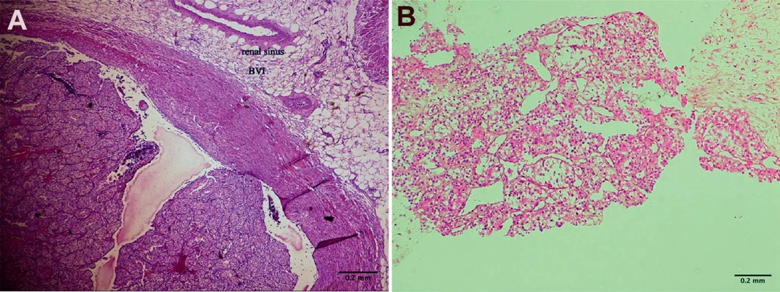
A 55-year-old male who underwent left radical nephrectomy,4 years back for a left renal mass,presented with swelling over left chest since 2 months.His initial histopathology report of the radical nephrectomy specimen mentioned a 7.2 cm×6 cm large renal mass involving the upperpole of the left kidney which was infiltrating the renal sinus and was reported as a stage pT3a clear cell carcinoma with Fuhrman’s grade III nuclei.On physical examination,non-tender,firm swelling approximately 10 cm×5 cm sized was seen and palpated in the left upper chest(Fig.1).His current blood biochemical workup including liver function tests was normal.Contract enhanced computed tomography(CECT)of abdomen showed empty left renal fossa with no evidence of recurrence.CECT of the chest showed two well defined,heterogenous enhancing lesions with central necrosis,involving anterior chest wall muscle and extended up to the left lateral cardiac border with the larger one being approximately 99 mm×47 mm in size(Fig.2).Subsequently,percutaneous trucut biopsy from the chest wall swelling was performed which on histopathological examination was confirmed as clear cell carcinoma similar to the previous radical nephrectomy specimen.He was started on tyrosine kinase inhibitors(TKI).At 2-month follow-up,the patient is doing well and there has been a marginal reduction in size of the lesion.We plan to re-examine the patient after another month,see for any further reduction in size and do a metastatic workup before counselling for local surgical resection,if there’s a good therapeutic response.
Renal cell carcinoma usually has a propensity to metastasize hematogenously to the lungs and bones[1,2].Unusual sites of metastasis include gastrointestinal,head and neck,cutaneous,muscular sites,and even the heart[1-6].According to literature,chest wall metastases originating from clear cell renal cell carcinoma especially presenting in ametachronous manner are very rare[1,2,5,6].When it comes to large chest wall metastasis like in our case,it’s difficult to pinpoint the site of origin(bone or subcutaneous tissue).Treatment options described for these rare lesions include systemic therapy along with aggressive local resection with chest wall reconstruction,immunotherapy alone and sometimes highly focused radiation in carefully selected patients[3,4].This case highlights the fact that such a possibility should be considered in patients presenting with chest wall lesions who have a history of ccRCC and who have had no recurrence or metastases for a considerable period of time,in order to ensure precise diagnosis and treatment for such rare ectopic metastasis[3].

Figure 1 Clinico-radiologic images.(A)Clinical photograph showing the large mass on the left chest wall;(B and C)Axial and sagittal cuts of contrast enhanced computed tomography images showing two well defined,heterogeneously enhancing soft tissue density lesions with central necrotic core involving ribs and anterior chest wall muscle.

Figure 2 Histopathology images.(A)Photomicrograph of old nephrectomy specimen showing clear cells with abundant cytoplasm suggestive of clear cell carcinoma with delicate vascular network with blood vessel invasion and renal sinus invasion(HE stain,100×magnification);(B)Photomicrograph of core biopsy specimen from chest wall mass showing similar clear cells with abundant cytoplasm suggestive of clear cell carcinoma(HE stain,100×magnification).HE,hematoxylin and eosin.
Author contributions
Study design:P.Ashwin Shekar.
Data acquisition:Hardik Patel,P.Ashwin Shekar,Dinesh Reddy,Sandhya Ramachandran.
Data analysis:Hardik Patel,P.Ashwin Shekar,Dinesh Reddy,Sandhya Ramachandran.
Drafting of manuscript:Hardik Patel,P.Ashwin Shekar.
Critical revision of the manuscript:P.Ashwin Shekar.
Conflicts of interest
The authors declare no conflict of interest.
 Asian Journal of Urology2021年4期
- Asian Journal of Urology的其它文章
- Radical cystoprostatectomy with orthotopic neobladder for a case of treatment emergent neuroendocrine prostate cancer presenting as bladder mass with hematuria-a rare instance of tumor remission after local control
- Late upper urinary tract urothelial carcinoma following radical cystectomy,presenting as page kidney
- Perioperative anticoagulation and open distal corpora cavernosa shunt in the management of a case of stuttering idiopathic persistent childhood ischaemic priapism
- Effect of tamsulosin versus tamsulosin plus tadalafil on renal calculus clearance after shock wave lithotripsy:An open-labelled,randomised,prospective study
- A novel spherical-headed fascial dilator is feasible for second-stage ultrasound guided percutaneous nephrolithotomy:A pilot study
- Impact of COVID-19 on endourology surgical practice in Saudi Arabia:A national multicenter study
