
Technological advancements in the analysis of human motion and posture management through digital devices
2021-07-22 06:52:18FedericoRoggioSilviaRavalliGraziaMaugeriAntoninoBiancoAntonioPalmaMichelinoDiRosaGiuseppeMusumeci
World Journal of Orthopedics 2021年7期
Federico Roggio, Silvia Ravalli, Grazia Maugeri, Antonino Bianco, Antonio Palma, Michelino Di Rosa, Giuseppe Musumeci
Federico Roggio, Antonino Bianco, Antonio Palma, Department of Psychology, Educational Science and Human Movement, University of Palermo, Palermo 90144, Italy
Silvia Ravalli, Grazia Maugeri, Michelino Di Rosa, Giuseppe Musumeci, Department of Biomedical and Biotechnological Sciences, Human Anatomy and Histology Section, School of Medicine, University of Catania, Catania 95123, Italy
Giuseppe Musumeci, Research Center on Motor Activities, University of Catania, Catania 95123, Italy
Giuseppe Musumeci, Department of Biology, College of Science and Technology, Temple University, Philadelphia, PA 19122, United States
Abstract Technological development of motion and posture analyses is rapidly progressing, especially in rehabilitation settings and sport biomechanics.Consequently, clear discrimination among different measurement systems is required to diversify their use as needed.This review aims to resume the currently used motion and posture analysis systems, clarify and suggest the appropriate approaches suitable for specific cases or contexts.The currently gold standard systems of motion analysis, widely used in clinical settings, present several limitations related to marker placement or long procedure time.Fully automated and markerless systems are overcoming these drawbacks for conducting biomechanical studies, especially outside laboratories.Similarly, new posture analysis techniques are emerging, often driven by the need for fast and non-invasive methods to obtain high-precision results.These new technologies have also become effective for children or adolescents with non-specific back pain and postural insufficiencies.The evolutions of these methods aim to standardize measurements and provide manageable tools in clinical practice for the early diagnosis of musculoskeletal pathologies and to monitor daily improvements of each patient.Herein, these devices and their uses are described, providing researchers, clinicians, orthopedics, physical therapists, and sports coaches an effective guide to use new technologies in their practice as instruments of diagnosis, therapy, and prevention.
Key Words: Motion capture; Gait analysis; Inertial measurement unit; Wearable devices; Rasterstereography; Posture
INTRODUCTION
Over the last decades, human movement research has made significant progress in responding to the growing medicine and sport demand for precise and accurate methods to capture human movement[1] and refine data collection[2].Motion and posture analyses are effective tools used in diagnosis, therapy, and prevention of musculoskeletal disorders.Notably, human motion assessment during functional activities also plays a crucial role in rehabilitative medicine and sports.Nevertheless, there is a solid need to diversify the use of each system in relation to specific contexts.In some instances, 2D biomechanical analysis can offer a quick and effective method of evaluation.Movements, such as walking or running, do not require sophisticated approaches, since they are easily analyzed in the sagittal plane.Otherwise, if the movement needs to be studied on multiple planes or forces investigation is required, it is more appropriate to use a 3D system, which requires in-depth expertise.Biomechanical researchers aim to standardize human movement parameters that can be understandable, comparable, and shareable with the entire scientific community.Quantitative analysis of human movements and posture is an effective tool used to analyze the correct movement execution, identify injury risk factors[3], help clinicians make the best decision to reduce patients' recovery time, and suggest a proper treatment plan[4].
Assessing walking speed through wearable systems could be a valuable indicator of adults' health and functional status[5,6].For example, low physical activity levels are associated with muscle weakness, decreased mobility function, and widespread pains[7].Fast return to play sports and exercise could trigger joint pains and musculoskeletal alterations; therefore, an accurate motion and posture analysis could help planning the right approach to resume physical activity.
Hence, technological devices can broadly be used to diagnose musculoskeletal disorders and plan a preventive strategy for returning to the sport practice.Similar advice is suggested for those who return to physical activity after surgery.Long periods of inactivity caused by surgery inevitably lead to loss of muscle mass and reduction of movements fluidity; therefore, movement analysis in the return-to-dailyactivities phase can be performed to detect dysfunctions and re-educate the patients.
Although marker-based and non-invasive systems are more commonly used to evaluate pathological patients,e.g., subjects with spinal cord damage, amputees, strokes and cerebral palsy, scoliosis…, instrumental biomechanics have the potential for reaching every subject, from the one who suffers from musculoskeletal pathologies to the one who reports only mild pain.Therefore, it is encouraged to use these devices to study every and unexplored aspect of movement science.This review aims to highlight the importance of new technologies in human movement and posture analysis, suggesting how they can strengthen orthopedics, rehabilitation, health prevention, sports science and guide the clinicians towards a personalized diagnostic process and treatment plan based on the patient’s characteristics.
FROM MARKER-BASED TO MARKERLESS MOTION ANALYSIS SYSTEMS
The optoelectronic stereophotogrammetric multi-camera capturing system is the gold standard for motion analysis, tracking reflective markers placed on the body[8,9].One of the most known, the Vicon system (Vicon Motion Systems, Oxford, United Kingdom), consists of multiple infrared cameras for kinetic, kinematic, and spatiotemporal movement analysis.The markers, positioned on anatomical landmarks in correspondence with the joints involved in the analysis, allow tracking all the human motion features with high accuracy[10,11].This system precisely evaluates each joint's movements in the space at any time, and it can define the level of functional limitation and disability resulting from the evolution of a disorder, including post-traumatic or surgical alterations.Vicon is commonly used in clinical gait analysis, amputee rehabilitation, lower limb movement studies, cerebral palsy research, motor control, and neurosciences.The company produced a platform for the life sciences community called Nexus, a powerful, all-inclusive modeling and processing tool for movement analysis.Operators can reduce the time spent processing the data by creating their workflow templates; the system automatically loads the data and produces the report in the simplest or most detailed way required.However, marker-based systems show several limitations, including long preparation times, soft tissue artifacts, or unfeasibility of specific movements due to the presence of the markers, which can hinder the correct execution of the movement[12].These systems are expensive and require a large setting in order to place all the cameras needed for the analysis.The placement of the markers on anatomical landmarks can be challenging since it depends on the clinician's ability to locate them correctly and, therefore, human error could incur.Particularly for transverse plane movements, there is an inevitable variability in the marker positioning between different days or different clinicians' hands, reducing the measurements' reliability[13].In the literature, several protocols can be found for locating joint centers or defining segment pose, as shown in Figure 1A; however, these different protocols produce variable results, especially for the sagittal plane, compared to the same gait cycles[13].These drawbacks can limit the use of marker-based systems within certain areas of motion analysis.For these reasons, nowadays, markerless systems are offering new opportunities to obtain similar results.
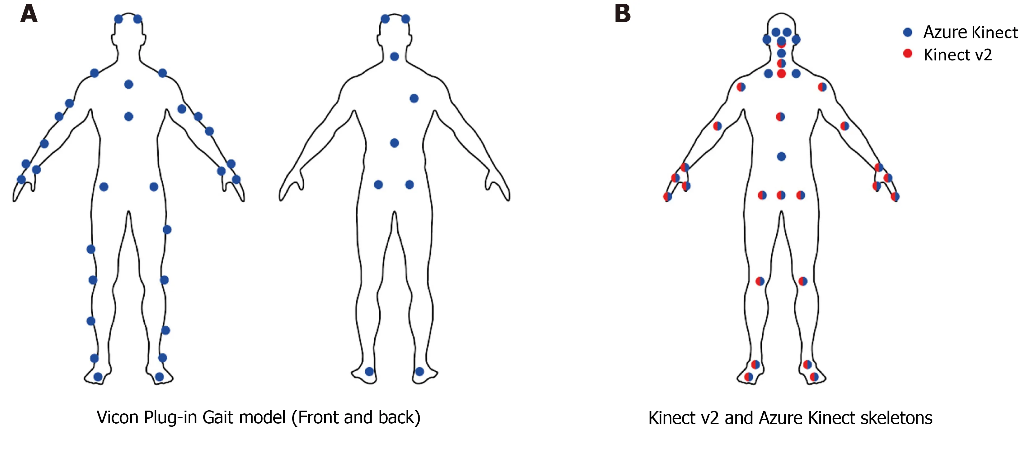
Figure 1 Markers setup for Vicon and markerless caption for Kinect cameras[28].
The markerless motion analysis system presents a fast, fully automatic, and noninvasive approach that can significantly improve research and practice within sports biomechanics and rehabilitation.For instance, motion can be analyzed during normal training in a common laboratory, without the long preparation times due to the markers placement and the laborious manual work.Furthermore, it can provide a reasonable solution for a common dilemma faced in biomechanics laboratories,i.e., the constant search for balance between accuracy and reproduction of motion without artifacts.Several researchers investigated the most common movements studied in biomechanics laboratories,e.g., walking, jumping, and jogging, by analyzing the accuracy of markerless systems compared to marker-based techniques[14-16].Therefore, a potential application of markerless systems in gait analysis in clinics is suggested, even though an experienced clinician should validate the results to ensure their reliability.The most popular markerless system, the Microsoft Kinect v1, is the first 3D camera whose affordable price made it accessible to almost all consumers.One of its innovations was that the sensor was suitable for gait assessment outside the laboratory, becoming a portable device[17-19].Soon after, Microsoft launched the Kinect v2 with improved hardware and skeleton tracking system; its software, Kinect for Windows SDK 2.0, allows tracking the 3D positions and orientations of 25 joints, up to 6 users simultaneously[20].This camera can track the skeleton through artificial intelligence (AI).Briefly, this feature allows recognizing joint centres and segment orientation, providing the ability to calculate joint kinematics and spatiotemporal aspects of the movement.The Microsoft Research team produced a specific algorithm to identify the anatomical landmarks, created by training a randomized decision forest algorithm using a subset of 100000 depth scans of a variety of movements, including running, dancing, driving, kicking[21].
In recent years, several studies investigated the reliability of Kinect v1 and v2 to evaluate if these devices could be used as an alternative to the multi-camera motion capturing systems.Several contexts were examined, such as walking on a treadmill or performing physical exercises at a static location[22].Wanget al[23] examined the differences in human pose assessment between Kinect v1 and Kinect v2 in relation to twelve different rehabilitation exercises.The authors reported that Kinect v2 has overall better accuracy in joint estimation and body rotation detection than Kinect v1.Capecciet al[24] investigated the accuracy of Kinect v2, in terms of joint positions and angles, during dynamic postures used in low back pain rehabilitation, reporting that this system can measure timing characteristic of physical exercises and reproduce dynamic features similar to a stereophotogrammetric system[25,26].Microsoft ended the production of the Kinect v2[27] in favor of new technology, the Azure Kinect DK.The latter offers significantly higher accuracy than other commercially available cameras[28].Unlike the Kinect v2, the Azure Kinect employs a Body Tracking SDK to track multiple users up to 32 joints, as shown in Figure 1B; it includes more joints,i.e., anatomical landmarks, in the face area, such as ears and eyes.Albertet al[28] collected data from Microsoft Kinect v2 and Azure Kinect in gait analysis and compared them to the Vicon system.The results showed high accuracy of both cameras, Microsoft Kinect v2 and Azure Kinect, but the latter showed better accuracy for spatial gait parameters.Figure 2 shows spatial agreement of Microsoft Kinect and Azure Kinect cameras with respect to the Vicon system.Walker View (Tecnobody?-Dalmine, Italy) is a treadmill whose base includes eight load cells that allow the system to detect the user's spatiotemporal parameters.The presence of a Microsoft Kinect 2 camera automatically identifies anatomical landmarks using AI.The system is connected to a 49" LCD Monitor for the biofeedback and the virtual reality.Its advantage is the fully automatic and non-invasive approach, which is also an improvement in sport and rehabilitation research and practice.During the testing phase the patient/athlete can auto select the preferred walking speed or choose the Speed Control feature that adapts the treadmill speed to user’s step velocity.In addition to the gait analysis, the Walker View can also perform run analysis, an especially useful evaluation for athletes.Regarding the training area, the patient can perform the Gait Trainer program where the software, through visive and acoustic feedback, helps him to improve his walking.Furthermore, the Walker View, thanks to the Smart Gravity system, can be used for patients with severe walking deficits unable to stand on their own.This system consists of a mechanical support to which a sling worn by the patient is connected.It can simulate a walk in the pool by selecting the appropriate weight reduction as if it were hydrokinesitherapy.
Miniaturized inertial measurement units (IMUs) are a new generation of lightweight, small, and inexpensive systems embedding 3D accelerometers, gyroscopes, and magnetometers, which may offer other opportunities for the biomechanical assessment of motor functions.IMUs can track the trajectory of anatomical segments in real-time and estimate the kinematic parameters of the gait cycle[29].Although evaluation protocols are not homogeneous, several studies estimated the possibility of assessing the gait analysis through IMUs[30-34], as reported in Table 1.Fuscaet al[35] recruited ten volunteers on which they placed markers for motion capture using Elite (BTS) System, and IMU sensor placed anteriorly and close to the body's center of mass.The authors stated that it is preferable to place the IMU sensor posteriorly between the superior anterior iliac spines since abdominal breathing could lead to artifacts.Four walking trials were simultaneously recorded at a self-selected speed by blindly comparing the two systems.The stride time and the cadence had a mean absolute percentage error of 5.7% and 4.9%, respectively, for IMU.The mean absolute percentage errors were 5.6% and 13.5% in the step length and step speed measurement, respectively.Therefore, results confirmed that the IMU is effective for a reliable assessment of human gait spatiotemporal parameters.The Italian Company Captiks Srl developed a novel inertial-sensor based system, Movit System[36], which allows measuring gait parameters by positioning the IMUs through elastic bands on the pelvis (between iliac crests), thighs, shanks, and feet, as shown in Figure 3.The company compared the results obtained from Movit System and Vicon optoelectronic system.According to the statistical analysis of the data on joint ROM reported by Cuesta-Vargaset al[37] and Poitraset al[38], the authors agreed on the excellent accuracy and test-retest reliability of IMUs on joint movement and walking tests.IMUs can encounter some drawbacks: these devices are placed on the human body through elastic bands, but unpredictable vibration artifacts can occur if the wearables are not firmly and adequately fixed.This represents an important issue since the artifacts are in the signal's frequency band, so not removable by filtering.Besides, misplacement of the sensors and movements that could cause the sensors to slip can lead to wrong measurements and make the analysis inconclusive[35].The main differences between the mentioned motion analysis systems can be found in Table 2.
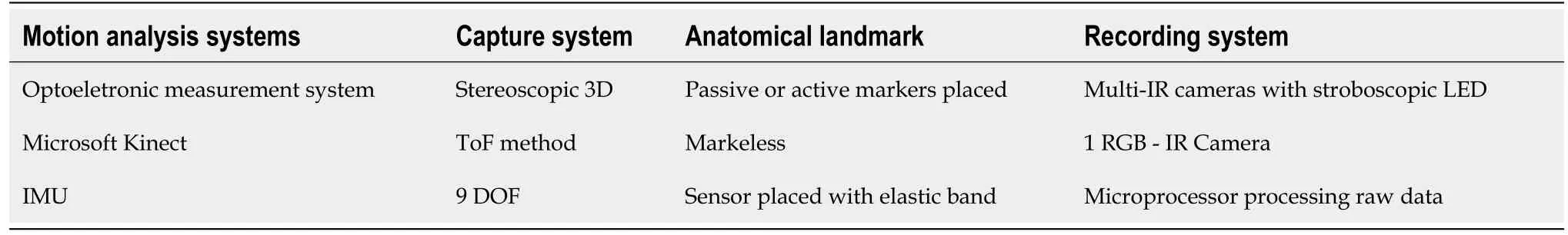
Table 2 Main features of motion analysis systems
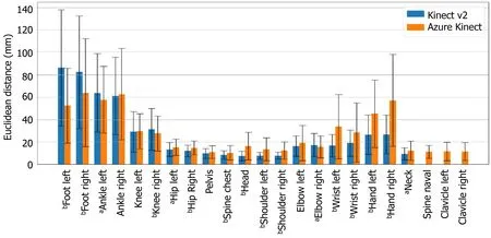
Figure 2 Spatial agreement of Microsoft Kinect and Azure Kinect cameras with respect to the Vicon system.
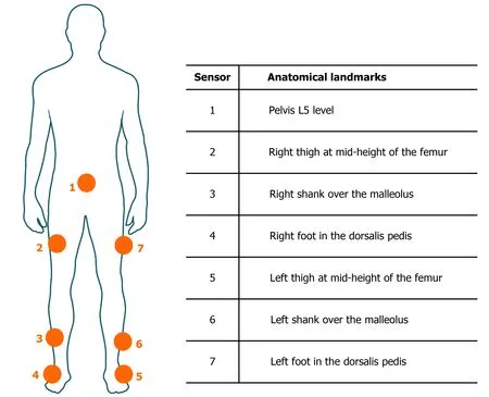
Figure 3 Body position of the seven inertial measurement units of Movit System.
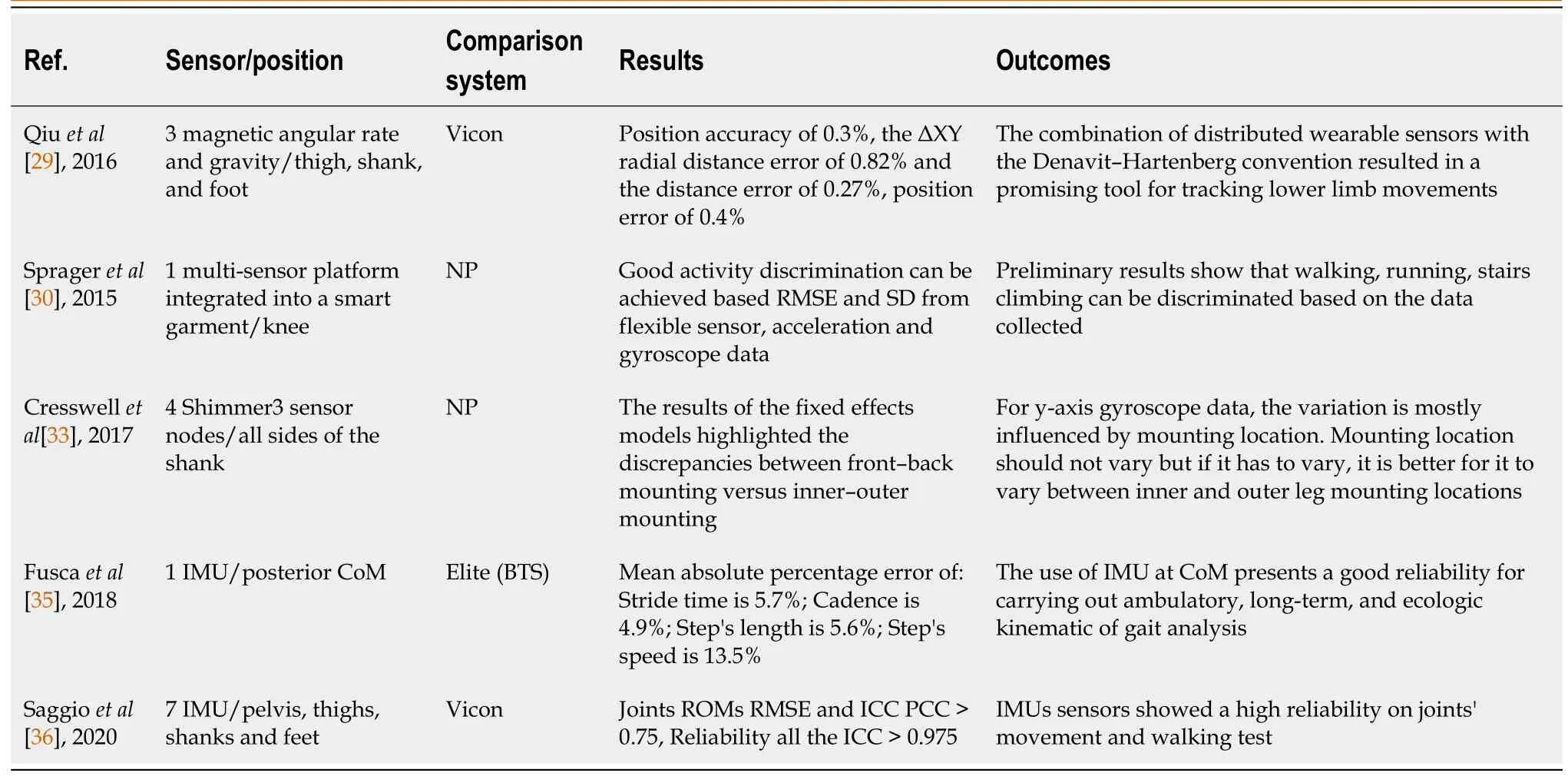
Table 1 Studies investigating the reliability of inertial measurement unit sensors
Considering that three-dimensional motion capture systems are rather expensive, different low-cost methods have been developed during recent years.For instance, Kinovea is a free 2D motion analysis software for computers that can measure kinematic parameters.This software allows analyzing video without markers, although its reliability may improve with marker positioning[39].Several studies tested Kinovea software[40-43] in different environments with good results: Damstedet al[39] investigated its ability to detect hip and knee joint positions in the initial contact phase during running; Elwardanyet al[44] analyzed the range of movement of the cervical spine in the sagittal plane while Mathewet al[45] used this software to study the ankle, knee, and hip joints at different gait cycle phases.Kinovea only needs a camera and eventually some markers, although it requires an experienced clinician to use it.Once the movement is recorded, the clinician, in post-production, places the virtual anatomical landmarks over the joint centers or the markers physically positioned on the user.Unlike Microsoft Kinect, Kinovea does not have the appropriate software for the skeleton tracking system, so accurate marker placement or precise location of virtual anatomical landmarks is required to evaluate the movement correctly, otherwise, the results may not be valid.As a matter of fact, the main limitations uncovered by the literature concern the lack of a standard video analysis protocol and the marker placement[44,45].The study conducted by Gonzálezet al[42] evaluated the inter- and intra-rater reliability of Kinovea and the agreement with a three-dimensional motion system (Vicon) to detect the lower limb's joint angles during walking.The results showed significant differences in the hip, knee and ankle angles reporting a ± 5° difference for the hip and ankle angles, ± 2.5° for knee angles.According to McGinleyet al[46], an error of 2° or less is considered acceptable in clinical evaluation.Errors between 2° and 5° are also reasonable, although the data should be interpreted cautiously.Errors over 5° could mislead the interpretation.To conclude, as reported by Littrellet al[47], the use of Kinovea could lead to high error for the pelvis and the foot during the stance period of the gait cycle.However, the software is reliable when analyzing other phases of walking and other kinematic parameters such as joint angles[48], especially for sports environments or dynamic conditions where sophisticated systems could be impossible to be used.
Recently, the use of smartphone applications (app) to measure gait parameters increased.Clinicians, researchers, and coaches can use these applications to analyze joint angles immediately.Unlike the previously mentioned systems, mobile applications are less expensive, portable, and easy to use.Although there is a lack of scientific studies investigating their reliability, the possibility of quickly measuring posture and joint angles in ordinary circumstances makes these applications compelling.Coach’s Eye (TechSmith Corp) is a 2D motion analysis mobile app able to evaluate gait analysis in patients and healthy individuals[49], although it was specifically designed for coaches and trainers to assess athletic performance.The app computes joint angles and their variations by a digitized goniometer without applying any marker on the body.The videos can be recorded on frontal and sagittal planes and analyzed, frame by frame, going forward or backward.An online video database allows comparing the recorded videos with those of other athletes.However, only a few studies compared the app's data with the 3D motion analysis systems during sports tasks.Mousaviet al[50] investigated the validity and reliability of Coach’s Eye app for selected lower-limb kinematics during treadmill running by comparing the results with those deriving from a traditional 3D motion analysis system (Vicon).The authors recruited 20 healthy female recreational runners who wore 16 reflective markers for the 3D comparison.The subjects were asked to run on a treadmill at a selfselected speed.Concerning the validity, Coach’s Eye showed only 1-2 degrees of difference compared to Vicon in kinematic measurements for the sagittal plane hip angles at touchdown and toe-off, knee angle at touchdown, ankle angle at toe-off, and rearfoot angles at touchdown and toe-off.Measures of ankle angle at touchdown and knee angle at toe-off were not as accurate, reporting a bias ranging from 4 to 20 degrees.Furthermore, Coach’s Eye demonstrated excellent test-retest reliability for all joint kinematic measures, in agreement with Krauseet al[51] who reported high reliability of the application during the squat execution.
The authors recommended the use of Coach’s Eye to record and assess sagittal plane lower-limb joint kinematics and rearfoot in/eversion at touchdown, hip, ankle, and rearfoot eversion at toe-off, but not for hip and ankle at touchdown or knee at toeoff.Nevertheless, given its ease of use and low cost, it would represent a manageable tool for sports coaches who frequently evaluate athletes.
Electromagnetic motion acquisition systems consist of a series of receivers that measure their position in space and transmit it to a nearby receiver.They are based on the electromagnetism principle: the emission source produces an electromagnetic field, and the sensors send the signalviacable to the processing unit, then the computerized system calculates sensors’ position and direction in space based on these signals.For example, Polhemus and Ascension are two of the most popular companies producing electromagnetic motion systems[52,53].This system finds application in analyzing a single fine movement, such as taking an object with the hand, which has high accuracy and a low margin of error compared to camera-based systems[54].However, complex movement analysis, such as walking, could be challenging.For this reason, the application is less suitable for clinical and sports movement analysis, such as gait analysis or technical sports gestures.Conversely, the entertainment industry exploits its high accuracy to reproduce the movement executed by a performer over a digital character[55].Recently, Polhemus enabled localization of medical instruments through the trackers, especially for image-guided therapy[56].The application of electromagnetic systems can be notably valid to enhance the medical students’ skills, such as the use of endoscope and surgical instruments, tissue manipulation, use of precision tools, and other procedural skills before operating on patients.
In summary, the optoelectronic system is undoubtedly the gold standard for motion analysis, although modern markerless options might overcome some disadvantages and offer a valid alternative for outdoor examination.The strength of markerless systems relies on testing more users in shorter times and less equipment than the marker-based system.The markerless approach is more suitable for sport and rehabilitation purposes rather than diagnostics.Another way to capture human motion is by IMUs, lightweight devices easy and comfortable to be used almost everywhere.Finally, software or mobile application is applied, especially in athletic contexts where sophisticated tools collide with sport practice.
GAIT ANALYSIS IN PREVENTION AND HEALTH PROMOTION
Gait analysis is recognized as a useful assessment tool in the field of human movement research, commonly used in biomechanical laboratories to assess walking ability in patients with specific motor disabilities[57-60], often due to conditions as severe developmental motor impairments[61], spinal cord damage[62], amputees[63], orthopedic surgery[64], strokes[65] and cerebral palsy[66].Specifically, clinical gait analysis can be classified into two levels of examination: a first level which deals with the clinical evaluation of the lower limb impairments by collecting data from spatiotemporal parameters, kinematics, and kinetics of locomotion; a second level which involves the use of dynamic electromyography, during gait, to evaluate the neuromuscular activity[67].For the purely medical use, the SIAMOC (Italian Society of Clinical Movement Analysis) proposes these guidelines: (1) in cerebral palsy, the use of gait analysis, combined with an expert clinical evaluation, can influence the planning of functional surgery; (2) in adult brain injuries, the use of gait analysis can influence the orthopaedic surgery, neuromuscular blocks or rehabilitation programs; and (3) in patients wearing lower limb prostheses, gait analysis might be useful for choices regarding the construction of the prosthesis and the planning of general models of rehabilitation[68].Information deriving from this evaluation allows to increase diagnostic accuracy, differentiate diagnosis and severity, and help in decisionmaking about the treatments.Evidence demonstrates the efficacy of 3D gait analysis in defining gait problems, their causes, and the appropriate treatments (e.g.,surgery against non-surgical treatment or type of surgery).However, gait analysis continues to be a helpful tool partially exploited.The literature is not yet robust regarding using this system even outside clinical contests.Therefore, human motion analysis is limited to clinical examination,i.e., orthopaedic, neurological, or surgical, not considering the possibilities deriving from daily life evaluation.Prevention and health promotion science could exploit gait analysis to avoid that a simple dysfunction develops into an actual disease.What would happen if orthopaedic patients performed gait analysis as a routine examination rather than in sight of surgery? For instance, according to Meireleset al[69], the early stages of knee osteoarthritis are challenging to detect, but it is known that biomechanical factors may contribute to its onset[70-73].This study calculated knee contact forces and their relations with external knee adduction and flexion moments and reported that mechanical loading was not significantly higher in early osteoarthritis subjects.These results highlight the possibility that other causes (e.g.,spatiotemporal parameters, hip or ankle kinematics) might occur to develop osteoarthritis and therefore, gait analysis might be considered in disease management since current treatments offer limited benefits[74].Several contests can benefit from an accurate gait analysis guiding clinicians towards the best decision based on patients' needs.Pathologies involving walking abnormalities that afflict the central nervous systems are highly variable, and several different motor patterns can be altered.In cerebral palsy subjects, gait analysis investigated the common altered gait patterns, including jump knee gait, toe-toe gait, crouch knee gait, stiff knee gait, and rotational abnormalities.However, each subject presents a unique combination of impairment and compensatory movements so, gait analysis can help clinicians recognizing the primary deformities and how they affect the musculoskeletal[75].While the use of this approach in cerebral palsy has been widely investigated, there is less evidence in other neurological conditions.Gait analysis among spinal cord-damaged populations can help establish individualized interventions to enhance walking, improving muscle strength, coordination, proprioception, and postural control.Specifically, Murphyet al[62] highlighted how gait analysis could provide specific information to clinicians to deal with spasticity management,i.e., botulinum toxin-a injection; best orthotic selection,i.e., hinged or rigid; surgery,i.e., joint fusion; or establish the well-suited rehabilitation program[76].In orthopedic contests, gait analysis can enhance understanding the subject's functional capacity reduction before surgery and, in the same way, indicate the elements that need to be improved following the surgery[77].The fear of moving after surgery is often present in patients[78]; digital support, in this case, can help the patient understanding that within a specific range of movement,e.g., walking for 500 m or walking at speed 2 km/h, will not suffer further pain and will not negatively affect recovery.
The use of gait analysis in the early stages of many pathologies could suggest reeducational intervention to reduce surgery later.Therefore, patients should be first examined through markerless systems and proceed to in-depth marker-based analysis when necessary, as reported in Figure 4, with improved time and cost-efficiency.
COMPUTERIZED ANALYSIS OF THE SPINE
Rasterstereography is a non-invasive method used to measure 3D spine deformities by analyzing the back's surface topography on triangulation principles[79,80].It was developed by Drerup and Hierholzer[81-83] in the 1980s as a valid alternative to radiography, and over the years, it has shown its high reliability in various studies[84-86], Table 3 shows the main differences among these studies.The system generates a 3D model of the spine by calculating specific deformities thanks to the precise convex shape of the spinous process of the vertebra prominence and the concavity of the lumbar dimples as fixed points.The rasterstereography is commonly used to assess the presence of scoliosis, but it can efficiently evaluate other parameters as pelvic obliquity, thoracic kyphosis and lumbar lordosis angles[87,88].Since this system is a non-invasive method, it can perform several analysis repeated over time with high reliability, which can minimize the use of X-rays[79].Krottet al[89] provided a metaanalysis of 19 eligible studies evaluating the reliability and validity of static rasterstereographic measurements in healthy subjects and patients with different spinal pathologies.The authors compared the results with the gold standard of radiological imaging techniques, reporting high validity levels in assessing patients' thoracic kyphosis angle, lumbar lordosis angle, and scoliosis angle.The easiest accessibility of rasterstereography can spread through evaluating common health problems among children, adolescents[90], and adults, such as non-specific back pain and postural insufficiencies[91-94].Several studies proved the validity and reliability during static upright standing in children and healthy adults, while recently, it was investigated the use of rasterstereography from static to a dynamic system.Michaliket al[86] evaluated the spinal posture and pelvic position under dynamic conditions,i.e.,walking, and compared it to static measurements of the spine using a dynamic rasterstereographic system.Significant differences were found between static and dynamic conditions about the trunk inclination (P< 0.001), kyphotic angle at 2 km/h (P= 0.003), lordotic angles (P< 0.001) and lordotic angles with increasing walking velocities (P< 0.001).No differences were found between static and dynamic measurements for the surface rotation.M?rzet al[95] used rasterstereography to determine the instantaneous influence of different occlusal positions on spine and body posture.After comparing ten spinal and body posture parameters (i.e.,trunk inclination, pelvic tilt, kyphotic and lordotic angles), in six different occlusal positions, only three parameters were found to differ.The authors concluded that a plausible explanation could be represented by neuromuscular compensation for body balance and posture on trigeminal proprioception[96].For instance, postural alterations provoked by changes in dental occlusion and masticatory muscle function suggest a neurophysiological link between the stomatognathic system and other body muscles[97,98].The rasterstereography field of application involves: screening programs,e.g.,early diagnosis and monitoring of scoliotic and scoliosis attitudes, lumbar hyperlordosis, dorsal hyperkyphosis, and all pathological conditions of the back; postural evaluation and musculoskeletal problems; design and verification of ergonomic and orthopaedic devices (ergonomic insoles, bites, prostheses, orthoses); support to therapeutic programs and postural reeducation.Formetric (DIERS Medical Systems, Chicago, IL, United States) is a rasterstereographic technology for analyzing the spine and posture that does not present any contraindications or side effects.It emits parallel lines of light across the posterior trunk surface and, based on the distortion of those lines, reconstructs a digital image of the back's surface and produces a digital model of the spine.The optical scan detects the anatomical landmarks (C7 or prominent cervical vertebra, sacrum, lumbar dimples), the symmetry of the spine and the rotation of each segment.Three different versions are currently available on the market: Formetric Basic, Formetric Basic 4D, and Formetric Basic 4D Motion.Formetric Basic produces a 3D analysis of the spine and posture, but it does not allow to perform a dynamic analysis.Formetric 4D can acquire image sequences, automatically processing average values, with a duration of the detection sequences even greater than 1 minute and the possibility to acquire up to 10 images per second.The newest version of this system is the Formetric 4D Motion, which can accomplish a dynamic analysis of the whole body and the skeletal system during a step execution or treadmill walking, due to the possibility to acquire up to 24 images per second.The high sampling rate allows excluding effects due to spontaneous postural oscillations or breathing.Once the exam has been performed, the system produces a report about physiological alterations of the spine both in the frontal and in the sagittal plane, degrees of vertebral rotation, pelvic tilt and antero-retroversion.
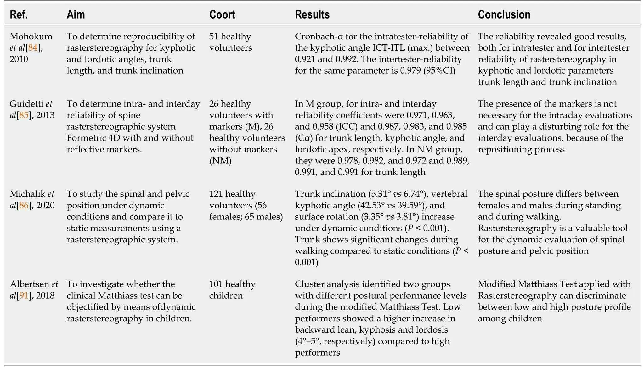
Table 3 Studies investigating the reliability of rasterstereography to evaluate the spine
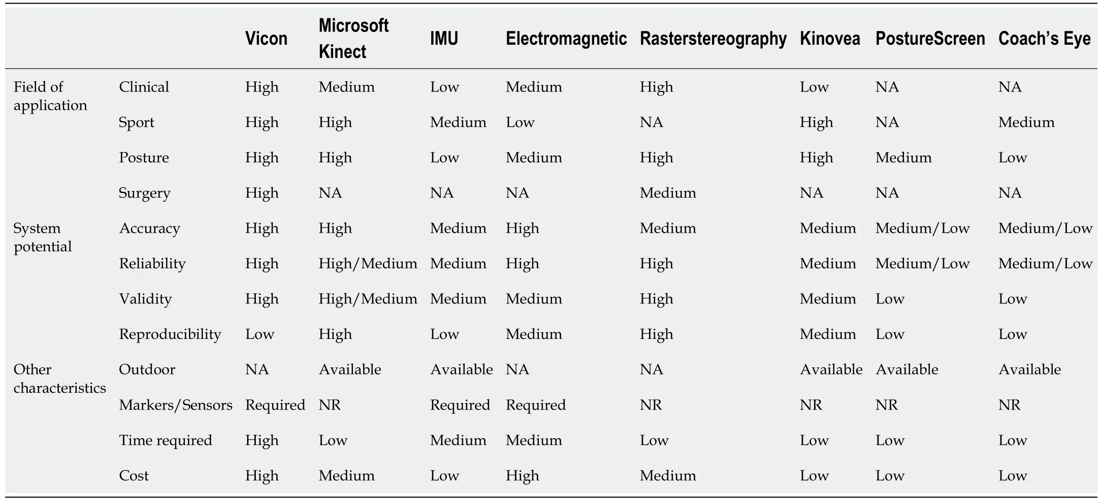
Table 4 Application outline of each mentioned system
MOBILE APPLICATION AND WEARABLE DEVICES FOR POSTURE MANAGEMENT
PostureScreen Mobile (PSM) (Trinity, FL, United States) is an app that guides clinicians in rapidly identifying anatomical landmarks and posture assessment.Without the use of reflective markers, the app calculates posture variables using digitized anatomical landmarks.The device camera is used within the app to take pictures of subjects from frontal and sagittal planes.Once the picture is taken, the clinician place digital anatomical landmarks on the picture to produce an evaluation of the misalignment of the landmarks on the coronal and sagittal planes, as reported in Figure 5.Therefore, the app provides a file report that indicates possible posture misalignments.In the frontal plane, it analyzes head, shoulders, hips tilt, and horizontal translation; in the sagittal plane, it analyzes the shift forward or rearward of the head, shoulders, hips and knees.Szucset al[99] tested the app in healthy young adults.Based on the results, the PSM app has been a reliable method for assessing posture within and across raters when using defined procedures and markers.For intra-rater reliability, most posture variables showed good to excellent reliability (> 0.75).Angulation variables showed moderate to good reliability range (0.50-0.75).In contrast with the previous study, Hopkinset al[100] analyzed the intraclass correlation coefficient of postural analysis between PSM app and Vicon 3D analysis.The results showed a significant bias in postural measurements in the frontal and sagittal with the PSM app, while the intraclass correlations were similar in most of the measurements between the two systems.These data suggest caution using the PSM app when highly accurate postural assessments are necessary.Instead, innovative use of this technology is proposed by Iacobet al[101], which tested the PSM app to evaluate dental occlusion anomalies.Both static and dynamic occlusion were evaluated.In the first part of the study, the subjects were divided according to normal or abnormal occlusion; statistically significant differences (P< 0.05) were obtained for only the head deviation angle.In the second part of the study, after examining the dental occlusion in each type of movement, subjects were grouped according to the presence or absence of premature contacts or interferences.Statistically significant differences regarding head deviation angle between the groups normal/abnormal occlusion were found, confirming a possible correlation between static occlusion and posture.Therefore, the PSM app is suggested to assess the impact of pathological dental conditions on posture.Furthermore, depending on the degree of postural deterioration, the clinician can direct the patient toward correction therapy before the postural abnormalities become definitive and harm the musculoskeletal system[101].
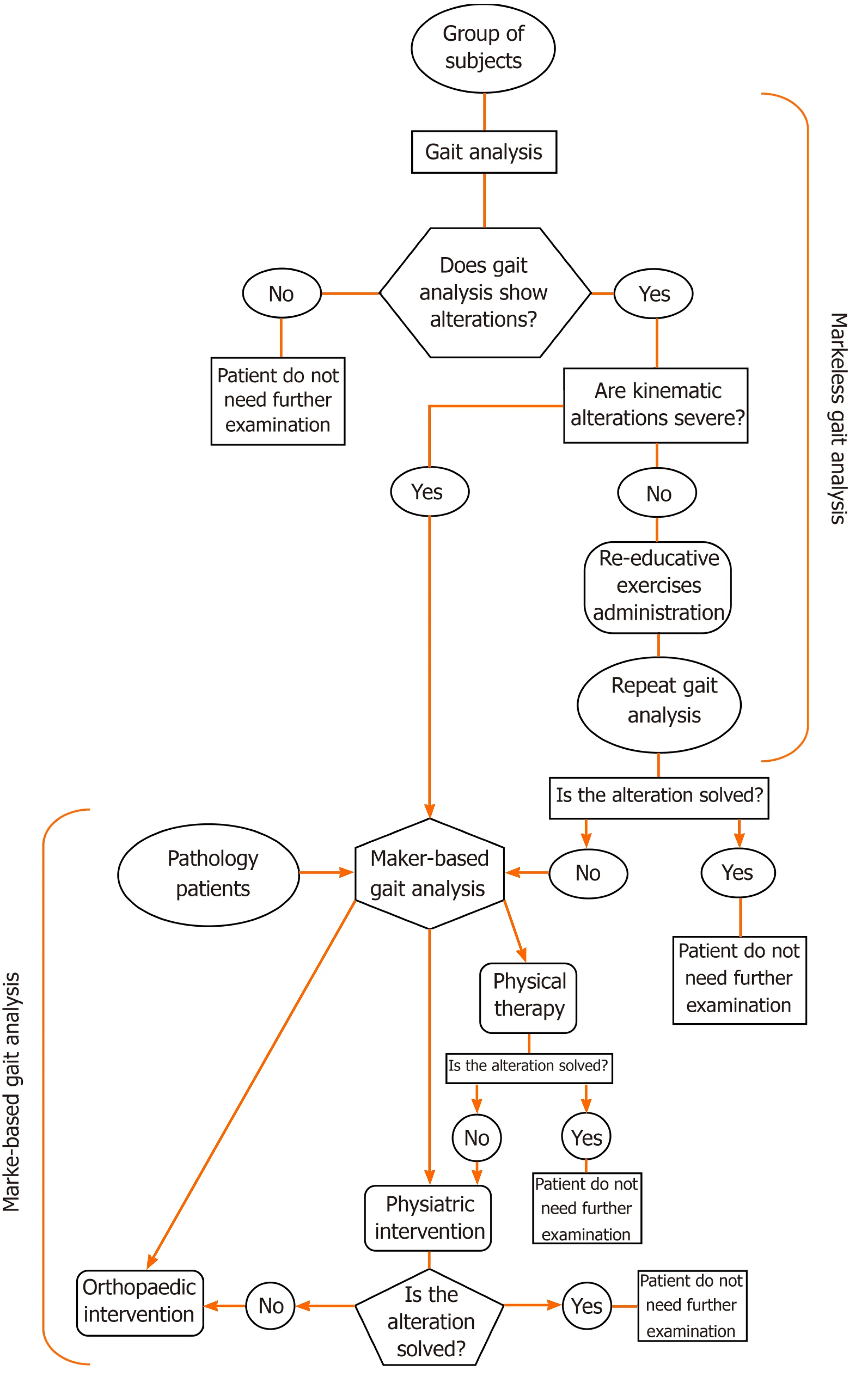
Figure 4 Flow chart of the gait analysis approach levels.
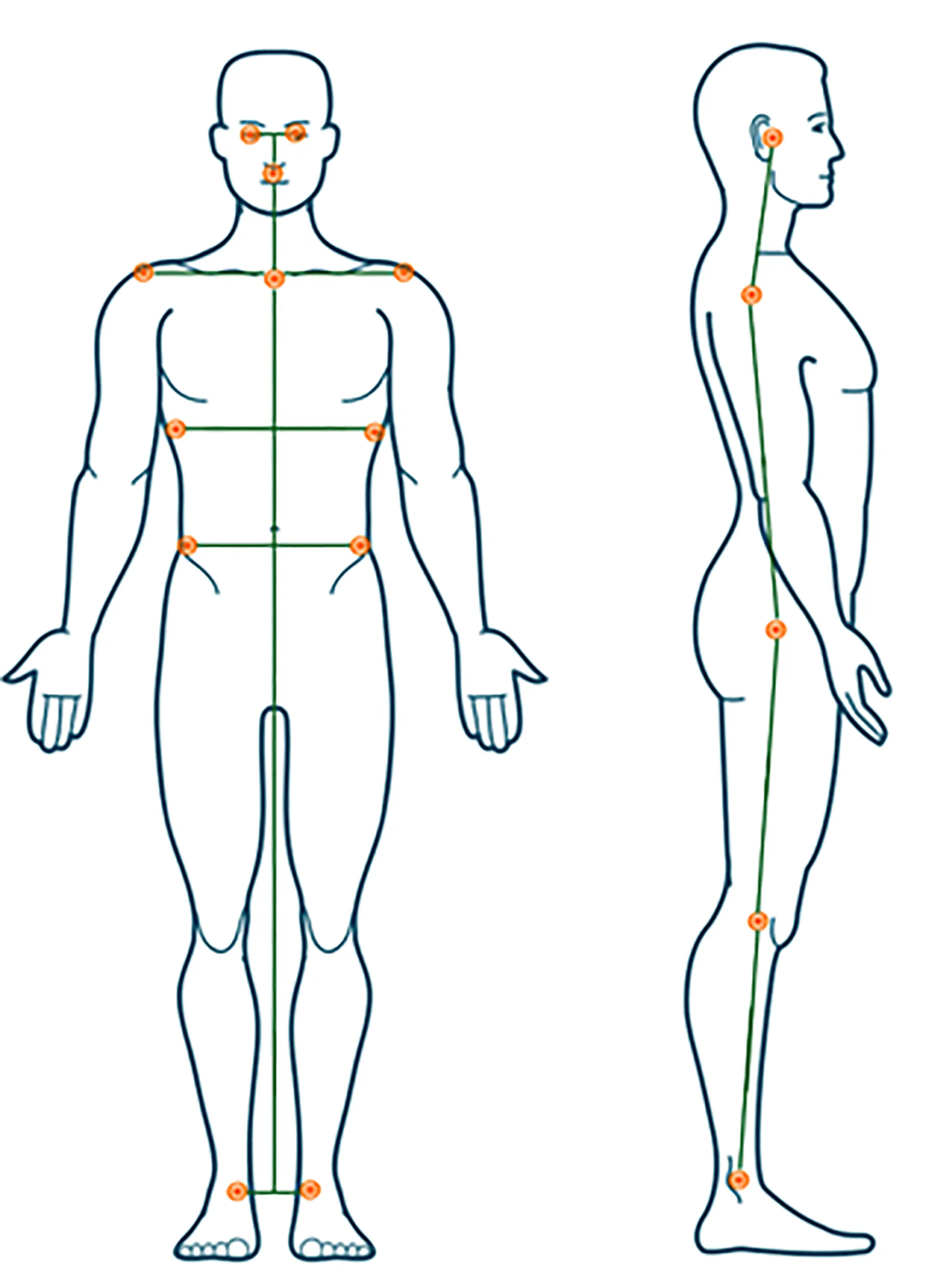
Figure 5 Body position of the digital landmarks of PostureScreen Mobile app.
Among innovative technologies, the growing reliability of wearable devices is noteworthy.Wearable devices have been welcomed into the daily lives of healthy individuals, older adults, and those with chronic illnesses[102,103].These devices represent an opportunity to quantify the movement patterns of all types of individuals in real-world settings.It is expected that the applicability of wearable devices to different populations will contribute to relevant advancements in understanding the daily gait patterns of walkers and runners[104].The use of wearables is constantly expanding, especially in the field of gait analysis[105], post-operative rehabilitation[106,107], diagnosis and treatment of musculoskeletal disorders (e.g.,scoliosis, kyphosis, lordosis)[108,109], and posture management in the workplace[110].In the context of posture analysis, wearables offer a low-cost and easy-to-use tool that provides real-time feedback for correcting workers' posture and reducing postural pain onset.Poor posture can lead to musculoskeletal disorders or spinal complications.It is well established that long sitting hours in front of computers cause pain, usually at the back.Abyarjooet al[111] proposed a wearable system for office workers that alarms the subject when he assumes a wrong posture.The Upright Go 2, following the success of its previous release (Upright Go), is a wearable device whose goal is to manage posture daily by promoting self-correction[112].The device is about 48mm large, lightweight, and with a battery that lasts up to 35 h.It can be applied to the back using hypoallergenic adhesives or worn with a unique necklace that ensures that the device stays on the back.It is equipped with multiple sensors that perceived if the subject slouches, and in this case, it will emit vibrations that stimulate the subject to regain a correct posture.The Upright Go 2 provides an app to monitor the progress over time, suggesting workouts to maintain the correct posture.Future studies about whether the subject would maintain a correct posture even after the device is no longer used would be an interesting prospect.
OVERALL CONSIDERATIONS
In recent years, a wide variety of technologies to study human movement has emerged, ranging from 3D visual software to wearable devices with almost imperceptible weight.The increase of technological devices has made it possible to expand the field of biomechanical assessment not only to the clinical environment but also to re-educational, sports and everyday life contexts.However, rapid technological development risks providing tools that are often not sufficiently validated.For example, the study conducted by Yoonget al[113], published just in November 2019, reports wearables that are no longer in production.For a device to be considered valid in the scientific field, it must comply with some strict parameters, and above all, it must maintain the reliability of its measurements constant.The evaluation of posture, movement, and gait and their deviations from physiological conditions are increasingly helpful in the clinical setting to diagnose musculoskeletal pathologies and in daily life to reduce the incidence of pain and disorders.As reported in Figure 4, this approach involves subdivisions based on the patient's criticality levels.Concerning gait analysis, a markerless system is a valuable tool for first-level screening as fully automatic and non-invasive, allowing to quickly analyze a large number of patients without stressing them with lengthy procedures.This first step streamlines the use of a marker-based system, making it available to more complex cases.
Similarly, the rasterstereography system, intended as a first-level approach, allows analyzing the back's topography leaving the second-level approach to radiographs, minimizing the need for repeat X-rays.In clinical practice, these approaches help in planning treatment, personalized rehabilitation programs, and surgical solutions.In everyday life, they give the possibility to remotely follow the patients, monitor progresses, and collect data on a large scale of users.As reported in Table 4, different systems serve different purposes, suggesting the need for a general scheme to direct the operator towards the most suitable analysis system to prefer.
CONCLUSION
This review highlighted the main applications of novel electronic devices in motion and posture analysis, describing their strengths and weaknesses.From the comparison of these systems, it is clear that some of the mentioned devices have the potential to be used in clinical practice, sports, and healthcare prevention.Therefore, it is suggested that the scientific community might embrace an improved biomechanical approach through these new currently available tools for a tailored evaluation of patient’s characteristics.The future of biomechanical research is a fast, fully automatic, noninvasive, and repeatable approach further away from human-dependent errors.
 World Journal of Orthopedics2021年7期
- World Journal of Orthopedics的其它文章
- Arthrodesis of the first metatarsophalangeal joint: The “when and how”
- Arthroscopic removal as an effective treatment option for intraarticular osteoid osteoma of the knee
- Strategies and outcomes in severe open tibial shaft fractures at a major trauma center: A large retrospective case-series
- Optimization of transdisciplinary management of elderly with femur proximal extremity fracture: A patient-tailored plan from orthopaedics to rehabilitation