Analysis of Characteristics and Risk Factors for Recovered COVID-19 Patients with Retest Positive for SARS-CoV-2 RNA
HU Wen-wen,WANG Mei-fang,CHANG Chan,LIU Yan,LEUNG Lai-han Elaine,DENG Ping-ji,GUO Gong-sun,TANG Yi-jun,*
(1.Postgraduate Training Basement of Jinzhou Medical University,Taihe Hospital,Shiyan 442000,Hubei,China;2.Department of Respiratory and Critical Care Medicine,Taihe Hospital,Shiyan 442000,Hubei,China;3.State Key Laboratory of Quality Research in Chinese Medicine,Macau Institute for Applied Research in Medicine and Health,Macau University of Science and Technology,Macau 999078,China;4.School of Public Health and Management,Hubei University of Medicine,Shiyan 442000,Hubei,China;5.The Health Commission of Shiyan,Shiyan 442000,Hubei,China)
Abstract:As the coronavirus disease 2019(COVID-19)is spreading around the world rapidly,recovered COVID-19 patients with retest positive(RP)for severe acute respiratory syndrome coronavirus 2(SARSCoV-2)RNA have been reported in many countries.However,the mechanisms of this re-positive test result remain unknown.A case-control study was performed on 153 COVID-19 patients,who had been admitted to three medical institutions in Shiyan City,Hubei Province,from January 24 to April 1,2020.The demographic features,epidemiological history,laboratory tests,clinical manifestations and chest computed tomography(CT)examination results were analyzed.Univariable and multivariable Logistic analyses were used to explore the characteristics and risk factors in RP patients.There were significant differences between the RP patients and patients with retest negative(RN)in white blood cell,neutrophil and red blood cell counts,serum hemoglobin,erythrocyte sedimentation rate(ESR),procalcitonin(PCT),lactate dehydrogenase,creatine kinase,albumin,D-dimer and fibrinogen levels,fever levels,length of hospitalization,viral shedding duration and length of absorption in CT image on admission and lymphocyte counts before discharge.Particularly,multivariate Logistic analysis showed that the fever level,D-dimer level,ESR and length of hospitalization were independent risk factors for RP patients.In addition,RP patients with low levels of albumin were more likely to have symptoms.The above results showed that clinicians should evaluate the probability of re-positive for discharged COVID-19 patients based on the clinical data on admission.It is important to conduct reduplicative nucleic acid tests in discharged COVID-19 patients during the 14-day quarantine and regular follow-up observations.Closer monitoring and more nutritional support should be given to those with higher fever,ESR and serum D-dimer levels,as well as longer hospital stays.
Key words:coronavirus disease 2019(COVID-19);severe acute respiratory syndrome coronavirus 2(SARSCoV-2);discharged patients;re-positive
In December 2019,a group of patients were diagnosed as unknown pneumonia,which broke out suddenly and spread around the world rapidly[1].The epidemic was later identified to be caused by a novel coronavirus[2],which was named SARS-CoV-2 by the International Committee on Taxonomy of Viruses(ICTV)[3].The disease caused by this virus was named COVID-19 by the World Health Organization(WHO)[4].As of November 1,2020,the number of confirmed cases reached a total of 45 968 799 globally,including 1 192 911 death cases,over 3.3 million new cases and nearly 45 000 new deaths reported over the past week[5].This has imposed a serious threat to global public health.Fortunately,as more and more studies on the clinical presentation,epidemiological features,laboratory examinations and imaging characteristics,as well as treatment and prognosis,COVID-19 has been effectively controlled in China.
Previous studies from various countries have reported that some patients,after recovering from COVID-19,could again test nucleic acid positive for SARS-CoV-2 by real-time polymerase chain reaction(RT-PCR)[6~8].The RP patients often had no or mild clinical symptoms;however,their health status,infectivity and the mechanisms behind this re-positive test result all remain unclear[9~10].Who is more susceptible to retest positive?What are the characteristics of these patients?Are the patients only detected re-positive for nucleic acid or do they have a recurrentinfection?Many problems are still unknown.Therefore,it is very important to carry out research on such problems.Our study aimed to analyze the characteristics and risk factors for RP patients recovered from COVID-19,which may be helpful to manage the discharged COVID-19 patients and reduce the probability of turning positive again.
1 Materials and methods
1.1 Patients and data collection
Fifty-one RP cases admitted to three medical institutions(Taihe Hospital,Xiyuan Hospital and the Second Treatment Hospital)in Shiyan City,Hubei Province,from January 24,2020 to April 1,2020,were studied in a case-control study.One hundred and two RN cases,the number of whom doubled the RP,were randomly selected from the same institutions and used as the control group.Before discharge,the COVID-19 patients must meet the following criteria:normal temperature for at least 3 days,obvious improvement in symptoms,stable organ functions,obvious absorption in CT image,and negative results for SARS-CoV-2 RNA twice(at an interval of at least 24 hours).After discharge,they were required to be quarantined for at least 14 days and had to get two negative SARS-CoV-2 RNA tests to complete their isolation period.Then regular follow-up observations were performed on them.There is no official definition of“retest positive”for novel coronavirus nucleic acids up to now.However,the well-recognized definition is as follows:when a previously confirmed and discharged COVID-19 patient tests positive for SARS-CoV-2 RNA(any testing time after discharge)[10].All collected cases met the diagnostic criteria of the COVID-19 Diagnosis and Treatment Program(Trial Version 7)[11].Exclusion criteria for COVID-19 patients were any one of the following:1)under the age of 18 years,2)dying from their infection during the disease course,and 3)incomplete key information in their medical records.
The demography,epidemiology,chronic medical history,length of hospitalization,clinical presentation,laboratory values on admission and before discharge,imaging characteristics,and the severity of COVID-19 were collected(including all data for RP patients during their first and second hospitalizations due to COVID-19).
1.2 RT-PCR method
Sputum and nasopharyngeal swab specimens collected from all patients were tested by RT-PCR for SARS-CoV-2 RNA within 3 hours.Laboratory confirmation of the virus was performed using RTPCR.All nucleic acid tests were conducted by Shiyan Center for Disease Control and Prevention,and all nucleic acid testing kits were supplied by DAAN Gene Co.,Ltd.of Sun Yat-sen University.
Viral nucleic acids were extracted from specimens,and detected by RT-PCR targeting two genes:open reading frame 1ab(ORF1ab)and nucleoprotein(N).The forward primer 5′-CCCTGTGGGTTTTACACTTAA-3′,the reverse primer 5′-ACGATTGTGCATCAGCTGA-3′and the probe 5′-FAM-CCGTCTGCGGTATGTGGAAAGGTTATGG-BHQ1-3′were used for detecting the ORF1ab gene.And the forward primer 5′-GGGGAACTTCTCCTGCTAGAAT-3′,the reverse primer 5′-CAGACATTTTGCTCTCAAGCTG-3′and the probe 5′-FAM-TTGCTGCTGCTTGACAGATT-TAMRA-3′were used for detecting the N gene.The RT-PCR assay was conducted using the following procedure:50℃for 15 minutes,95℃for 15 minutes,45 cycles of 94℃for 15 seconds and 55℃for 45 seconds.The RT-PCR results were defined negative if the Ct value>40 or no amplification,and positive if the Ct value<37.For suspected cases with the Ct value between 37 and 40,repeated experiments were conducted on them until a clear diagnosis was made.
1.3 Statistical analysis
Continuous variables were expressed as mean±standard deviation if they were normally distributed or as median(inter-quartile range,IQR)if they were not.Independent group t-test was used to compare means for continuous variables when the data were normally distributed;otherwise,Mann-Whitney U test was used.Categorical variables were expressed as numbers(%).Proportions for unordered categorical variables were compared by χ2-test or Fisher’s exact test.Proportions for ordered categorical variables were compared by Wilcoxon ranksum test.To explore the risk factors associated with RP,univariable and multivariable Logistic regression models were used.Considering the total number of RP group(n=51)in our study and to avoid multicollinearity in the model,five variables(ESR,D-dimer and fever levels on admission,length of hospitalization and lymphocyte count before discharge)were chosen for multivariable analysis based on the previous findings and clinical constraints.For unadjusted com-parisons,a two-sided α of less than 0.05 was considered statistically significant.Statistical analyses were done using the SPSS 19.0 software.
2 Results
2.1 Demographic and epidemiological characteristics
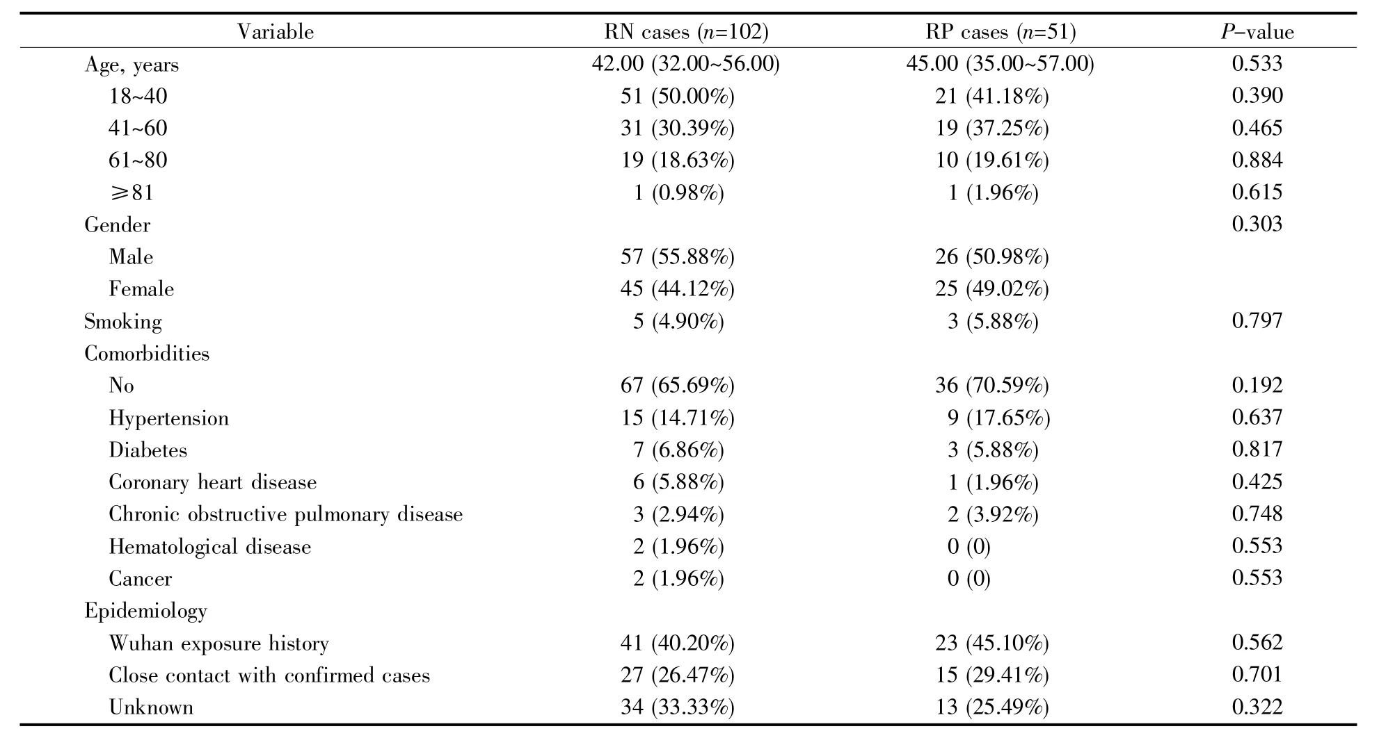
The demographic and epidemiological characteristics of 153 discharged COVID-19 patients from the three medical institutions are shown in Table 1.The median age of RN patients,including 57(55.88%)males and 45(44.12%)females,was 42.00 years(IQR 32.00~56.00).The number of males and females in RP group was almost equal,and their median age was 45.00 years(IQR 35.00~57.00).Among the 51 RP patients,1(1.96%)was aged over 81.There was no significant difference between the two groups in the age and gender distribution.Only a few patients,5(4.90%)in the RN group and 3(5.88%)in the RP group,were current or former smokers for at least 10 years.
Less than half of the 153 COVID-19 patients had comorbidities(32.68%).Of the 51 RP patients,15(29.41%)had comorbidities,including 9(17.65%)hypertension,3(5.88%)diabetes,and 2(3.92%)chronic obstructive pulmonary disease.The proportion of chronic obstructive pulmonary disease was higher in the RP cases than that in the RN cases.However,there was no significant difference in comorbidities between the two groups.Also,a recent history of exposure in Wuhan or close contact with confirmed cases showed no significant difference.
2.2 Clinical characteristics
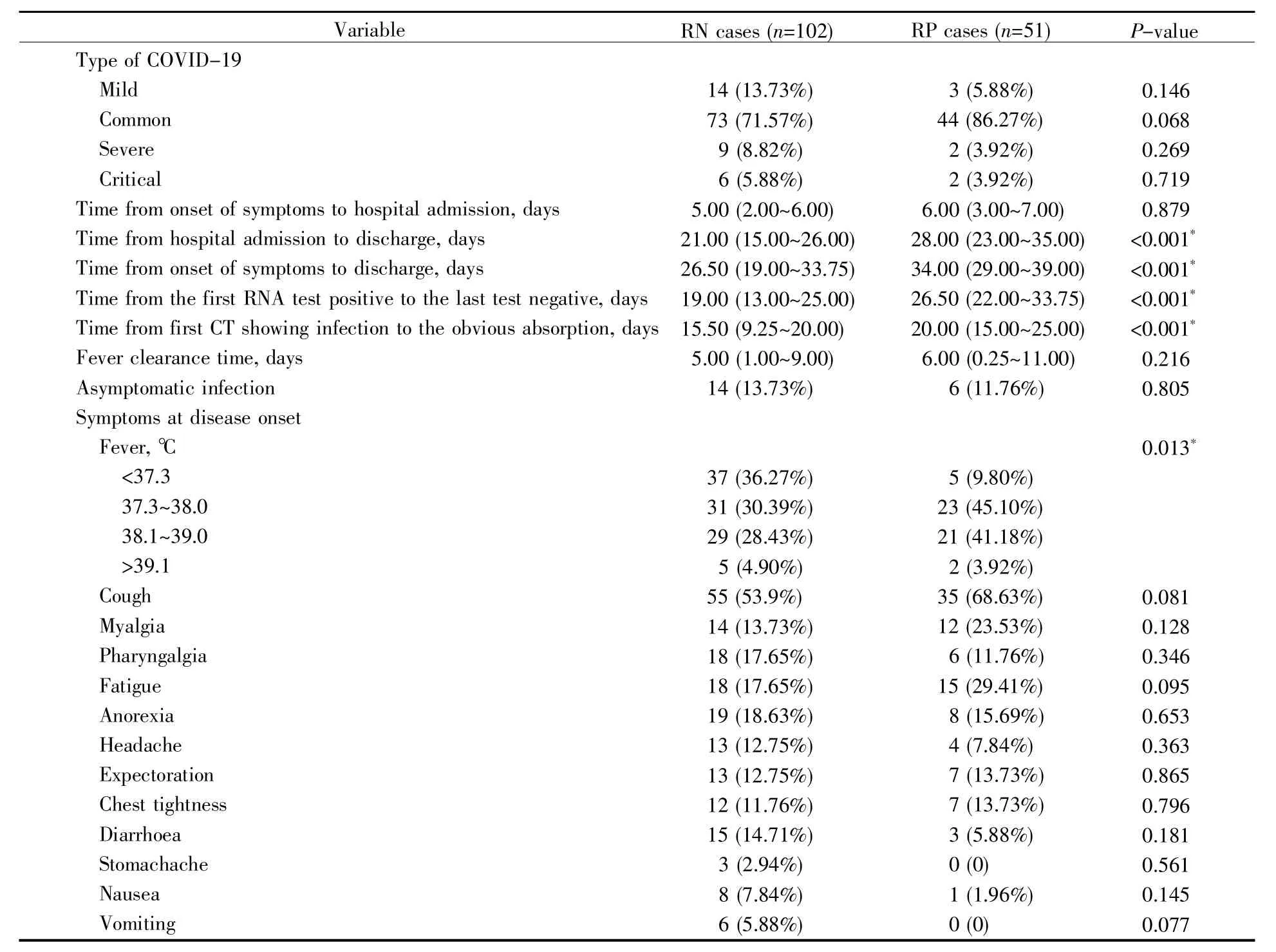
The clinical characteristics of the two groups are shown in Table 2.According to the protocol,all collected cases were divided into four types:mild,common,severe and critical.Most of the 153 patients were common type,73 (71.57%)in RN group and 44(86.27%)in RP group.Compared with the RN group,the proportion of the severe and critical patients in RP group was not higher,which showed that the occurrence of RP had no obvious relationship with the four types.

Table 1 Demographic and epidemiological characteristics of COVID-19 patients
Among the 153 patients,20(13.07%)were asymptomatic.The most common symptoms were fever,cough,and fatigue,while less common symptoms were headache,nausea,vomiting and stomachache.One hundred and eleven(72.55%)patients had a fever,90(58.82%)had a cough and 33(21.57%)had fatigue.Seventeen(11.11%)patients had a heada-che,9(5.88%)had nausea,6(3.92%)had vomiting and 3(1.96%)had a stomachache.There was a statistically significant difference in the fever level between the RP and RN groups(P<0.05).
Among the RP patients,the median time from admission to discharge(length of hospitalization)was 28.00(IQR 23.00~35.00)days,which tended to be longer than 21.00(IQR 15.00~26.00)days in the RN patients(P<0.05).The median time from initial symptom to discharge in RP patients was 34.00(IQR 29.00~39.00)days,while 26.50(IQR 19.00~33.75)days in the RN patients(P<0.05).The median time from the first positive RNA test to the last negative test before discharge in the RP cases was 26.50(IQR 22.00~33.75)days,also longer than 19.00(IQR 13.00~25.00)days in the RN cases(P<0.05).Similarly,the median time from the first chest CT showing infection to obvious absorption in image in the RP cases was 20.00(IQR 15.00~25.00)days,which was longer than 15.50(IQR 9.25~20.00)days in the RN cases(P<0.05).Because there was a high correlation between the above variables,the length of hospitalization was chosen for multivariable analysis based on the fact that it might be the variable we aimed to study mostly.
2.3 Laboratory and imaging characteristics
As shown in Table 3,14(27.45%)RP patients on admission showed leucopenia(white blood cell count lower than 4.00×10⁹L-1)and 7(13.72%)neutropenia(neutrophil count lower than 2.00×10⁹L-1),and the values of leukocytes and neutrophils had significant difference(P<0.05).The lymphocyte count before discharge,red blood cell count and hemoglobin level on admission in RP patients were lower than those in RN patients(P<0.05).The ESR,PCT and D-dimer levels were significantly higher in RP patients than in RN patients(P<0.05).In addition,the albumin and fibrinogen concentrations also showedsignificant difference between the two groups.

Table 2 Clinical characteristics of COVID-19 patients
Most COVID-19 patients were confirmed lung infections by chest CT.Fifty-five(53.92%)RN patients and 42(82.35%)RP patients presented bilateral lung lesions,while 14(13.72%)RN patients and 3(5.88%)RP patients had normal chest CT images.No significant difference was found in imaging signs on admission between the two groups.
2.4 Logistic regression analysis
There were 153 patients with complete data for all variables(51 RP and 102 RN patients)in the binary Logistic regression model by stepwise regression method.The results showed that the fever level(OR=2.083,95%CI:1.382~3.140,P<0.001),length of stay(OR=1.095,95%CI:1.057~1.135,P<0.001),ESR(OR=1.066,95%CI:1.044~1.088,P=0.004)and D-dimer level(OR=1.049,95%CI:1.031~1.075,P=0.007)were independently associated with RP(Table 4).

Table 3 Laboratory and imaging characteristics of COVID-19 patients
2.5 Characteristics of RP patients
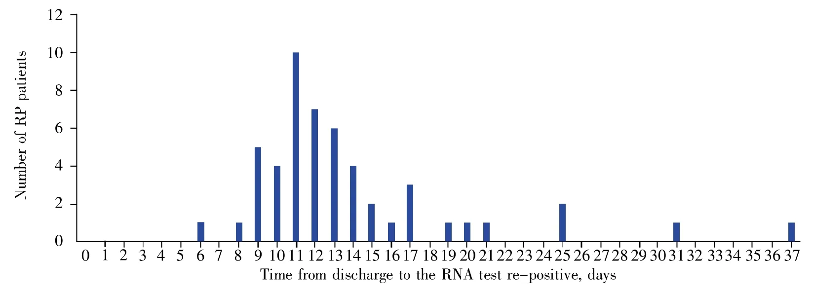
Among the 51 RP patients,the median time from discharge to re-positive nucleic acid test was(13.49±4.24)days.The shortest duration of re-positive detection was 6 days,while the longest was 37 days(Fig.1).Twenty-four(47.06%)RP patients were asymptomatic when they tested re-positive.There was no significant difference in demographic and epidemiological characteristics of RP patients(Table 5).The values of white blood cell count before discharge,neutrophil count,hypersensitive C-reactive protein(h-CRP)and albumin on admission showed significant differences between asymptomatic and symptomatic patients(Table 6).Multivariable regression showed that the albumin level(OR=0.793,95%CI:0.650~0.966,P=0.021)was an independent factor to predict RP patients with symptoms(Table 7).

Table 4 Binary Logistic regression analysis for the related factors predicting RP

Fig.1 Time from discharge to retest positive in the RP group

Table 5 Demographic and epidemiological characteristics of RP patients

Table 6 Laboratory and imaging characteristics of RP patients

Table 7 Binary Logistic regression analysis for the related factors predicting RP patients with symptoms
3 Discussion
From January 24,2020,to May 20,2020,672 patients with COVID-19 had been discharged from the three hospitals in Shiyan,Hubei Province.Sixty-five patients(9.67%)showed positive nucleic acid in the 14-day isolation period or the follow-up observations,which was almost consistent with a previous study[12].However,some studies showed higher re-positive rate:38 out of 262 discharged COVID-19 patients(14.50%)tested positive for SARS-CoV-2 RNA[13],and 17(17.35%)of 98 convalescent patients tested positive[14].In our study,no significant difference was found in age and comorbidities.Additionally,it also showed that the occurrence of RP patients had little relationship with mild,common,severe and critical types.However,another retrospective study showed that RP patients were characterized by younger age and minor symptoms[13].
We confirmed several risk factors for RP patients in this study.In particular,high levels of ESR and D-dimer on admission,longer hospital stays and fever were associated with higher incidence of nucleic acid re-positive patients.In addition,leucopenia,neutropenia,anemia on admission,lymphopenia before discharge,high levels of PCT,prolonged viral shedding and absorption in CT image were also related to higher re-positive risk.A high D-dimer level on admission was a significant predictor of increased mortality for patients with COVID-19[15~17].The elevated D-dimer levels may result from the inflammatory response to infection with SARS-CoV-2 and subsequent activation of coagulation,especially a cytokine storm[18].Thus,D-dimer testing would contribute to tracking with severity of disease and inflammation[19].Interestingly,we found that fever is a risk factor of RP patients,but the underlying reason is unknown.Previous studies confirmed that there was a positive correlation between the incidence of fever and interleukin-6(IL-6)level which is a typical multifunctional cytokine regulating the inflammatory response in immune regulation[20~21].IL-6 is an independent factor to predict progression of COVID-19[22~23].
In addition,ESR,PCT and CRP are indicators of inflammation,so we suspect that SARS-CoV-2 recurrence is related to the inflammatory response.The inflammatory response plays a crucial role in COVID-19,and the inflammatory cytokine storm can cause more complications or even death[24~25].Previous studies showed that ESR,PCT and CRP were independentfactorsto predictthe severity of COVID-19[22,26~27].Wang et al.[28]showed that inflammatory indicators(ESR,CRP,and IL-6)were negatively correlated with CD8+T-cell levels,but positively correlated with the CD4+/CD8+ratio.ESR was negatively correlated with total lymphocyte and CD4+T-cell levels.Another study confirmed that CD4+T lymphocytes would affect the duration of SARSCoV-2 RNA detection[29].Inflammatory cytokines established a pro-inflammatory feedback loop with monocytes,macrophages and T cells,which could enhance SARS-CoV-2 infection[30].Furthermore,hyperinflammation proved relevant to immune dysregulation[31].Therefore,we speculate hyperinflammation may lead to RP occurrence through mediating immune response.
Several previous studies have shown that lymphopenia is common in severe COVID-19 patients compared to the patients with mild disease,along with increased neutrophil count and the neutrophilto-lymphocyte ratio(NLR)[26,32~33].The decline of lymphocytes including CD4+and CD8+T cells,B cells,and natural killer(NK)cells have been found in patients with severe COVID-19[34].Lymphopenia often occur in chronic viral infections[35].In this study,leucopenia,neutropenia on admission and lymphopenia before discharge were found to be associated with RP patients.COVID-19,a subacute infection,can cause T cell depletion,inactivation,and exhaustion,which increase the possibility of immunosuppression,and then lead to COVID-19 viral persistence[36].Lymphopenia before discharge may increase the risk of RP patients.However,the cause of leucopenia and neutropenia on admission need more evidence.
Unexpectedly,our study showed that the low values of red blood cell count and hemoglobin were linked to RP patients.It was reported that human immunodeficiency virus(HIV)protease inhibitors lopinavir and ritonavir were suitable for COVID-19 patients with serious complications[37],and that erythrocyte-associated indicators were markers for prognostic factors and clinical stage[38~39].Another research also confirmed that the erythrocyte-associated indicators were related to COVID-19 recurrence,and may result from erythrocytes feeding SARS-CoV-2[40],which is similar to HIV infection.
The durations of hospitalization and viral shedding are also factors affecting the occurrence of RP patients.Several studies suggested that prolonged viral RNA detection was associated with duration of illness and hospitalization[41~42],and prolonged viral shedding was a risk factor for death of COVID-19 patients[15].Future studies should be conducted on the mechanisms of the relationship between viral shedding duration and RP patients.Previously,Bian et al.[43]conducted an autopsy in a ready-for-discharge COVID-19 patient who provided the first pathological evidence for residual virus in the deep lung tissues.It was found that the RP for SARSCoV-2 might be the RNA fragments,not active virus.This finding revealed the existence of remaining SARS-CoV-2 in the lung from the readyfor-discharge patient,suggesting that the negative results of nasopharyngeal swab-PCR test might not reflect the virus in lung tissue.Liu et al.[44]suggested that RP may be due to the biological characteristics of SARS-CoV-2,clinical status of patients,and even related to the sampling methods and nucleic acid testing kits.There are studies showing that virulence of SARS-CoV-2 may be weakened in the process of transmission.However,persistent positive RNA in feces after negative nasopharyngeal swabs suggests a possible prolonged transmission period,which challenges current quarantine practices[45].
In our study,the median time from discharge to recurrence of positive SARS-CoV-2 RNA was found to be(13.49±4.24)days,and the longest duration among the RP patients was 37 days.The most asymptomatic COVID-19 patients on admission were only nucleic acid positive without any symptom.These findings may be helpful for our prevention and controlservicesfordischarged COVID-19 patients.Moreover,our study suggested that the albumin level was an independent factor to predict asymptomatic or symptomatic RP patients.A low value of serum albumin was a risk factor for mortality in patients with sepsis or community-acquired pneumonia[46~47].Based on the above findings,COVID-19 patients with higher levels of pre-hospital body temperature,D-dimer,and ESR and longer duration of hospital stays should be paid more attention to avoid the possibility of being re-positive.Nutritional support should be enhanced in all patients to strengthen their antiviral immunity.
4 Conclusion
To sum up,several risk factors for RP patients were identified in our study,which may be very useful for improving management of COVID-19 patients.Although COVID-19 has been controlled in China,the number of infected people worldwide is still increasing.Therefore,it is necessary to conduct reduplicative nucleic acid tests in discharged COVID-19 patients during the 14-day quarantine and regular follow-up observations,in order to prevent possible spread of COVID-19.Furthermore,it is also crucial to strengthen the management and nutritional support of patients who are susceptible to re-infection.Due to the limitations of our study,larger sample size and more specific research would be needed to confirm the potential risk factors of RP patients.
Ethical approval and consent to participate
This study was approved by the Human Ethics Committee and the Research Ethics Committee of Taihe Hospital,Hubei,China.Data records were de-identified and completely anonymous,so informed consent was waived.
Competing interest
The authors declare that there are no competing interests.
Acknowledgments
We acknowledge all health-care workers involved in the diagnosis and treatment of patients.

