Vesicular carriers containing phenylethyl resorcinol for topical delivery system;liposomes,transfersomes and invasomes
Thnporn Amnuikit,Tunyluk Limsuwn,Psrt Khongkow,Prpporn Boonme,c
aDepartment of Pharmaceutical Technology,Faculty of Pharmaceutical Sciences,Prince of Songkla University,
Songkhla 90112,Thailand
bInstitute of Biomedical Engineering,Faculty of Medicine,Prince of Songkla University,Songkhla 90112,Thailand
cNanotec-PSU Center of Excellence on Drug Delivery System,Songkhla 90112,Thailand
Keywords:Phenylethyl resorcinol Transfersomes Invasomes Tyrosinase inhibition?Corresponding author.Department of Pharmaceutical Technology,Faculty of Pharmaceutical Sciences,Prince of Songkla University,Songkhla 90112,Thailand.Tel.:+66896588838.
A B S T R A C T Topical administration of phenylethyl resorcinol(PR)has attracted much attention as skin lighteningagent with potent anti-tyrosinaseactivity.Twonovel types of elastic carrierswere developed to overcome the limitation of PR as topical delivery by increasing the solubility,stability and decreasing skin irritation compared to conventional liposomes.In addition,it also promotes skin penetration of PR to reach deep skin layer at the target site.The lead formulations were obtained from the invasomes containing 1%(w/v)D-limonene mixed with 10%(v/v)absolute ethanol as the skin enhancer,and transfersomes containing 15%(w/w)sodium deoxycholate(SDC)as edge activator.All formulations gave a vesicle size<500nm,polydispersity index(PDI)<0.3,high zeta potential,entrapment efficiency>50%,and good stability on storage at 30°C at 75%RH for 4 months.Transfersomes have a lower degree of deformability(6.63%)than invasomes(25.26%).In contrast,the liposomes as rigid vesicles do not show a deformable property.This characteristic affects the skin permeation,and thus,transfersomes with high elastic property provided a significantly higher cumulative amount,steady state flux(Jss)and permeability coefficient(Kp)compared to other formulations.However,in vitro PR accumulation in full-thickness newborn pig skin demonstrated that the application of elastic carrier formulations gave significantly higher accumulation than liposomes,and gave anti-tyrosinase activity up to 80%.These results are straightforwardly related to the results of cellular level study.Transfersomes and invasomes showed higher tyrosinase inhibition activity and melanin content reduction when compared to liposomes in B16 melanoma cells.In addition,acute irritation test in rabbits confirmed that these formulations are safe for skin application.Therefore,elastic vesicle carriers have the efficiencytodeliverPRintothedeepskin inbothquantityandeffectivenesswhicharebetter than conventional liposomes and appropriate for a skin lightening product.
1. Introduction
Melanin is a complex polymer derived from the amino acid tyrosine and produced by the melanogenesis pathway in melanocytes which is located in the basal layer of the epidermis.The light absorption of melanin in skin plays an important role for photo-protection from the ultraviolet(UV)radiation[1].However,overproduction of melanin causes skin darkening and abnormal distribution of melanin in different and specific parts of the skin which in fact causes hyperpigmentation disorders including melasma,freckles,lentigines,etc.[2].Tyrosinase inhibition is an effective strategy for treating hyperpigmentation because tyrosinase controls the ratelimiting steps of melanin synthesis.
Phenylethyl resorcinol(4-(1-phenylethyl)1,3-benzenediol,PR)is phenolic compound developed by Symrise(Holzminden,Germany)undertradenameasSymWhite?377.PR,which is a new skin lightening agent,inhibits the tyrosinase activity by obstruction of the conversion of tyrosine to L-3,4-dihydroxyphenylalanine(L-DOPA)[3].It reduced tyrosinase activity approximately 22 folds more effectively than kojic acidinvitromushroomtyrosinaseandinvitroepidermalmodel(MelanoDermaTM),respectively[4].It can increase the fairness of Asian human skinin vivoat a concentration of 0.5%[5].In addition,PR has antioxidant property and improved UV protection activity when incorporated with inorganic titanium dioxide(TiO2)hybrid composites rather than bare TiO2[6].Recently,PR showed potential in antifungal activity.It was effective against the nine dermatomycoses:Microsporum gypseum,Microsporum canis,Trichophyton violaceum,Arthroderma cajetani,Trichophyton mentagrophytes,Epidermophyton floccosum,Nannizzia gypsea,Trichophyton rubrumandTrichophyton tonsuranswhichishighlyactivethanantifungalagent fl uconazole[7].However,the poor aqueous solubility and low stability in light are the limitations of PR for the formulation as cosmetic and pharmaceutical dermal products.The color changes from white to pink tone when the formulation is exposed to light which may cause the application of PR to be ineffective whereas the poor aqueous solubility may reduce its absorption when used[8].In addition,it has been reported that,a 1%PR in the formulation,causes skin irritation,and may lead to consumer rejection of the product[9].
Nanoencapsulation technique was used in liposomes[10],transfersomes[11],invasomes[12]and ethosomes[13]to increase the solubility and photo-stability,and decrease skin irritation of PR by protecting the encapsulated actives from external environment and from directly contact to the skin.Flexible or elastic liposomes are the type of phospholipid vesicles which were modified for improving skin drug delivery systems.Although it is similar to conventional liposomes in morphology,the permeability across skin is different.Transfersomes or ultra- fl exible vesicles are the first generation of elastic liposomes.They were achieved by incorporating edge activators(EA)to the lipid bilayers.An edge activator is often a single-chain surfactant such as sodium cholate,sodium deoxycholate,dipotassium glycyrrhizinate,Spans and Tweens.A single-chain surfactant with a high radius of curvature and mobility,are able to destabilize the lipid bilayers of vesicles,and increase the elasticity and flexibility of the lipid bilayer which makes the vesicles have ultra-deformable property[14].Several studies reported that transfersomes are efficient for transdermal and topical drug delivery[11].Ethosomes are the fluid vesicles which contain ethanol at relatively high concentration(20%–45%)as skin enhancer.The elastic vesicle of ethosomes is an important feature related to skin permeability enhancement,which may be due to the synergistic mechanism between high concentration of ethanol,phospholipid vesicles,and lipid bilayers in stratum corneum.Therefore,they can penetrate into the skin and allow enhanced delivery of various drugs to deeper skin strata[15].Invasomes are soft flexible liposome vesicles with very high membrane fluidity.They contain ethanol and terpenes which play the role of penetration enhancer.The mechanism of skin permeation of invasomes is similar to ethosomes[12].
Traditional or conventional liposome vesicles are of large size,more than 400nm in diameter and have a rigid structure.Cholesterol is added in the formulation to stabilize the structure and generate more rigid liposomes.These are too large to fit within the intercellular lipid domains of the stratum corneum,and the liposomes are generally reported to accumulate in the stratum corneum,upper skin layers and in the appendages,with minimal penetration to deeper tissues or the systemic circulation[16].Conventional liposomes remain near the skin surface,dehydrate and fuse with the skin lipids,whereas deformable transfersomes squeeze through stratum corneum lipid lamellar regions penetrating deeper by following the osmotic gradient to permeate to the deeper layers of the epidermis.Due to the flexibility conferred on the vesicles by the other vesicle compositions molecules such as ethanol and surfactant,elastic vesicle compositions have been investigated to develop systems that are capable of carrying drugs and macro-molecules to deeper tissues.
These vesicles have a unique skin permeation mechanism.Transfersomes can pass through the intact skin and can retain their shape,because of the presence of edge activators in the formulation,whereas the ethosomes have ethanol,and invasomes have combination of ethanol and terpenes in the formulation which play the role of penetration enhancer by disturbing the organization of the stratum corneum lipid bi-layer.Therefore,the aim of this study was to develop and characterize phenylethyl resorcinol-loaded transfersome and invasome drug delivery systems for overcoming the limitation of PR as topical delivery,by increasing the solubility and light stability,and decreasing skin irritation.The stability,in vitroskin permeation,and skin irritation were assessed to evaluate the suitability of the formulations for skin lightening applications.
2. Materials and methods
2.1. Materials
PR was purchased from Starchem Enterprises Limited(Nanjing,China).L-α-phosphatidylcholine from soybean(SPC)and cholesterol(CHOL)were purchased from Sigma-Aldrich(Missouri,USA).Absolute ethanol was bought from RCI Labscan Limited(Bangkok,Thailand).Tween20andTween80werepurchased from P.C.Drug Center Co.,Ltd.(Bangkok,Thailand).Span 20 and Span 80 was purchased from S.Tong Chemicals Co.,Ltd.(Bangkok,Thailand).Kojic acid,tyrosinase enzyme and dimethyl sulfoxide(DMSO)were purchased from Sigma-Aldrich(WGK,Germany).Citral,(R)-(+)-Limonene and(1R)-(-)-Fenchone were purchased from Sigma-Aldrich(WGK,Germany).Sodium deoxycholate were purchased from Homedia?(Mumbai,India).Milli-Q water was used throughout the experiment.All chemicals were used as received,without any modification.
2.2. Methods
2.2.1.Preparation of PR-loaded vesicle carrier
Preparation of PR-loaded transfersomes and invasomes was followed and modified from the method of Limsuwan et al.[13].The PR in the formulations was fixed as 0.5%(w/v).The 0.5%(w/v)CHOL,3%(w/v)SPC and water up to 100%(v/v)were the main composition of liposomes.The skin enhancers were varied in terms of concentrations and ratios as shown in the Table 1.The invasome formulations used fenchone,citral and D-limonene mixed with 10%(v/v)ethanol as skin enhancers.The transfersome formulations used Tween 80,Tween 20,Span 80,Span 20 and SDC as skin enhancers.All compositions were prepared by thin- fi lm hydration method as follows:First,the oil phase which included SPC,CHOL,PR and skin enhancer were dissolved in absolute ethanol.The aqueous phase used was water for liposomes and transfersomes;ahydro-ethanolicsolutionconsistingofwaterand10%(v/v)absolute ethanol for invasomes.Second,the oil phase and aqueous phase were separately sonicated at 60°C for 30min until homogeneity.Then,the absolute ethanol was removed from oil phase via evaporation by a rotary evaporator(Model Eyela N-1000 series,Tokyo Rikakikai Co.,Ltd.,Japan)while the other components formed the thin lipid film.Afterward,the lipid film was hydrated with 10ml of aqueousphase,followed by shaking for 5min.Finally,the mixtures were sonicated at 60°C for 30 min to obtain the complete formulations.
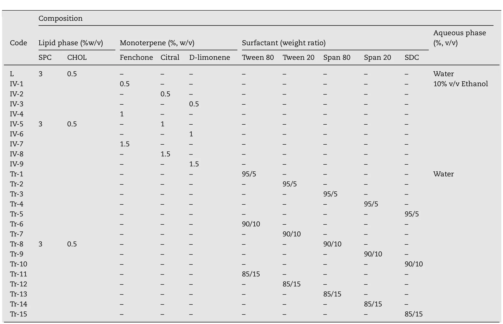
Table 1–The composition of liposome,invasome and transfersome formulations.
2.2.2.Characterization of the vesicle formulations containing PR
2.2.2.1.Evaluation of the physical appearances.The color,phase separation,and precipitation of all formulations were visually observed.The formulations with good physical properties were selected to evaluate the other properties.
2.2.2.2.Determination of vesicle size,size distribution and zeta potential.The vesicle size,size distribution and zeta potential of the formulations were evaluated at 25°C using a Zeta Potential Analyzer(Model ZetaPALS,New York,USA)with a scattering angle(θ)of 90°.In all cases,10 μl of samples were dispersed with 4ml of Milli-Q water before measurement.All determinations were performed in triplicate and presented as mean±standard deviation.
2.2.2.3.Chromatographic conditions for quantitative determination of PR.Quantitative determination of PR in all studies was performed by the method described by Limsuwan et al.[17].The instrument was coupled with the Agilent 1100 series photodiode-array detector(PDA)(Waldbronn,Germany),autosamplers and Agilent Chemstation for LD 3D software.The reverse-phase chromatography was carried out with a BDS HYPERSIL C18column(150mm x 4.6mm,5μm)at 25 °C.The mobile phase was a mixture of acetonitrile/methanol/milli-Q water(40:20:40,v/v/v)and maintained at 0.8ml/min.The 20μl sample solution was injected and the absorbance was detected at 254nm.
2.2.2.4.Measurement of entrapment efficiency.The ultracentrifugation technique was used to determine the percent entrapment efficiency(%EE)of PR in all formulations using Ultracentrifuge(OptimaTML-100XP,Beckman,USA)equipped with SW60 Ti Rotor at 60 000rpm at 4°C for 1.5h.The free PR was determined from supernatant solution after centrifugation and dilution with milli-Q water.The total PR amount in the formulation was measured after breaking the vesicles with methanol at ratio of 1:1 of sample and methanol.All samples were filtered through a 0.45μm syringe filter membrane and PR content was analyzed using HPLC.All determinations were performed in triplicate and the%EE was calculated according to the Eq.(1):

where,%EE is the percent entrapment efficiency,T is the total amount of PR in the formulation and F is the free PR amount.
2.2.2.5.Measurement of elasticity and deformability.The elasticity and deformability measurements were carried out by extrusion method which was modified from Singh et al.[18].Amongst the transfersome and invasome vesicles extruded at 8.5×10⁻⁴bar through the polycarbonate filter membrane(Sigma Chemicals,UK)having a pore diameter of 200nm,the amount of vesicles which were extruded during 5 min was measured.The vesicle size before and after filtration was monitored by Zeta Potential Analyzer(Model ZetaPALS,New York,USA)at 25 °C with a scattering angle(θ)of 90°.Afterward,values of degree of deformability were calculated[19].All determinations were performed in triplicate.
2.2.2.6.Visualizationofstructureandsurfacemorphology.The surface morphology of PR-invasome and-transfersome vesicles was observed by SEM(Quanta 400,FEI,Czech Republic)at an accelerating voltage of 20kV.100μl of sample was diluted with 3ml Milli-Q water,dropped on cover slip and dried.Then,the sample was stained with 2–3 drops of crystal violet solution and Gram’s iodine solution.After that,the dried sample was gold coated in a sputter coater(Leica EMD005,Czech Republic)under an argon atmosphere(50Pa)at 50mA for 70 sec and observed at optimized magnification.The structure of PR loaded ethosome,invasome and transfersome vesicles were observed by TEM(TEM,JEM-2010,JEOL,Japan).The 10μl of the formulation was diluted with 3ml milli-Q water and dropped on the holey film grid.The dried coated grid was observed by TEM at optimized magnification with an acceleration voltage of 160kV.In this study,the optimized composition was selected on the basis of a vesicle size less than 500nm,narrow size distribution(pdI<0.3),high absolute value of zeta potential and%EE>50%[13,20].
2.2.3.Evaluation of storage stability
ThephysicochemicalstabilityofPR-loadedtransfersomesand invasomes was evaluated after storing at 4±1°C,30±1°C and 45±1°C under controlled humidity of 75%RH in a Constant Climate Chamber Model HPP260(Memmert,Schwabach,Germany)for 0,1,2 and 4 months.The physicochemical stability was evaluated in term of the physical appearance,vesicle size,size distribution,zeta potential,total PR content and entrapment efficiency.Testing was carried out using triplicate samples at each storage condition.
2.2.4.In vitro skin permeation study
In vitroskin permeation and deposition of PR in the skin of transfersome and invasome systems were evaluated using modified Franz-diffusion cell(Model Hanson 57-6M,California,United States)with an effective diffusion area of 1.77 cm2.Newborn pig skin was used as the diffusion membrane because of its similarity to human skin in terms of general structure,thickness,hair follicle content and lipid component[20].The methodology of this study was followed from the method of Limsuwan et al.[13,20].The skin diffusion membrane was placed between the donor and receptor compartments with the stratum corneum toward the donor compartment.The donor part was filled with 1ml of the vesicle formulation whereas the receptor part was filled with 11ml of a mixture of 0.44%w/v sodium chloride(NaCl)in phosphate bufferpH7.4:propyleneglycol(80:20,v/v)asreceptormedium.In addition,water jacket of diffusion cell was maintained at 37±1°C to control the skin temperature to 32±2°C and a magnetic stirrer at 500rpm was used throughout the experiment.At time intervals at 0.5,1,2,4,6,8,12 and 24h,1ml of receiver medium was withdrawn and an equal volume of fresh medium at 37±1°C was replenished into the chamber to maintain sink conditions.The experiments were repeated fi ve times,PR contents in receiver medium were analyzed in triplicate by HPLC and permeation data analysis was evaluated.The cumulative amount(Qt,μg/cm2)of PR that permeated through the pig skin per unit area of skin was calculated according to Eq.(2):

where,Qtis cumulative amount of PR permeated per unit area of skin(μg/cm2),Cnis concentration of PR determined at No.n sampling interval(μg/ml),Ciis concentration of PR determined at No.i sampling interval(μg/ml),Vis volume of individual Franz diffusion cell(ml),Sis volume of sampling aliquot andAis effective diffusion surface area,1.77 cm2
TheQtamount was plotted as a function of time and the steady state flux(Jss)was calculated from the slope of the linear portion of the plot.The lag time(Tlag)of the PR to permeate through the pig skin before reaching the receptor fluid was calculated from the X-intercept of the plot.The value of permeation coefficient(Kp)for PR was calculated using the Eq.(3):

whereC0is the initial concentration of PR in the donor compartment.
2.2.5.Residual PR in the donor compartment
At the end of experiment,the excess formulation was removed from the membrane,cleaned with methanol(10ml)and sonicated at room temperature(32±2°C)for 30min.The clear supernatant that was obtained from centrifugation using a Hermle Z323K Centrifuge(Wehingen,Germany)at 6000rpm,4°C for 30min was lysed with methanol in the ratio of 1:1(v/v)and analyzed using HPLC to determine the amount of residual PR in the donor compartment.All samples were analyzed in triplicate.
2.2.6.In vitro skin deposition study
The skin membrane after removal of residual PR in the donor compartment was cleaned,cut into small pieces and homogenized in methanol(5ml)using homogenizer(Model PT 1200 E,Polytron,Switzerland)at room temperature(32±2°C)for 2min.Then,the sample was sonicated at room temperature for 30min and centrifuged at 6000rpm at 4°C for 30min to separate the skin lipid.The clear supernatant was analyzed using HPLC to determine the amount of PR which had accumulatedintheskindiffusionmembrane.Allsampleswereanalyzed in triplicate.
2.2.7.Evaluation of anti-tyrosinase activity
The extracted PR from skin diffusion membrane after permeation testing as described above(2.2.6)was evaluated for tyrosinase activity using the dopachrome method.Mushroom tyrosinase was assayed with L-dopa as substrate and Kojic acid as a positive control.The reaction mixture contained 20 μloftestsamplesorpositivecontrolsinDMSO,140??lofphosphate buffer(pH 6.8),20 μl of mushroom tyrosinase(203.3 unit/ml)and 20 μl of 0.85mM L-dopa.After the addition of L-dopa the reaction was measured for optical density with microplate reader at 492nm wavelength,before incubation.Then,the reaction mixture was incubated at 25°C for 20min and the OD of dopachrome formation after incubation was measured.The%tyrosinase inhibition was calculated according to the Eq.(4):

where:Aisthe difference ofOD before and after incubation at 492nm (containing all reagents and enzyme except the test sample).
Bis the difference of OD before and after incubation at 492nm(containing all reagents except the test sample and enzyme).
Cis the difference of OD before and after incubation at 492nm(containing all reagents,test sample,and enzyme).
Dis the difference of OD before and after incubation at 492nm(containingallreagentsandthetestsampleexceptenzyme).
2.2.8.Cell culture
B16 melanoma cell line was a kind gift from Assistant Prof.Dr.Sukanya Dej-adisai,Faculty of Pharmaceutical Sciences,Prince of Songkla University(Thailand).Cells were cultured in Dulbecco’s modified Eagle’s medium(DMEM)high glucose,supplemented with 10%fetal bovine serum(Gibco,USA),2mM glutamine,1%penicillin/streptomycin and maintained at 37°C in a humidified incubator with 10%CO2.
2.2.9.Quantification of cellular tyrosinase activity in B16 melanoma cells
Cellular tyrosinase activity was measured by adapting the assay established by Tomita et al.[21]with some modification.Brie fl y,cells(5×10⁴ cells/well)were stimulated with α-MSH(100nM)and then incubated in 24 well plates with different formulations containing 5 μM of PR for 72h.After treatment,the cells were collected with trypsin-EDTA and centrifuged at 6000rpm for 5min to obtain cell pellets.The cell pellets were freeze-thaw lysed in 200 μl of 2%Triton X-100 in 0.1M phosphate buffered saline(pH 7.2).After protein quantification by BCA method,100 μl of cell lytic solution(100μg/ml)were mixed with 100 μl of 0.2%L-Dopa in phosphate buffer solution(pH 7.2),and incubated for 1 h at 37°C.The optical densities were measured as 475nm using a microplate spectrophotometer.The tyrosinase activity of each formulation was presented as percentage against that of the control cells.
2.2.10.Determination of cellular melanin content in B16 melanoma cells
Cells(5×10⁵ cells/dish)were stimulated with α-MSH(100nM)and then incubated in 60mm dishes with different formulations containing 5μM of PR for 72h.After treatment,the cells were washed twice with ice-cold phosphate-buffered saline,and lysed in 200 μl of 1N NaOH for 1h at 95 °C and then vortexed to solubilize the melanin.Lytic solution(100 μl)was added in a 96 well plate.The total amount of melanin was measured at 405nm wavelength.The melanin content was determined based on the absorbance divided by the protein concentrationintheextractfromeachcellpellet.Themelanin content of each formulation was then calculated and presented as percentage against that of the control cells.
2.2.11.Investigation of acute dermal irritation/corrosion
The effects of PR in transfersome and invasome formulations on skin irritation were evaluated using the Guidelines 404 of the OECD(Acute Dermal Irritation/Corrosion)by TISTR(Thailand Institute of Scientific and Technology Research).
Three healthy adult white albino rabbits,in range of 2–3kg body weight were used in this study.One day before experimentation,the epidermal hair on dorsolumbar region was removed into a 10×10 cm2area.The 2.5×2.5 cm2of two hairless areas were selected for testing.0.5ml of the test sample and distilled water(negative control)were applied to each gauze patch.Then,the patch was applied on the selected skin sites and wrapped with adhesive tape to avoid movement.After 4h,all patches were removed and the residual sample was removed gently with cotton wool.The skin irritation in terms of erythema and edema was subjectively examined at time 1,24,48,and72h,or,morethanthesetimeintervals,whentheirritation still occurred,but not over 14 d of observation periods.The skin reactions were scored accounting for the numerical scoring system from 0 to 4,ranging from,none to severe signs,by two professional inspectors.
2.2.12.Statistical analysis
Theresultsobtainedin thestudywerepresentedas mean±standard deviation(SD)or mean±standard error of mean(SEM).One-way analysis of variance(ANOVA)followed by post hoc analysis was used to test the statistical significant of differences among groups.p<0.05was considered statistically significant.hancer,bothhighandlowconcentrationsgaveayellowcloudy formulation with no precipitate of drug and other components.
Invasomesizewasdistributedintherangeof208.10±12.00 to 819.30±101.90nm.It was reported that the vesicle size increases with the increasing concentration of penetration enhancer[12].The size of invasomes containing 0.5%,1%and 1.5%(w/v)citral was 312.00±10.30,651.00±161.80 and 782.60±75.60nm,respectively.The pdI of all invasomes was less than 0.3,indicating homogeneous population of the vesicles.The zeta potential of all invasome formulations showed high negative charge(-48.43±4.64 to-71.36±3.54)which representsthestabilityoftheformulations(Table2)duetolow possibility of vesicle aggregation,and normally considered as a stable formulation[22].
The%EE of all invasomes was found in the range of 88.08%±0.02%to 98.23%±0.00%.It is related to log P value.The formulation containing monoterpene with the low log P gave more%EE than the monoterpene with the high log P.Table 2 showed that the formulation containing fenchone(logP=2.13),citral(logP=3.45)and D-limonene(logP=4.58)gave a%EE value of 92.29%±0.02%(IV-1),88.08%±0.02%(IV-4),and 84.34%±0.04%(IV-7),respectively.This result may be effect of the solubility of PR in the lipid bilayers.Since the log P of PR is 2.11,it may be soluble in the composition with nearly the same value.In this study,the optimized PR-invasome formulation was obtained from the formulation no.IV-6 with the 341.17±54.11nm vesicle size,0.188±0.040 PDI,-59.47±2.66mV zeta potential and 91.32%±0.00%entrapment efficiency.
3. Results and discussion
3.1. Characterization of vesicle carrier
3.1.1.Characterization of PR-liposomes
The main formulation was obtained from 0.5%(w/v)PR,0.5%(w/v)CHOL,3%(w/v)SPC and water up to 100%(v/v).In this study,the main composition was used to represent the liposome formulation which is the rigid vesicle,to compare with the developed vesicles.The physicochemical properties of developed vesicles,with other skin enhancers added in each formulation(Table 1)were evaluated.Fig.1 and Table 2 show the physical appearance and the vesicle size,PDI,zeta potential and entrapment efficiency of all formulations.The liposome formulation(formulation code.L)showed a large vesicle size(641.52±19.02nm)with PDI(0.267±0.22),absolute value of zeta potential(-27.32±1.00mV)and entrapment efficiency(53.55%±4.9%).
3.1.2.Characterization of PR-invasomes
In case of invasomes,they contain fenchone,citral and D-limonene which are monoterpenes mixed with 10%(v/v)absolute ethanol as the skin enhancer.At a high concentration of fenchone(1%–1.5%w/v)it gave the formulation with precipitate(IV-4 and IV-7),while the formulation using low concentration(0.5%w/v)gave a good physical appearance.On the other hand,citral gave an unstable formulation when low concentration was used(IV-2 and IV-5).The good physical property results were obtained from using D-limonene as skin en-
3.1.3.Characterization of PR-transfersomes
Transfersome formulation had a yellowish turbid appearance with good physical property when prepared using Tween 20 and 80,Span 20 and 80,and sodium deoxycholate as edge activator,whereas the formulation containing Span 80 precipitated after preparation.
The size decreased together with the formulation containing surfactants with high HLB value.In this study it was found that the surfactant with high HLB(Tween 80,HLB=15;SDC,HLB=16;Tween 20,HLB=16.7)gave smaller size than the surfactant with low HLB(Span 20,HLB=8.6)as shown in Table 2.Transfersomes containing Tween 80,SDC and Tween 20 gave a mean vesicles size of 350.40±4.60,353.70±10.90 and 387.50±4.80nm,respectively,while the vesicle size of 991.30±330.60nm was obtained from the formulation containing Span 20 when prepared by the same concentration.The surfactant with low HLB might be attributed to enhance surface free energy through increasing hydrophobicity,by interacting with lipid head groups of the membrane,which increase packing density of phospholipid layer leading to increase surface free energy that causes fusion between lipid bilayers to form a large vesicle[23].However,Span 80 has low HLB with strongest hydrophobicity.That may cause the aggregation between vesicles,to form larger vesicle and tend to precipitate.In this study,the formulation containing Span 80 as edge activator becomes precipitated after preparation.The pdI,zeta potential and%EE of all transfersomes are as demonstratedinTable2.ThePDIofallformulationswereintherange of 0.093±0.046 to 0.285±0.020 which were within 0.3 indi-cating that the population of the vesicles was homogeneous.The zeta potential of all transfersome formulations were high negative charge(-41.86±0.73 to-68.55±2.56).High negative zeta potential represents a good stability of the formulations.
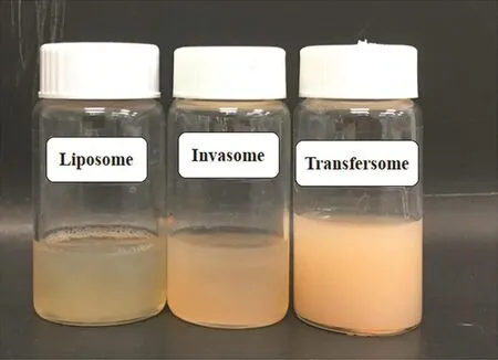
Fig.1–The physical appearance of liposome,invasome and transfersome formulations.
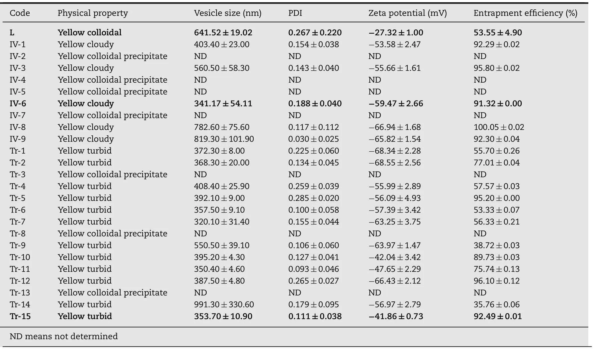
Table 2–The physical appearance,particle size,size distribution,zeta potential and entrapment efficiency of liposomes,invasomes and transfersomes.

Table 3–The deformability results of all formulations before and after filter through a 200nm polycarbonate filter.
The%EE of all transfersomes demonstrated in range of 35.76%±0.06%to 95.20%±0.00%.The highest%EE of transfersomes was observed in the formulation using SDC(95.20%±0.00%)when compared with other formulations.Since SDC is anionic surfactant,it may cause an increased electrostaticattraction with thepositivechargeofthe drug,which causes an increased%EE.The optimized PR-transfersome formulation which matched with criteria set was the formulation no.Tr-15.It gave the small vesicle size(353.70±10.90nm)with low PDI(0.111±0.038),high absolute value of zeta potential(-41.86±0.73 mv)and high entrapment efficiency(92.49%±0.01%).
3.2. Elasticity and deformability of vesicle carrier
Table 3 shows the deformability results of all vesicle carriers before and after filtering through a 200nm polycarbonate filter.The results demonstrated that invasomes(25.26%)have a higher degree of deformability than transfersomes(6.63%),while liposomes cannot pass 200nm pore size.The results confirmed that all developed vesicles have elastic property,whereas,the liposome formulation is a rigid vesicle.
In addition,the size of transfersome vesicles that was filtered through a polycarbonate filter is larger than the pore sizeof fi lter(200nm).Thesizesoftransfersomevesiclesbefore and after filter were 398.37±9.82nm and 371.97±8.72nm,respectively.ThepdIoftransfersomesinbothbefore(0.06±0.08)and after(0.13±0.09) fi lter were less than 0.3,indicating homogeneous population of the vesicles.These results confi rm that the transfersome vesicle has elastic and flexible properties because of retaining vesicle size and shape after filtrationd=6.63%.This characteristic of transfersomes is achieved by incorporating suitable edge activators to the lipid bilayers.In contrast,the invasomes which passed through 200nm pore size show deformable property.The size of invasomes after filtration was smaller than the original vesicles size before filtration,but still larger than pore size of filter.The sizes of invasomes before and after filter were 327.83±25.19nm and 245.03±2.44nm,respectivelyd=25.26%.Therefore,these results showed that invasomes only have deformability property.This property could be explained by effect of ethanol concentration,which reduced the interfacial tension of the vesicle membrane,leading to provide ability for small vesicles to extrude through the membrane[23].
3.3. Visualization of structure and surface morphology of vesicle carrier
Fig.2 shows the surface morphology of PR liposome,invasome and transfersome vesicles when observed by SEM and TEM.SEM photographs showed that all formulations revealed spherical structure with smooth surface(Fig.2A–2C).TEM photographs indicated the surface morphology of liposomes and invasomes showed unilamellar vesicle structure(Fig.1D and 1E),whereas,unilamellar to multilamellar was shown in case of transfersomes(Fig.1F).
3.4. Storage stability of vesicle carrier
The storage stability of all vesicle carriers was evaluated in term of vesicle size,zeta potential,total active content and%EE after storage at 4±1°C,30±1°C and 45±1°C under controlled humidity of 75%RH for 0,1,2 and 4 months as shown in Fig.3.
Stability results indicated that the physical property of transfersome formulation unchanged over 4 months when stored at 4±1°C and 30±1°C.In contrast,invasome formulation shows only to be stable at 30±1°C.The zeta potential values of all systems were decreased when time increased(Fig.3C to 3D).The zeta potential of invasomes and transfersomesdecreasedto-47.84±13.20and-40.76±8.11mVwhen stored at 30±1°C for 4 months,respectively.However,all zeta potential values were above 30mV over 4 months which demonstrated the good stability of the formulation.
The vesicle size of all formulations showed that it increased with time in all conditions(Fig.3A to 3B).The vesicle sizes of invasomes and transfersomes increased,by around 16.89%and,8.87%after storage at 30±1°C over 4 months.It increased about 8.91%when the transfersome formulation was stored at 45±1°C for 3 months.In addition,it slightly increased approximately 6.28%when the transfersomes were kept under 4±1°C for 3 months.However,all formulations showed narrow size distribution(PDI<0.3).The increase of size might be caused by the aggregation and fusion of vesicles itself.
However,the transfersome formulation showed higher percentage of total active content(Fig.3F)and EE(Fig.3H)at 4±1°C and 30±1°C,than at 45±1°C when tested after storing for 1,2,and 4 months.It gave percentage of total active content more than 85%at 4±1°C and 30±1°C.This may be due to higher fluidity of lipid bilayers which leads to higher drug leakage when stored at higher temperature[24].Therefore,the transfersome formulations in this study should be stored at 4±1°C and 30±1°C,whereas,the invasome formulation needs to be stored only at 30±1°C.
3.5. In vitro skin permeation and deposition study
Table 4 illustrates the percent recovery of PR in each compartment of the diffusion cell and skin permeation parametersafterapplicationofliposome,invasomeandtransfersome formulations.The amount of PR in the three compartments was analyzed following experimentation for 24h and expressed as a percentage of the applied dose(0.5mg/ml).The results showed that the liposome,invasome and transfersomeformulationsgave%recoveryas88.48±4.29,85.38±3.86 and 92.47±6.02%respectively,which were close to 90%–110%,which is the acceptable limit of OECD TG428[25].Thus,it was recognized that this method has performance and trustworthiness.In the percent recovery results,most of PR would not penetrate the skin.It accumulated at the donor compartment.The low penetration of PR may be because of the excellent barrier properties of the outermost skin layer named stratum corneum which had “the brick and mortar”structure[26].
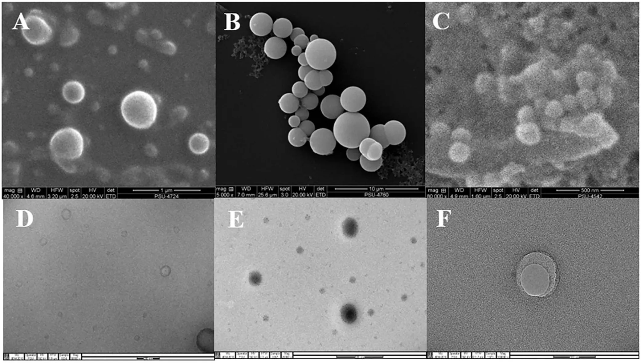
Fig.2–SEM photographs of liposome(A),invasome(B),and transfersome formulations(C),and TEM photographs of liposome(D),invasome(E)and transfersome formulations(F).

Table 4–The percent recovery of PR in each compartment of the diffusion cell and skin permeation parameters after application of liposome,invasome and transfersome formulations.
Table 4,Figs.4 and 5 show the skin accumulation and permeation results.Comparing with liposomes,all elastic carriers demonstrated that the application of them gave the PR accumulation,and permeation,higher than the application of liposomes.The skin permeation of PR after using liposomes was 20.65±1.72μg/cm2while the elastic carriers showed the skin permeation of PR ranging from 47.99±3.73 to 72.66±4.66μg/cm2.In addition,the skin accumulation in full-thickness newborn pig skin also showed that the elastic carriers gave higher PR accumulated than that of liposomes.The PR accumulations of liposome,invasome and transfersome formulations were 28.18±2.59,54.43±4.62 and 71.21±8.40μg/cm2,respectively.The good results may be due to the special mechanism of permeation across skin of flexible or elastic carrier.
In case of ethosomes,they have deformability property which is an important feature related to enhancement of skin permeability.It penetrates skin using the synergistic mechanism between high concentration of ethanol,phospholipid vesicles and lipid bilayers in stratum corneum.Ethanol interacts with skin lipid molecules in the polar head group region,resulting in reducing the rigidity of the stratum corneum lipids,and increasing their fluidity,which may finally lead to an increase in skin permeability.In addition,the ethanol may provide the vesicles with soft and flexible characteristics which enable them to squeeze through the pores in stratumcorneum which are much smaller than their diameter size[15,27].
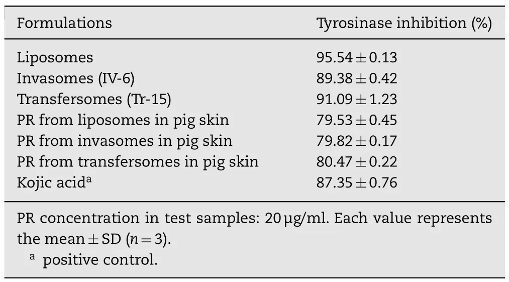
Table 5–Tyrosinase inhibitory activity of PR in liposomes,invasomes and transfersomes and pig skin following topical application in liposomes,invasomes and transfersomes.
In case of invasomes,they are soft flexible carrier with very high membrane fluidity.The mechanism of skin permeation of invasomes is similar to ethosomes,by ethanol and terpenes disturbing the organization of the stratum corneum lipid bilayer.Ethanol interacts with polar head group region of the skin lipid molecules,terpenes break the interlamellar hydrogen bonding network of stratum corneum lipid bilayer which may finally lead to the openings creating new polar pathways.In addition,ethanol may provide the vesicles with soft and fl exible characteristics which may finally lead to increase in skin permeability[12].
In case of transfersome carrier,it has ultra-elastic property[14]which allows it squeeze itself through the pores of the intact skin which is much smaller than its own diameter,into or through the deep skin layer.In addition,it penetrates across skin under the influence of a trans-epidermal water activity gradient in hydrophilic pathways of the skin.Moreover,it is able to retain its shape because of the presence of edge activators in the formulation[11].
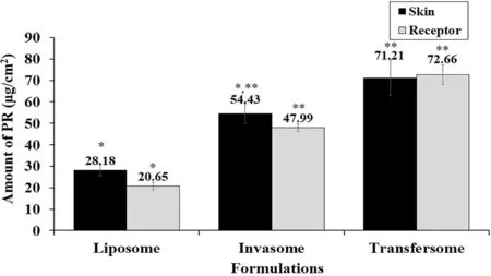
Fig.4–In vitro accumulation of PR in full-thickness newborn pig skin and receptor compartment after testing liposomes,invasomes and transfersomes for 24h.Each bar represents the mean±SEM(n=5).?-???:means in the same color bar with different asterisk differ significantly(P<0.05).Statistical significance was determined by one way ANOVA.
Comparison within the flexible or elastic carriers,all the results showed that the highest amount of PR that permeate across pig skin membranes was obtained from the application of transfersome formulation(Figs.4 and 5).In addition,the transfersomes with high flexible property provided a significantly higher cumulative amount,steady state fl ux(Jss;3.26±0.45μg/cm2/h)and permeability coefficient(Kp;0.65±0.09×10⁻³cm/h)compared to other formulations.These results may be linked with the degree of deformability,since,invasomes(25.26%)have a higher degree of deformability than transfersomes(6.63%)(Table 3).In contrast,the liposomes as rigid vesicles did not show deformable and elastic property.
Moreover,TEM photographs indicated the surface morphology of transfersomes showed multilamellar structure(Fig.2F),so they can entrap the more amount of PR for the topical delivery.Therefore,transfersomes with high elastic property[28]could penetrate across the skin more than liposomes and invasomes in both terms of the depth of skin layer,and amount of PR.
3.6. Anti-tyrosinase activity of vesicle carrier
Table5showedthat allcarriers exhibited tyrosinase inhibition activity in the range of 89.38%±0.42 to 95.54%±0.13%which is greater than kojic acid(87.35±0.76),a potent tyrosinase inhibitor.Since skin accumulation study(Fig.4)showed that the elastic vesicle formulations resulted in higher PR retention in the skin compared with liposomes,but tyrosinase inhibitory activity was similar.This result may be the effect of a shielding effect of vesicle systems.However,the tyrosinase inhibition activity of PR after application of liposomes,invasomes and transfersomes showed approximately 80%,which is estimated to result in highly efficient skin lightening activities.
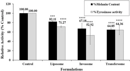
Fig.6–Effects of vesicle carriers containing PR on melanin production and tyrosinase activity in B16 melanoma cells.Cells were stimulated with 100nM α-MSH and then treated with indicating formulations(each formulation containing 5μM PR)for 72h.Cellular melanin content and tyrosinase activity were measured and analyzed.Bars represent the means±S.D.of three independent experiments(n=3).???? P<0.01,??? P<0.05 compared with the control.
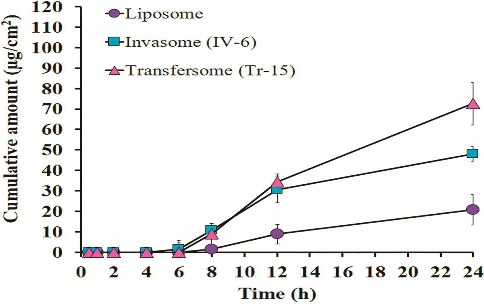
Fig.5–Cumulative permeation of PR across pig skin membranes following application of liposomes,invasomes and transfersomes.Each point represents the mean±SEM(n=5).
3.7. Effect of vesicle carrier on cellular melanin production and tyrosinase activity
Fig.6 shows that the PR formulated in liposomes,invasomes,and transfersomes significantly inhibited the tyrosinase activity as well as melanin production in B16 melanoma cells compared with the control.Interestingly,both tyrosinase inhibition activity and melanin production of elastic vesicle carriers showed higher activity than liposomes.Consistent with skin accumulation study(Fig.4.)shows that the elastic vesicle formulations exhibited high PR retention in the skin.The study in B16 melanoma cells also revealed that the elastic vesicle formulations displayed the higher tyrosinase inhibition activity and melanin content reduction when compared to liposomes.These findings confirmed that elastic vesicle carriers represent suitability for the PR delivery system,which proves it has highly effective skin lightening properties.
3.8. Acute dermal irritation testing of vesicle carrier
An acute irritation test in rabbits were carried out,based on erythema and edema formation systems after application PR in liposomes,invasomes and transfersomes in 72h.
The slightly erythema formation was observed in case of the application of transfersomes at 1 h.The recovery of this skin reaction occurred within 24 h of the observation period.However,the skin area which was treated with the liposomes and invasomes gave no significant differences from the skin area which was treated with distilled water(controlled area).Therefore,the short-term exposure suggests that PR at 0.5%(w/v)was safe for using as topical products.
4. Conclusions
The main formulation was composed of 0.5%(w/v)CHOL,3%(w/v)SPC and water up to 100%(v/v).The optimized vesicles formulations were obtained from adding skin enhancer into the main composition.The invasomes contained 1%(w/v)D-limonene mixed with 10%(v/v)absolute ethanol as the skin enhancer,and transfersomescontained15%(w/w)sodiumdeoxycholate(SDC)as edge activator.All carriers gave a vesicle size<500nm,polydispersity index(PDI)<0.3,high zeta potential,entrapment efficiency>50%,and good stability on storage at 30°C at 75%RH over 4 months.In addition,all elastic vesicles also showed a higher flexible property than liposomes.Moreover,transfersomes have a higher elasticity than invasomes and liposomes.It leads to provide a higher cumulative amount,steady state flux(Jss)and permeability coef ficient(Kp),compared to other formulations.Moreover,the skin accumulation results exhibited that the elastic vesicle formulations which have the higher PR retention in the skin,showed the higher tyrosinase inhibition activity,and melanin content reduction when compared to conventional liposomes in B16 melanoma cells.Therefore,elastic vesicle carriers such as transfersomes and invasomes,presented suitability for the PR delivery system,which is highly effective for skin lightening products.
Declaration of interest
The authors report no conflicts of interest.The authors alone are responsible for the content and writing of this article.
Acknowledgment
We are grateful to the Graduate School,Faculty of PharmaceuticalSciencesandPrinceofSongklaUniversityforproviding financial support.Tunyaluk Limsuwan acknowledges the grant of the Prince of Songkla University Scholarship for her Ph.D.study.
 Asian Journal of Pharmacentical Sciences2018年5期
- Asian Journal of Pharmacentical Sciences的其它文章
- Special issue:Pharmaceutical innovation
- Effect of silicone oil on the microstructure,gelation and rheological properties of sorbitan monostearate–sesame oil oleogels
- Design and characterization of monolaurin loaded electrospun shellac nanofibers with antimicrobial activity?
- Formulation and evaluation of gels containing coconut kernel extract for topical application?
- The effect of surfactant on the physical properties of coconut oil nanoemulsions?
- Preparation and characterization of nanoparticles from quaternized cyclodextrin-grafted chitosan associated with hyaluronic acid for cosmetics
