Advances and challenges of gastrostomy insertion in children
Rana Bitar, Amer Azaz, David Rawat, Mohamed Hobeldin, Mohamad Miqdady, Seifeleslam Abdelsalam
Abstract When oral feeding cannot provide adequate nutritional support to children, enteral tube feeding becomes a necessity. The overall aim is to ultimately promote appropriate growth, improve the patient’s quality of life and increase carer satisfaction. Nasogastric tube feeding is considered appropriate on a short-term basis. Alternatively, gastrostomy feeding offers a more convenient and safer feeding option especially as it does not require frequent replacements, and carries a lower risk of complications. Gastrostomy tube feeding should be considered when nasogastric tube feeding is required for more than 2-3 wk as per the ESPEN guidelines on artificial enteral nutrition. Several techniques can be used to insert gastrostomies in children including endoscopic, image guided and surgical gastrostomy insertion whether open or laparoscopic. Each technique has its own advantages and disadvantages. The timing of gastrostomy insertion, device choice and method of insertion is dependent on the local expertise, patient requirements and family preference, and should be individualized with a multidisciplinary team approach. We aim to review gastrostomy insertion in children including indications, contraindications, history of gastrostomy, insertion techniques and complications.
Key Words: Laparoscopic gastrostomy; Percutaneous endoscopic gastrostomy; Laparoscopic-assisted gastrostomy; Laparoscopic-assisted percutaneous endoscopic gastrostomy; Radiologic gastrostomy; Open gastrostomy
INTRODUCTION
When oral feeding becomes compromised, nasogastric tube feeding is considered appropriate for the support of fluid and nutrition in children on a short-term basis; however, in the long term this type of feeding has many limitations and carries a reduced survival rate[1]. Gastrostomy tubes (GTs) are inserted with the aim to support the long-term nutritional needs of children. Gastrostomy insertion has evolved over the last century to involve multiple feeding devices and various insertion techniques. The device chosen and the insertion technique are selected after careful consideration of patient background medical history, patient and family needs, and available facilities. Prior to insertion, adequate counselling of parents and a multi-disciplinary team (MDT) review is recommended. We aim to present the indications, contraindications, history, advances, insertion techniques, challenges and complications of gastrostomy insertion in children.
INDICATIONS AND CONTRAINDICATIONS
Indications
Gastrostomies should be considered when enteral feeding is required for more than 2-3 wk[2] with the aim of correcting significant nutritional deficiencies, promoting growth in children, and avoiding further body weight loss. Weight per age should be interpreted using disease-specific growth chart centiles when available. The ESPGHAN recommendation on gastrostomy insertion in children recommends consideration of gastrostomy insertion when enteral feeding is required for more than 3-6 wk[3]. Gastrostomies are generally considered in children with underlying chronic nutritional needs such as patients with oncological, metabolic, renal, neurological and gastrointestinal tract disorders in which oral intake is insufficient to sustain growth.
While the specific indications for gastrostomy placement are many and variable, the most frequent indications are related to inadequate oral fluid and nutrition intake and/or impaired swallowing in disorders of the central nervous system, either as a primary cause or in conjunction with chromosomal or metabolic disorders. In addition, renal disorders, congenital cardiac disease, oncological disorders, chronic respiratory diseases such as cystic fibrosis, and gastrointestinal disorders such as Crohn’s disease and intestinal failure may require gastrostomy feeding to correct nutritional deficiencies. Gastrostomies are also inserted in congenital or acquired conditions such as esophageal atresia and craniofacial surgery, when oral intake may be anatomically impeded. Moreover, they may be necessary in children who require nutritional restitution to attain recommended weights advisable for certain surgical interventions, for example, in infants with congenital cardiac disorders[4,5]. Gastrostomies are also sometimes indicated in children with unsafe swallowing, at risk of recurrent aspiration from oral feeding and when gastric drainage and decompression is required in cases of foregut dysmotility. Another rare but recognized indication is to deliver therapeutic formulae in patients with certain metabolic disorders, which are usually unpalatable. Finally, it can be offered to patients who require many medications due to other organ diseases to improve compliance and effectiveness of medications[6-8].
Contraindications
Contraindications are relative and can typically be overcome. They include lack of a safe tract for percutaneous insertion due to adhesions, congenital anomalies, severe kyphoscoliosis, distorted anatomy due to multiple abdominal surgery, and interposed organs (liver, colon). In this case, surgical gastrostomy may be the only option whether it is laparoscopic or open. Significant coagulation disorders should be corrected, and placement should be deferred until full recovery if the patients suffer from hemodynamic instability, sepsis, significant ascites, infectious peritonitis, and abdominal wall infection at the placement site. Gastrostomy insertion in patients undergoing peritoneal dialysis is high risk and may be considered a relative contraindication by some centers[9].
HISTORY AND ADVANCES
The initial use of enteral nutrition in the gastrointestinal tract to nourish patients dates back to 1500 BC[10]. Over the centuries, research has evolved and contributed to better understanding of nutritional needs, methods to access the gastrointestinal tract, development of new tubes and equipment, with better understanding of digestion, absorption, and use of macro- and micronutrients.
The very first gastrostomy was used with the purpose of alimentation in obstruction at the gastric cardia or above in adults. It was initially proposed by Egeberg in 1837, and after multiple attempts and failures, it was not performed successfully until 1876 by Verneuil[11]. At that time gastrostomies were inserted by conventional open surgery[12]. In pediatrics, the procedure has been a mainstay in the early stage treatment of esophageal atresia; the first survivors of this condition were reported by Leven[13] and Ladd[14] in the early 1940’s, and gastrostomy insertion was part of the therapy. Reports by Martin and Fultzl[15] in 1959, Holder and Gross[16] in 1960, Meeker and Snyder[17] in 1963, and others widened the popularity and indications for gastrostomy to include many pediatric surgical conditions. The less invasive percutaneous endoscopic gastrostomy (PEG) tube placement was introduced in 1980 by Gaudereret al[18]. Glow in the stomach of a newborn infant undergoing endoscopy inspired the development of this procedure. The first PEG was inserted in the pediatric operating room on June 12, 1979 in the University Hospital of Cleveland, United States, on a four-month-old with inadequate oral intake[19] under local anesthesia with sedation. Although PEG was originally described in children, it has become a popular method of enteral nutrition in all ages. In 2001, 20 years after it was invented, over 216000 were performed annually in the United States[20].
The application of gastrostomy later extended to cover non-surgical indications, such as supporting the nutritional requirements of patients with severe neurologic impairments and developmental delay. As these two patient groups had a higher risk for general anesthesia, the open operation solely to place a gastrostomy promptly changed to the less invasive approach, the PEG. It was among the first innovations that expanded endoscopy from a diagnostic tool to a therapeutic instrument. It was not until 1991 when laparoscopic gastrostomy application was first cited in the literature[21]. It had the advantage of direct visualization of the peritoneal cavity during placement to protect from inadvertent bowel injury and optimize gastrostomy location while being less invasive than open gastrostomy. Although PEG and laparoscopic-assisted gastrostomy (LAG) are the two most frequently used procedures for gastrostomy placement, to date, there is no agreement as to which procedure, is superior. Many centers prefer to insert a PEG owing to its simplicity and low cost[22]. PEG has since become widely accepted in both the adult and pediatric populations.
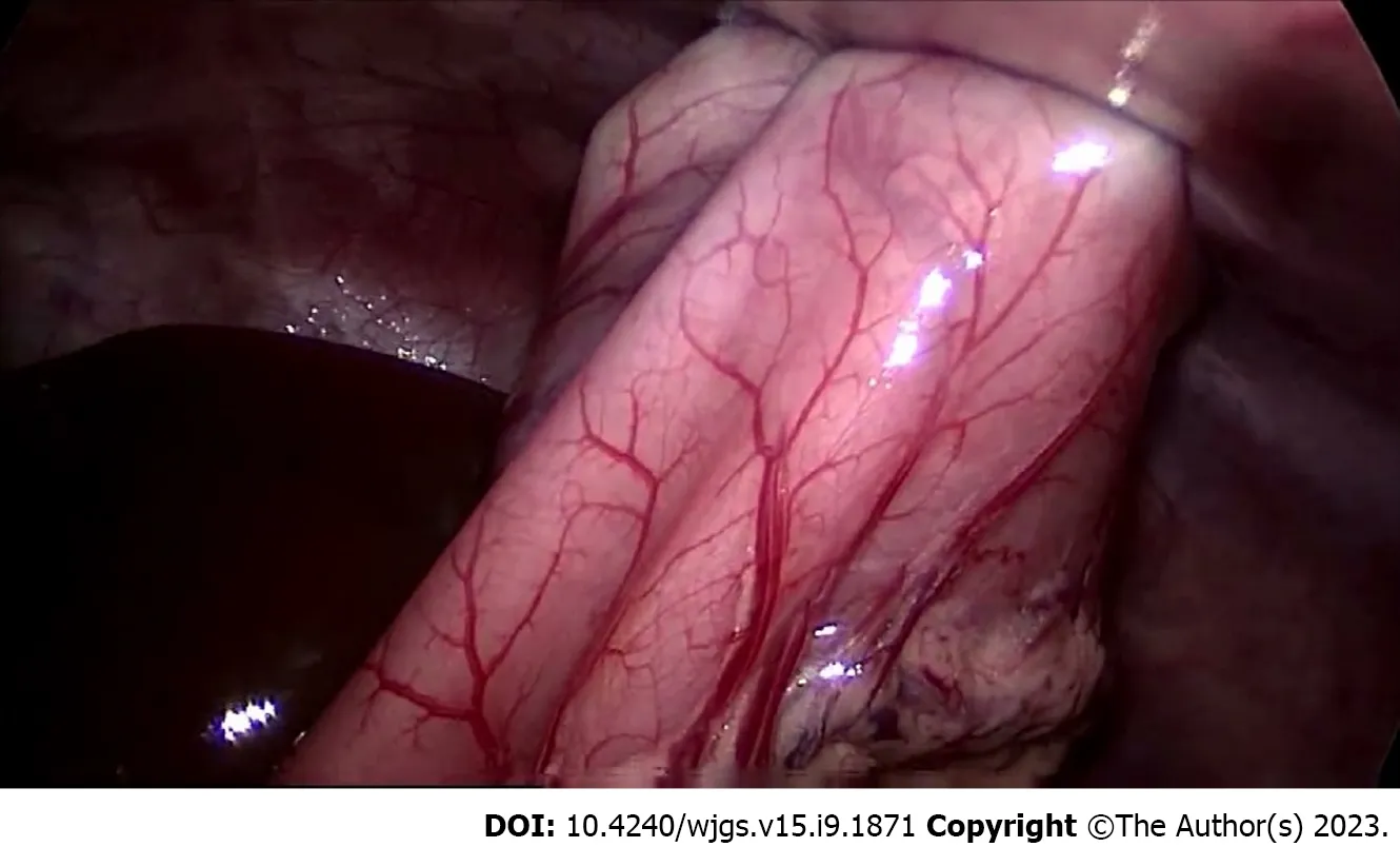
Once the concept of a minimally invasive procedure for gastrostomy was introduced, further modifications were introduced to reduce complication rates and facilitate the operative technique. Techniques such as the push gastrostomy technique has the advantage of avoiding the step of pulling the GT through the oropharynx and esophagus and preventing the carriage of microorganisms to the peristomal site[4]. Although the push technique is associated with a lower peristomal infection rate than the pull technique in adults[23], this has not been demonstrated in pediatric patients[24,25]. Another technique modification to avoid the need for frequent general anesthesia with its associated risks, was the one-step gastrostomy device. The one-step gastrostomy device was an appealing, low-profile gastrostomy introduced in pediatric patients which uses a balloon device[26]. The one-step gastrostomy is being increasingly used. As the balloon device does not offer the as secure fixation of the stomach to the abdominal wall as an internal bumper, gastropexy was introduced so that the stomach is fixed to the abdominal wall by sutures or T-fasteners[27], as demonstrated in Figure 1. Gastropexy is performed to ensure adequate apposition of the stomach and the anterior abdominal wall[26,28]. The onestep PEG/LAG placement with the push technique and T-fastener gastropexy[24] gained popularity due to its unique advantages. Regardless of tube insertion technique, GTs are generally changed to a low-profile button after 6 wk to 8 wk to allow for tract maturation[29]. Recently more and more centers have started to insert primary gastrostomy button feeding tubes.
A new technique, combining the use of endoscopy and laparoscopy in gastrostomy insertion was described in 1995 by Stringelet al[30], where laparoscopic-assisted PEG (LAPEG) was performed on 2 children when attempts at simple PEG had failed. This technique has been used particularly in difficult cases where PEG was felt to be high risk or impossible. LAPEG combines both endoscopy and laparoscopy for gastrostomy insertion, while using a single umbilical incision to insert the laparoscope to assist in gastrostomy placement. Using the laparoscope permits accuracy in the placement of the PEG, allows identification and subsequent lysis of adhesions, and safe completion of the PEG. In some centers LAPEG is performed routinely[31], in others it is used when PEG is felt to be unsafe or impossible, in other centers it is used if the abdominal wall is > 2 cm, making it technically difficult to perform a laparoscopic gastrostomy[32].
GASTROSTOMY INSERTION OPERATIVE TECHNIQUE
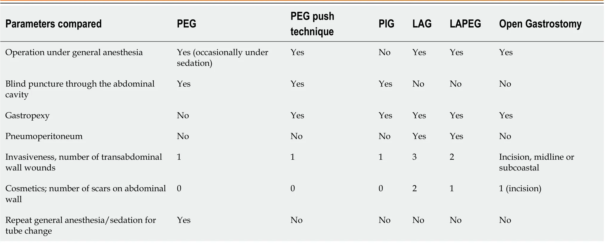
Parents should be given detailed information on the benefits, principles and decision making behind the choice of technique for gastrostomy insertion by the professional undertaking the procedure. Table 1 demonstrates the characteristics of different gastrostomy placement techniques. After MDT involvement, alternative methods of gastrostomy insertion should also be discussed including the pros and cons of each. Procedural as well as intermediate and long-term risks of GT insertion should be discussed with the parents/carers well in advance of the procedure to enable adequate time to process the information, consider any questions and make an effective well-informed decision before giving consent. Regardless of the technique, at the time of gastrostomy insertion, it is recommended that patients are given antibiotics preoperatively[33-35]. Most centers will allow immediate use of the GT for medications, and commencement of feeds may be variable depending on institutional consensus and is no later than one postoperative day.

Table 1 Characteristics of different gastrostomy placement techniques

Figure 1 Gastropexy of the stomach to the abdominal wall using three trans-gastric tuckers.
Open gastrostomy
Open gastrostomy has been in use for more than 100 years and has remained the standard until the introduction of less invasive insertion techniques. Nowadays, open gastrostomy is reserved for cases where the anatomy does not allow for a safe LAG or PEG insertion or the child cannot tolerate the pneumoperitoneum; as in cases of scar tissue formation from previous surgery. It should also be considered if the patient requires other surgical procedures at the same time.
There are different techniques described for open gastrotomy tube insertion, the most common technique used includes an incision made in the upper abdomen either midline or left subcostal and the abdominal cavity is entered. The stomach is identified, and an appropriate location for GT insertion selected, on the anterior wall of the body of the stomach, an opening is made on the stomach. The GT is passed through the abdominal wall ideally with the rectus sheath away from the umbilicus and costal margin and then inserted into the stomach. The tube is secured to the stomach with purse string sutures placed around the tube. The stomach is then anchored to the abdominal wall from the inside with sutures. Finally, the surgical incision is closed with sutures.
PEG, pull through technique
Under endoscopic guidance, the stomach is inflated and a position for gastrostomy insertion on the anterior abdominal wall is identified using transabdominal impulse/finger indentation and transillumination. The abdominal wall and skin are injected with local anesthesia. A puncture cannula is inserted through the anterior abdominal wall into the stomach cavity under endoscopic control while the stomach is inflated to allow opposition of the stomach wall to the abdominal wall. The needle is removed from the cannula and an introducer device containing a double thread is inserted through the cannula. The thread is pushed through the cannula until it is visible endoscopically in the stomach cavity. The thread is then caught and secured through the endoscope with forceps or a snare. The endoscope with the biopsy forceps/snare and adherent thread are pulled out through the mouth as one unit. The thread is then interlocked with the PEG, the PEG is lubricated with lubricant jelly, and the guide thread outside the abdominal wall is pulled through the cannula while the PEG is pulled through the mouth, esophagus and into the stomach. The PEG tube is pulled through the abdominal wall with the inner disk fitting snug along the gastric mucosa. Finally, the PEG is fixed to the anterior abdominal wall by adjusting the external bumpers that are provided with the gastrostomy device used.
Percutaneous image guided gastrostomy
The GT is inserted into the stomach using the Seldinger technique. A nasogastric tube is placed shortly before the procedure to allow air insufflation. Gastric puncture is performed under fluoroscopic guidance with an 18G puncture needle in the left upper abdominal quadrant. To confirm insertion of the needle through the stomach lumen, the radiologist will aspirate air into a syringe or flushes the needle with contrast medium, the gastric and abdominal wall are securely fastened together (gastropexy). Gastropexy is usually performed using introducer needles preloaded with anchors. The abdominal wall and gastric wall are approximated, the gastric wall and stomach wall are punctured near the anchors. The tract is dilated using serial dilators, after adequate dilatation, a balloon type gastrotomy is pushed into the gastric lumen through a peel away sheath. The retention balloon is inflated with water, and the procedure is completed with contrast injection through the GT to confirm correct tube position and to exclude extravasation or other complications.
LAG
Multiple modifications have been described for laparoscopic gastrostomy. In general, the procedure starts with insufflation of the peritoneum. Pressures are maintained between 6 to 12 mmHg based on patient comorbidities and size. A 5-mm telescope is placed through an umbilical port. An extra 5-mm port site is placed in the left upper quadrant below the costal margin. The stomach is then visualized and grasped along the greater curvature.
This small portion of the stomach can be delivered through the abdominal wall, at that time the port is removed and sutures are placed between the stomach and the abdominal wall. A small opening is made in the stomach and the tube is placed into the stomach.
Another technique to perform LAP gastrostomy is to fix the stomach to the abdominal wall by T fasteners or stitches then access the stomach by a needle followed by introduction of a guidewire through the needle. In this case the GT is inserted using the Seldinger technique; serial dilatation of the tract is performed using dilators, after adequate dilatation of the tract, a balloon type gastrotomy is pushed into the gastric lumen through a peel away sheath. Finally, the tube position is checked by infusing and aspirating saline solution under laparoscopic control.
Push one-step PEG
Under endoscopic control, gastropexy is performed using 3 fasteners. At the center of the gastropexy a puncture site is identified and a trocar is inserted into the gastric lumen under direct visualization by the endoscopist. A guidewire is passed through the trocar which is later used to pass a dilator. After serial dilatation of the future gastrostomy track, a feeding tube is inserted into the stomach and the dilator is peeled away. The balloon is inflated
LAPEG
The procedure requires both endoscopic and laparoscopic techniques, and therefore, both an endoscopist, and pediatric surgeon are required.
A 5-mm optical is placed through the umbilicus for the laparoscope. Pneumoperitoneum is recommended at 8-12 mmHg. At the same time, a gastroscopy is performed and the stomach lumen is visualized. After insufflating the stomach, the optimal site for gastrostomy is chosen using external finger indentation and direct visualization. Gastropexy is performed using 3 fasteners, and a needle is inserted into the gastric lumen, a guidewire is passed through the needle which is later used to guide the dilator. After serial dilatation of the track, the GT is inserted and the balloon inflated and the tube is fixed to the skin at an appropriate length. Gastrostomy is inserted under direct laparoscopic and endoscopic visualization.
COMPLICATIONS AND CHALLENGES
GT insertion carries a procedural risk and is also associated with intermediate and long-term post-operative complications. Complications, can be classified as minor or major. Major complications involve failure of GT placement, gastrostomy peritoneal leak causing peritonitis, tube dislodgement, buried bumper syndrome, adjacent bowel injury, major bleeding, esophageal tear, and gastrocolic fistula formation. Minor complications, on the other hand, include minor skin infection, granulation tissue formation, tube leak, and tube occlusion. There is also the possibility of the development or aggravation of gastroesophageal reflux disease[8]. A large meta-analysis looked at complication rates and mortality in association with different gastrostomy insertion operative techniques in children, and data from 18 articles with 4631 patients were analyzed. Techniques compared were that of PEG (pull, single stage, or introducer, percutaneous image guided gastrostomy) and LAG insertion. The overall complications encountered were; minor (33% of patients)-granulation (10.30%), local infection (8.30%) and leakage (6.00%), major (10.00% of patients)-systemic infection (3.50%), cellulitis (1.00%) perforation (< 0.30%) and lethal (0.15%). Interestingly, prematurity or young age did not affect complication rate[4].
Laparoscopic techniques have been reported to be safer than endoscopic gastrostomy insertion procedures. In a retrospective comparative study between PEG, LAG and open gastrostomy including 236 children; the overall rates of major complications were 9.2% in the endoscopic gastrostomy, 8.9% in the laparoscopic, and 8.1% in open gastrostomy groups[36]. In a larger meta-analysis, which included 8 studies and 1550 pediatric patients; LAG technique was found to be associated with only 1% chance of major complications compared to 5.4% in the PEG technique[37]. Laparoscopic gastrostomy has unique advantages, the surgeon has a better visual intraperitoneal field thereby lowering the risk of perforation of hollow viscous and vascular injury, and particularly the formation of gastrocolic fistula which has been reported in children following PEG. A study examining endoscopic gastrostomy placement in infants less than one year of age found that despite successful placement in a healthier cohort, PEG had more morbid and more costly complications, specifically a 3.8% risk of gastrocolic fistula, compared to laparoscopic gastrostomy[38]. Interestingly, in a systematic review, 8.4% (2.1%-19.4%) of children who underwent PEG and 2.5% (0.0%-8.6%) of children who underwent LAG required re-intervention under general anesthesia with a reported significant difference[39].
Considering the various techniques used to insert GTs in children, the magnitude of the challenges faced with the procedure and the likelihood of complications is highly dependent on the technique used for GT insertion. The higher the blinded components of the technique the more likely are the challenges and major complications. The more the directly visualized components of the technique the less likely are the challenges and major complications. Fixation of the stomach to the abdominal wall is another factor that reduces the likelihood of major complications, it reduces the occurrence of tube dislodgement and possible subsequent intraperitoneal leak. Gastropexy is feasible during laparoscopic, radiologic and push one step endoscopic gastrostomy[26,28]. Based on the above principles, LAPEG in children is associated with a high safety profile due to direct endoscopic and laparoscopic visualization of the entire GT insertion process. In a retrospective review of 76 pediatric patients, LAPEG was performed and completed safely with no recognizable peri-operative complications, despite 26% of the cohort being considered high risk with significant preexisting comorbidities. The safety and use of LAPEG has also been supported by previous reports[40-48]. A retrospective review comparing LAPEG to LAG demonstrated both procedures to be comparable in reducing the major complication rate but with the added advantage of significantly shorter procedure time in the LAPEG group[48]. In the past, 10 kg of weight was considered the lower limit of body weight for insertion of PEG tubes, below which the procedure was deemed to be more technically challenging[49]. However, PEG insertion is reported to be safe in infants with weight as low as 2.3 kg[50]. Minaret al[51] described successful PEG in 39/40 infants with a mean weight of 3.25 kg at the time of procedure. The only major complication reported was esophageal injury. There exists the hypothesis from scholar peers that younger children may be at higher risk of complications at the time of PEG placement as they have thinner tissues which may be easier to transilluminate the gastrocolic omentum or the transverse colon. Thus, resulting in accidental penetration and traversing of the colon, and resultant gastrocolic fistula which can go undetected. Hence, the recommendation by some in using the laparoscopic technique or the LAPEG technique in small patients[52]. In the LAPEG report of 76 patients, one third of the patients were < 7 kg in weight and one third were < 7 mo of age. Therefore, LAPEG is a potential option for this subset of patients.
CONCLUSION
Although gastrostomy insertion has become a common procedure in children, the best method of placement still needs to be determined. The method of placement can vary significantly according to patient age, local expertise, and available healthcare facilities[53]. Therefore, more research is still needed including the best insertion technique for individual patient groups, the timing and type of best enteral feeds to be initiated after placement, and identifying specific risk factors for the development of complications. In our current climate of health economics, reduction in the cost and local availability of resources required for gastrostomy placement in children should be considered. The ideal gastrostomy procedure is a one-step procedure, performed under minimal anesthesia, with no complications, at reduced cost and optimal resources utilization as long as patient safety is considered first and foremost. As technology improves, advanced minimally invasive robotic surgical procedures are likely to expand. This can only succeed if we continue to challenge and improve our current practice by continued collaboration between pediatric surgeons and gastroenterologists with more good quality multi-center research of novel practices and modifications of gastrostomy techniques and perioperative management.
ACKNOWLEDGEMENTS
The authors would like to knowledge Mr. Emad Bashir Sharfi, Retired Medical Librarian in Sheikh Khalifa Medical City for his dedication and support in obtaining all required articles to support the completion of the minireview.
FOOTNOTES
Author contributions:Bitar R made substantial contributions to the conception and design of the work, interpretation of data, drafting, writing and revising the manuscript critically for important intellectual content and approved the final version to be published, and agrees to be accountable for all aspects of the work; Azaz A, Rawat D, Hobeldin M, Miqdady M, and Abdelsalam S made substantial contributions to the design of the work, acquisition and interpretation of data and revising the manuscript critically for important intellectual content, and approved the final version to be published and agree to be accountable for all aspects of the work.
Conflict-of-interest statement:There is no conflict of interest to declare.
Open-Access:This article is an open-access article that was selected by an in-house editor and fully peer-reviewed by external reviewers. It is distributed in accordance with the Creative Commons Attribution NonCommercial (CC BY-NC 4.0) license, which permits others to distribute, remix, adapt, build upon this work non-commercially, and license their derivative works on different terms, provided the original work is properly cited and the use is non-commercial. See: https://creativecommons.org/Licenses/by-nc/4.0/
Country/Territory of origin:United Arab Emirates
ORCID number:Rana Bitar 0000-0002-2852-7707; Amer Azaz 0000-0003-2303-0846; David Rawat 0000-0003-1788-8758; Mohamad Miqdady 0000-0001-9089-9424.
S-Editor:Chen YL
L-Editor:Webster JR
P-Editor:Wu RR
 World Journal of Gastrointestinal Surgery2023年9期
- World Journal of Gastrointestinal Surgery的其它文章
- Quantitative evaluation of colorectal tumour vasculature using contrast-enhanced ultrasound: Correlation with angiogenesis and prognostic significance
- Risk factors for myocardial injury during living donor liver transplantation in pediatric patients with biliary atresia
- Value of enhanced computed tomography in differentiating small mesenchymal tumours of the gastrointestinal from smooth muscle tumours
- Multifactor analysis of the technique in total laparoscopic gastric cancer
- Clinical significance of serum oxidative stress and serum uric acid levels before surgery for hepatitis Brelated liver cancer
- Prediction model of stress ulcer after laparoscopic surgery for colorectal cancer established by machine learning algorithm
