KIFC3 promotes proliferation, migration and invasion of esophageal squamous cell carcinoma cells by activating EMT and β-catenin signaling
INTRODUCTION
Esophageal cancer is one of the most dangerous tumors affecting humans, ranking seventh in incidence and sixth in mortality worldwide[1]. The main histological type of esophageal cancer in western countries is adenocarcinoma, while that in Asian countries is squamous cell carcinoma, where it has high incidence and mortality[2-4]. Although clinicians and researchers have made great efforts to unravel the pathophysiology of esophageal cancer, the mechanism of esophageal squamous cell carcinoma (ESCC) is still unknown[5-7]. Recent research has shown that dysregulation of
and cell cycle regulators are prominent characteristics of ESCC, which may also be detected in precursor lesions,but the molecular progression from dysplasia to invasive ESCC remains unclear[3,8]. Further studies are urgently needed to reveal the underlying molecular mechanisms and discover effective treatment targets to improve ESCC survival.
The Prince s next idea for Potentilla s amusement was to cause a fleet of boats exactly like those of Cleopatra, of which you have doubtless read in history, to come up the little river, and upon the most gorgeously decorated of these reclined the great Queen herself, who, as soon as she reached the place where Potentilla sat in rapt attention, stepped majestically37 on shore and presented the Princess with that celebrated38 pearl of which you have heard so much, saying: You are more beautiful than I ever was
The kinesin superfamily proteins (KIFs) are a group of proteins that function as microtubule-based motors for transporting cellular cargo and playing crucial roles in chromosomal and spindle movements during mitosis and meiosis[9]. There are 45 KIFs identified in humans; among which, several have demonstrated varied functions in tumor pathobiology
different mechanisms, including regulation of cell cycle progression and metastasis[10-12]. Among these members, KIFC3 has negative end-directed microtubule motor activity and plays roles in Golgi positioning and integration, as well as in apical transport, in epithelial cells[13,14]. Studies of null mice have revealed that KIFC3 is dispensable for normal development and reproduction[15]. Overexpression of KIFC3 may mediate docetaxel resistance in breast cancer cells[16]. KIFC3 expression was positively associated with cell invasion and migration,and overexpressed KIFC3 was associated with shorter overall survival in hepatocellular carcinoma[17].However, the role of KIFC3 in ESCC has not yet been reported. In view of the importance of cell cycle regulators in the development of ESCC and the involvement of KIFC3 in mitosis, we speculated that this protein might be involved in the occurrence and development of ESCC.
In this study, we aimed to examine KIFC3 expression in ESCC and further explore its role and the relevant mechanisms in ESCC tumor progression, in an attempt to provide promising insights into the mechanism of this disease and possible therapeutic targets.
Then he was just the kind of man they liked, said they, and he might easily earn a good penny, before he was a day older, for the king paid a hundred dollars to anyone who would stand as sentinel in the church all night, beside his daughter s chest
MATERIALS AND METHODS
Human tissue specimens
Primary ESCC tumor tissues and corresponding nontumor tissue specimens were collected from the First Affiliated Hospital of Zhengzhou University. All experiments were carried out in accordance with the Declaration of Helsinki (amended in 2013). All procedures were supervised and approved by the Ethics Committee of the First Affiliated Hospital of Zhengzhou University (No. 2021-KY-0446-001).
But I ended by ceasing to covet20 that gift more than any of the others I have seen, for, like the gift of pleasing, it cannot really give satisfaction
Cell lines and cultures
The human ESCC cell lines KYSE30, KYSE150, KYSE450, KYSE510 and Eca109 and human normal esophageal epithelial cell line Het-1A were obtained from the Type Culture Collection of the Chinese Academy of Sciences and cultured in RPMI-1640 medium supplemented with fetal bovine serum (FBS)(10%) at 37°C in 5% CO
.
Western blotting
A Transwell assay was used to detect cell migration and invasion. For the invasion assay, 100 μL Matrigel (serum-free medium diluted 1:8) was added to the upper chamber of the Transwell chamber(Corning, USA). After shaking well, the gel was incubated at 37°C and solidified for 2–4 h. Cells were seeded at 10
per well in the upper chamber with 100 μL serum-free medium. For the lower chamber,500 μL medium (20% FBS) was added. After 24 h, the cells that passed through the filter were fixed with 4% paraformaldehyde for 30 min, stained with 0.5% crystal violet for 30 min, and washed with PBS.Finally, the cells were examined under a fluorescence microscope (BX51; Olympus, Japan). The migration assay was similar to the invasion assay, with the difference that no Matrigel was added to the upper chamber.
Transfection
pLVX-Puro-KIFC3-shRNA- and pLVX-Puro-KIFC3-expressing lentiviruses were designed and provided by Genechem (China). KYSE150 cells were transfected with pLVX-Puro-KIFC3-shRNA- expressing lentiviruses and KYSE450 cells with pLVX-Puro-KIFC3-expressing lentiviruses, while control cells were transfected with empty vectors. After transfection with lentiviruses, 2 μg/mL puromycin dihydrochloride (Beyotime) was added for 6–8 d to establish stable cell lines. Knockdown and overexpression efficiency were validated using western blotting, and the validated cell lines were recorded as KYSE150shNC, KYSE150shKIFC3, KYSE450oeNC and KYSE450oeKIFC3.
Trembling with anticipation verging28 on anxiety, I looked around with searching eyes. Nothing had changed. The scene was exactly the same as I had left it ten years ago. There was only one thing missing -- she wasn t there! I had drawn29 out the short straw! I felt crestfallen30. My mind went blank and I stood motionles overcome with gloom, when suddenly, I felt that familiar electrifying31 touch, the same shiver and the familiar thrill. It jolted32 me back to reality, as quick as lighting33. As she softly put two promfret fish in my hand I was feeling in the seventh Heaven.
Colony formation assay
The cancer cells were seeded in a six-well plate at 1000 cells/well and cultured for 14 d. The colonies were washed once with phosphate-buffered saline (PBS), fixed with 1 mL 4% paraformaldehyde for 15 min, and stained with 1 mL 0.5% crystal violet for 30 min. After washing with deionized distilled water,the images were captured using a digital camera. The number of colonies was then counted.
Cell cycle analysis
The cancer cells were seeded in a six-well plate at 2.5 × 10
cells per well. When the cell density was 60%,cells were collected, washed with cold PBS twice, and then fixed in 70% ethanol at 4°C overnight. The cells were washed with cold PBS twice, and then incubated with RNaseA (0.1 mg/mL) and propidium iodide (0.02 mg/mL) at 37°C for 30 min in the dark. The cell cycle was detected using flow cytometry.
Transwell assay
Total proteins were extracted using RIPA buffer supplemented with 1% phenylmethylsulfonyl fluoride and 1% Protease Inhibitor Cocktail. Protein concentration was measured using the BCA kit (Beyotime,China). Proteins were separated using SDS-PAGE and transferred onto a polyvinylidene difluoride membrane. After blocking with 5% fresh milk for 1 h, the membranes were incubated with a specific primary antibody overnight at 4°C, followed by incubation with the corresponding secondary antibody for 1 h. Finally, the bands on the membranes were detected using the ECL detection system (Thermo)and quantified using Quantity One Software version 4.3. Primary antibodies for KIFC3, E-cadherin, Ncadherin, vimentin, proliferating cell nuclear antigen (PCNA), cyclin D1, β-actin, and secondary antibodies were purchased from Proteintech (China), matrix metalloproteinase (MMP)7 and c-Myc were purchased from Abcam (Cambridge, MA, USA), and β-catenin was purchased from Cell Signaling Technology (Danvers, MA, USA).
Immunofluorescence
Based on the
data above, we further investigated the role of KIFC3 using xenograft tumors
. All animals were in a fit state during the experiment and all
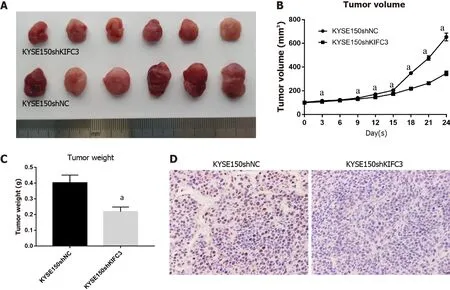
data were included in the analysis. The transplanted tumors grew rapidly in the KYSE150shNC group but were suppressed in the KYSE150shKIFC3 group (Figure 4A and 4B), and the weight of tumors in the KYSE150shKIFC3 group was significantly lower (Figure 4C). Immunohistochemistry of tumors showed that the expression of Ki-67 was decreased significantly in the KYSE150shKIFC3 group (Figure 4D), proving that KIFC3 knockdown inhibited the proliferation of ESCC
at the molecular level. Taken together, KIFC3 promotes ESCC proliferation
.
Xenograft tumor experiment using nude mice
All animal research procedures were approved by the Ethics Committee of the First Affiliated Hospital of Zhengzhou University (No. 2021-KY-0446-001). All animal experiments were carried out in accordance with the National Institutes of Health Guide for the Care and Use of Laboratory Animals(NIH Publications No. 8023, revised 1978). Male BALB/c nude mice (age 4 wk) were purchased from Beijing Life River Experimental Animal Technology Co. Ltd. and kept in a temperature-controlled specific pathogen free environment with a regular light/dark cycle and provided with adequate rodent diet and water. Ten mice were randomly divided into KYSE150shNC and KYSE150shKIFC3 groups.There were five mice in each group. KYSE150shNC cells (10
) and KYSE150shKIFC3 cells (10
) were collected, washed with PBS, suspended in 200 μL PBS, and subcutaneously implanted into the right flank of the dorsal region of nude mice. A Vernier caliper was used to measure tumor sizes every 3 d,and the tumor volume (TV) was calculated using the following formula:TV (mm
) = 0.5 × d
× D, where d is the shortest diameter and D is the longest diameter. After nine measurements, the longest diameter of tumor reached 15 mm. All mice were killed and the tumor specimens were collected and weighed.
Immunohistochemistry
Immunohistochemical staining was performed using an UltraSensitive TM SP kit and DAB kit (Maixin,China). Tumor tissues were embedded in paraffin and cut into 4-μm sections. The deparaffinized sections were incubated with a primary antibody against KIFC3 (1:200 dilution, Abcam, #ab154419) or Ki-67 (1:100 dilution, Abcam, #ab16667) overnight at 4°C, and normal goat serum was used as a negative control. After washing, the tissue sections were then incubated with a biotinylated anti-rabbit secondary antibody (1:200 dilution, Aspen, #AS-1107) for 1 h at 25°C. The sections were subsequently incubated with horseradish-peroxidase-conjugated streptavidin (Beyotime, #A0303) and developed using 3, 3-diaminobenzidine. An optical microscope (BX51; Olympus) was used to observe the specimens. Two observers, who were blinded to the data of the samples, evaluated, counted and analyzed the positive cells. The proportion of positive tumor cells was scored as follows:1 (< 10%positive tumor cells), 2 (10%–50% positive tumor cells), 3 (50%–75% positive tumor cells), and 4 (> 75%positive tumor cells). The intensity of staining was graded according to the following criteria:0 (no staining), 1 (weak staining = light yellow), 2 (moderate staining = yellow brown), and 3 (strong staining= brown). The staining index was calculated as the product of the proportion of positive cells times the staining intensity score (range:0–12). The median of staining index was used as the cut-off value; the staining index higher than the cut-off value was identified as high expression, while that less than the cut-off value was identified as low expression.
Statistical analysis
Graphpad Prism 7 was used for all analyses. Data are expressed as the mean ± SD. To analyze the differences between groups,
test and nonparametric tests were used.
< 0.05 was considered statistically significant.
RESULTS
KIFC3 expression is upregulated in ESCC and is associated with poor prognosis
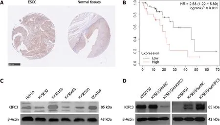
To investigate KIFC3 expression, ESCC tissues and adjacent nontumor tissues were collected from 34 patients (26 male and 8 female, aged 47–72 years) with ESCC. Immunohistochemical assays showed that KIFC3 expression was lower in ESCC tissues than in adjacent nontumor tissues (Figure 1A). Moreover,data from Kaplan–Meier Plotter (https://kmplot.com/analysis) revealed that lower expression of KIFC3 was associated with better overall survival in patients with ESCC (Figure 1B). These results indicate that KIFC3 plays an important role in ESCC development. To explore the role of KIFC3 in ESCC cells, its expression level was evaluated in ESCC cell lines Eca109, KYSE30, KYSE150, KYSE450 and KYSE510, as well as normal esophageal epithelial cell line Het-1A. KYSE450 and KYSE150 cells displayed the lowest and highest expression levels of KIFC3, respectively (Figure 1C). Therefore,KYSE450 cells were used for subsequent overexpression assays, while KYSE150 cells were used for knockdown assays. Western blotting verified that the corresponding cell lines were successfully constructed (Figure 1D).
KIFC3 promotes cell proliferation in ESCC cells
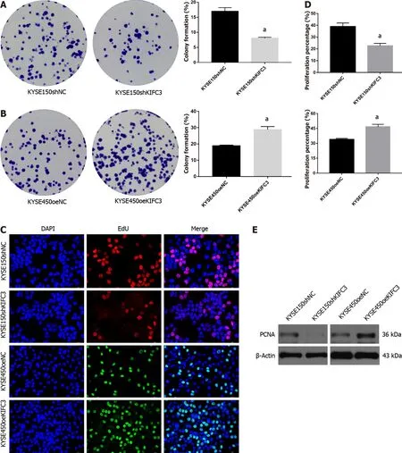
To investigate the effect of KIFC3 in ESCC, colony-formation and EdU assays were performed to observe the proliferation of KYSE150 and KYSE450 cells. The results showed that the colony-formation capacity was decreased after knockdown of KIFC3 in KYSE150 cells, while KIFC3 overexpression promoted colony-formation capacity in KYSE450 cells (Figure 2A and 2B). EdU assay showed that KIFC3 knockdown inhibited the proliferation of KYSE150 cells, while its overexpression promoted proliferation of KYSE450 cells (Figure 2C and 2D). In addition, KIFC3 knockdown inhibited the expression of PCNA, while its overexpression promoted PCNA expression (Figure 2E), which indicated that KIFC3 promoted proliferation in ESCC cells at the molecular level.
How delighted the witch was when she found the clothes all finished! The next time Prince Ring came to see her she gave them to him, and he paid her many compliments on her skilful23 work, after which he took leave of her in the most friendly manner
KIFC3 promotes cell cycle progression in ESCC cells
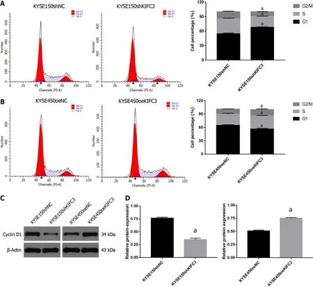
To further investigate the role of KIFC3 in cell cycle progression, cell cycle analysis was conducted. The percentage of G0/1 phase in KYSE150shKIFC3 group was significantly higher, and percentage of S and G2/M phase was lower than that in KYSE150shNC group (Figure 3A), while the percentage of G0/1 phase in KYSE450oeKIFC3 group was significantly lower, and percentage of S and G2/M phase was higher than that in KYSE450oeNC group (Figure 3B). At the molecular level, KIFC3 knockdown caused a decrease while KIFC3 overexpression promoted the expression of cyclin D1, which plays an important role in the transition from G1 to S phase (Figure 3C and 3D). These results indicate that KIFC3 positively regulates cell cycle in ESCC cells.
KIFC3 promotes tumor growth in vivo
KYSE150 and KYSE450 cells were seeded onto glass coverslips in 24-well plates overnight. The cells were then washed with PBS and fixed with 4% paraformaldehyde for 15 min at 25°C. Next, the cells were permeabilized for 15 min with 0.3% Triton X-100 in PBS. The cells were then blocked with 5% BSA in PBS for 1 h at 25°C and then incubated overnight at 4°C with the primary antibody against β-catenin(1:100 dilution) overnight; normal goat serum was used as a negative control. The treated cells were incubated with an FITC-labeled or Cy3-labeled goat anti-rabbit IgG (H+L) highly cross-adsorbed secondary antibody (Beyotime) in the dark for 1 h. The nuclei were visualized after staining with 2 μg/mL DAPI (Biosharp, China) for 10 min at 25°C. The glass coverslips were sealed with antifade reagent (Biosharp) and examined under a fluorescence microscope (BX51; Olympus).
53. Pearls and precious stones: Hansel and Gretel feel no guilt73 for taking the witch s treasure, similar to Jack with the Giant s treasure in Jack and the Beanstalk. The witch s attempt to kill them and subsequent death is implied as justification for taking the jewels.Return to place in story.
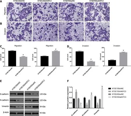
To detect whether KIFC3 exerts effects on the migration and invasion of ESCC cells, Transwell migration and invasion assays were used. Accelerated cell migration was observed in the KYSE450oeKIFC3 group compared with the KYSE450oeNC group, while it was less active in the KYSE150shKIFC3 group than in the KYSE150shNC group (Figure 5A and 5C). Transwell invasion showed similar results (Figure 5B and 5D). To further explore the molecular mechanism of KIFC3-promoted migration and invasion, we detected the expression of E-cadherin, N-cadherin and vimentin,which are key molecules of epithelial–mesenchymal transition (EMT). The results indicated that Ncadherin and vimentin, which are associated with the mesenchyme phenotype and indicate a higher possibility of tumor migration and invasion, were decreased, while E-cadherin, which is associated with the epithelial phenotype, increased after KIFC3 knockdown. In KIFC3-overexpressing cells, the expression of N-cadherin, vimentin and E-cadherin showed the opposite results (Figure 5E and 5F).These results indicate that KIFC3 promotes migration and invasion through EMT in ESCC cells.
Hao WW designed and performed experiments, analyzed data, and wrote the article; Xu F conceived the study, analyzed the data, and revised the article; all authors have read and approved the final manuscript.
KIFC3 promotes expression of β-catenin in ESCC cells
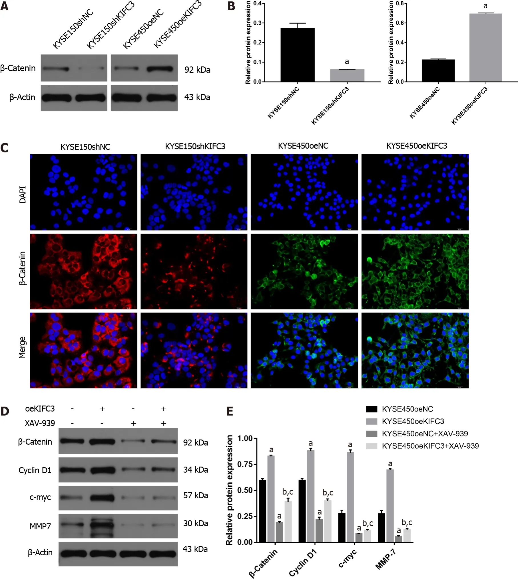
Previous studies have demonstrated that β-catenin plays an essential role in the proliferation, migration,and invasion of ESCC cells. In our study, western blotting showed that β-catenin protein expression in KYSE150shKIFC3 group was decreased significantly compared with that in the KYSE150shNC group(Figure 6A and 6B), while expression of β-catenin was increased significantly in the KYSE450oeKIFC3 group compared to that in the KYSE450oeNC group (Figure 6A and 6B). Immunofluorescence showed the same results; β-catenin expression was decreased after KIFC3 knockdown and increased after KIFC3 overexpression (Figure 6C). These results suggest that KIFC3 promotes β-catenin expression in ESCC cells.
1872FAIRY TALES OF HANS CHRISTIAN1 ANDERSENDELAYING IS NOT FORGETTINGby Hans Christian AndersenTHERE was an old mansion2 surrounded by a marshy3 ditch with adrawbridge which was but seldom let down:- not all guests are goodpeople. Under the roof were loopholes to shoot through, and to pourdown boiling water or even molten lead on the enemy, should heapproach. Inside the house the rooms were very high and had ceilingsof beams, and that was very useful considering the great deal of smoke which rose up from the chimney fire where the large, damp logs of wood smouldered. On the walls hung pictures of knights4 in armour5 and proud ladies in gorgeous dresses; the most stately of all walked about alive. She was called Meta Mogen; she was the mistress of the house, to her belonged the castle.
KIFC3 promotes the progression of ESCC via β-catenin signaling
To explore the role of β-catenin signaling in KIFC3-promoted proliferation, migration and invasion, we used XAV-939, an inhibitor of β-catenin. KYSE450oeNC and KYSE450oeKIFC3 cells were treated with XAV-939, and the expression of proteins downstream of β-catenin, which play an important role in proliferation, migration, and invasion was evaluated. Although the expression of c-myc, cyclin D1 and MMP7 was increased after KIFC3 overexpression (
< 0.05), when β-catenin was inhibited by XAV-939,the levels of c-myc, cyclin D1 and MMP7 were decreased even when KIFC3 was overexpressed (
<0.05) (Figure 6D and 6E). These results suggest that KIFC3 promotes the progression of ESCC
βcatenin signaling.
DISCUSSION
ESCC is one of the most malignant tumors that impose a significant medical burden on the health system. Although several gene mutations that could increase the susceptibility to ESCC have been identified[8], the exact mechanism that induces tumor progression is still unclear. KIFC3 has been reported to be overexpressed in a number of cancers and may be involved in cell cycle and cell proliferation, which suggests that KIFC3 is an oncogene, thus arousing our interest in its possible role in ESCC progression.
In the present study, we found that KIFC3 was upregulated in ESCC tissues compared to adjacent nontumor tissues. Data from the Kaplan–Meier Plotter website indicated that high levels of KIFC3 expression were correlated with poor ESCC prognosis. These findings indicate that KIFC3 may be involved in the development of ESCC, and that high expression of KIFC3 may be a prognostic factor in ESCC. Functional assays showed that knockdown of KIFC3 suppressed proliferation in KYSE150 cells,and the cell cycle was arrested compared with that in KYSE150shNC cells. Migration and invasion were inhibited in KYSE150shKIFC3 cells. Compared with KYSE450oeNC cells, cell proliferation, migration,and invasion were activated in KYSE450oeKIFC3 cells, further demonstrating that KIFC3 promoted the progression of ESCC cells. Furthermore, KIFC3 knockdown inhibited ESCC proliferation
. Given the above results, we may consider KIFC3 as a tumor marker associated with the progression and prognosis of ESCC.
EMT plays a potential role in the promotion of tumor invasiveness, metastasis and resistance to apoptotic stimuli, and it is marked by downregulation of epithelial biomarkers, such as E-cadherin, and the upregulation of mesenchymal biomarkers, such as N-cadherin and vimentin. Cells undergoing EMT can acquire greater mobility and become prometastatic[18-20]. EMT is known to play an important role in the metastasis of ESCC[21-23]. In addition, members of the KIF family are involved in the regulation of EMT and thus affect the migration and invasion of tumors[24-26]. In our study, KIFC3 knockdown suppressed the migration and invasion of KYSE150 cells, accompanied by the upregulation of Ecadherin and downregulation of N-cadherin and vimentin, while KIFC3 overexpression showed the opposite results in KYSE450 cells. These results indicate that KIFC3 promotes the migration and invasion of ESCC cells by inhibiting EMT.
β-Catenin signaling has been shown to be associated with the regulation of processes such as proliferation and invasion, which are involved in the occurrence and progression of cancers[27,28]. Moreover,the deactivation of β-catenin has been a potential treatment target in ESCC[29-31]. Thus, regulation of βcatenin may play an important role in the development of ESCC. It has been reported that in HEK293 cells depleted of centrosomes, normal accumulation of β-catenin in response to Wnt signaling is attenuated[32]. KIFC3 regulates centrosome cohesion in a microtubule-dependent manner[14].
Collectively, the relationship between KIFC3 and β-catenin is worth exploring. In the present study,KIFC3 overexpression promoted the expression of β-catenin and downstream molecules of β-catenin signaling, such as cyclin D1, c-myc and MMP7. However, after treatment with XAV-939, an inhibitor of β-catenin, KIFC3-induced upregulation of cyclin D1, c-Myc, and MMP7 was blocked, which means that β-catenin plays a vital role in KIFC3-induced tumor progression. Although further investigation is required to clarify the exact mechanism underlying the effect of KIFC3 on β-catenin, our research demonstrated that KIFC3 promotes tumor progression
β-catenin signaling in ESCC.
To elucidate the role of KIFC3 in ESCC and the underlying mechanisms.
CONCLUSION
KIFC3 is upregulated in ESCC, and KIFC3 overexpression is associated with poor prognosis in ESCC patients. KIFC3 promotes the proliferation, migration, and invasion of ESCC cells by activating EMT and β-catenin signaling. Overall, our study provides a comprehensive understanding of KIFC3 in ESCC and its underlying mechanisms, which strongly suggests that KIFC3 may be a potential new therapeutic target for ESCC treatment.
The study was reviewed and approved by the Institutional Review Board at the First Hospital of Zhengzhou University (No. 2021-KY-0446-001).
ARTICLE HIGHLIGHTS
Research background
Esophageal squamous cell carcinoma (ESCC) is one of the most common malignancies. The mechanism of ESCC is still unclear.
Research motivation
Kinesin family member (KIF)C3 has microtubule motor activity and may be involved in mitotic progression. KIFC3 was also shown to be involved in cell invasion and migration, as well as in survival,in hepatocellular carcinoma.
Research objectives
Observe a child; any one will do. You will see that not a day passes in which he does not find something or other to make him happy, though he may be in tears the next moment. Then look at a man; any one of us will do. You will notice that weeks and months can pass in which day is greeted with nothing more than resignation, and endure with every polite indifference1. Indeed, most men are as miserable2 as sinners, though they are too bored to sin-perhaps their sin is their indifference. But it is true that they so seldom smile that when they do we do not recognize their face, so distorted is it from the fixed3 mask we take for granted. And even then a man can not smile like a child, for a child smiles with his eyes, whereas a man smiles with his lips alone. It is not a smile; but a grin; something to do with humor, but little to do with happiness. And then, as anyone can see, there is a point (but who can define that point?) when a man becomes an old man, and then he will smile again.
Research methods
The expression of KIFC3 was evaluated in ESCC tissues and normal tissues. In addition, KIFC3 knockdown and KIFC3-overexpressing cell lines were constructed and then colony formation, EdU assays, cell cycle analysis, Transwell assays, and western blotting were performed to explore the underlying mechanisms of action. A xenograft tumor model in nude mice was used to verify the role of KIFC3 in tumorigenesis.
Research results
We showed that KIFC3 was upregulated in ESCC tissues and was associated with poor prognosis.KIFC3 promoted cell proliferation, mitosis progression, migration and invasion. In addition, KIFC3 knockdown suppressed ESCC tumorigenesis in an
model. Mechanistically, we validated the involvement of KIFC3 β-catenin signaling and epithelial–mesenchymal transition (EMT) in ESCC progression.
Research conclusions
We found that KIFC3 was overexpressed in ESCC, and the expression of this protein was associated with prognosis in ESCC patients. Furthermore, KIFC3 promoted proliferation, migration, and invasion of ESCC
β-catenin signaling and EMT.
Research perspectives
Although the detailed mechanism underlying the effect of KIFC3 in promoting ESCC progression should be studied more carefully in our next research, we believe our research strongly suggests that KIFC3 may be a potential new therapeutic target for ESCC treatment.
She rang the bell, and scarcely had she touched it before she found herself in a chamber2 where a bed stood ready made for her, which was as pretty as anyone could wish to sleep in
When Grannonia heard these words, and saw how deeply rooted the Prince s love for her was, she felt very happy, and blushing rosy36 red, she said: But should I get the other lady to give up her rights, would you then consent to marry me? Far be it from me, replied the Prince, to banish37 the beautiful picture of my love from my heart
Then you can rot at the bottom of the sea, both of you, said the old woman; and perhaps it may be the case that your mother would rather keep the two sons she has than the one she hasn t got yet
All animal research procedures were approved by the Ethics Committee of the First Affiliated Hospital of Zhengzhou University (No. 2021-KY-0446-001). All animal experiments were carried out in accordance with the National Institutes of Health Guide for the Care and Use of Laboratory Animals (NIH Publications No. 8023, revised 1978).
The authors declare no conflicts of interest.
Suddenly he asked the waiter, Would you please give me some salt? I d like to put it in my coffee. Everybody stared at him, so strange! His face turned red but still, he put the salt in his coffee and drank it. She asked him curiously3, Why you have this hobby? He replied, When I was a little boy, I lived near the sea, I liked playing in the sea, I could feel the taste of the sea, just like the taste of the salty coffee. Now every time I have the salty coffee, I always think of my childhood, think of my hometown, I miss my hometown so much, I miss my parents who are still living there. While saying that tears filled his eyes. She was deeply4 touched. That s his true feeling, from the bottom of his heart. A man who can tell out his homesickness5() , he must be a man who loves home, cares about home, has responsibility6 of home... Then she also started to speak, spoke7 about her faraway hometown, her childhood, her family.
No additional data are available.
When he had quite done he climbed up on the hut, and, blowing his flute, he chanted Pii, pii, fall rain and hail, and directly the sky was full of clouds, the thunder roared, and huge hailstones whitened the roof of the hut
The authors have read the ARRIVE guidelines, and the manuscript was prepared and revised according to the ARRIVE guidelines.
This article is an open-access article that was selected by an in-house editor and fully peer-reviewed by external reviewers. It is distributed in accordance with the Creative Commons Attribution NonCommercial (CC BYNC 4.0) license, which permits others to distribute, remix, adapt, build upon this work non-commercially, and license their derivative works on different terms, provided the original work is properly cited and the use is noncommercial. See:https://creativecommons.org/Licenses/by-nc/4.0/
China
Weiwei Hao 0000-0002-5156-6878; Feng Xu 0000-0002-5205-781X.
Wu YXJ
Kerr C
Yuan YY
1 Siegel RL, Miller KD, Fuchs HE, Jemal A. Cancer Statistics, 2021.
2021; 71:7-33 [PMID:33433946 DOI:10.3322/caac.21654]
2 Fan J, Liu Z, Mao X, Tong X, Zhang T, Suo C, Chen X. Global trends in the incidence and mortality of esophageal cancer from 1990 to 2017.
2020; 9:6875-6887 [PMID:32750217 DOI:10.1002/cam4.3338]
3 Smyth EC, Lagergren J, Fitzgerald RC, Lordick F, Shah MA, Lagergren P, Cunningham D. Oesophageal cancer.
2017; 3:17048 [PMID:28748917 DOI:10.1038/nrdp.2017.48]
4 Thrumurthy SG, Chaudry MA, Thrumurthy SSD, Mughal M. Oesophageal cancer:risks, prevention, and diagnosis.
2019; 366:l4373 [PMID:31289038 DOI:10.1136/bmj.l4373]
5 Cao W, Lee H, Wu W, Zaman A, McCorkle S, Yan M, Chen J, Xing Q, Sinnott-Armstrong N, Xu H, Sailani MR, Tang W,Cui Y, Liu J, Guan H, Lv P, Sun X, Sun L, Han P, Lou Y, Chang J, Wang J, Gao Y, Guo J, Schenk G, Shain AH, Biddle FG, Collisson E, Snyder M, Bivona TG. Multi-faceted epigenetic dysregulation of gene expression promotes esophageal squamous cell carcinoma.
2020; 11:3675 [PMID:32699215 DOI:10.1038/s41467-020-17227-z]
6 Triantafyllou T, Wijnhoven BPL. Current status of esophageal cancer treatment.
2020; 32:271-286[PMID:32694894 DOI:10.21147/j.issn.1000-9604.2020.03.01]
7 Yang YM, Hong P, Xu WW, He QY, Li B. Advances in targeted therapy for esophageal cancer.
2020; 5:229 [PMID:33028804 DOI:10.1038/s41392-020-00323-3]
8 Liu X, Zhang M, Ying S, Zhang C, Lin R, Zheng J, Zhang G, Tian D, Guo Y, Du C, Chen Y, Chen S, Su X, Ji J, Deng W,Li X, Qiu S, Yan R, Xu Z, Wang Y, Cui J, Zhuang S, Yu H, Zheng Q, Marom M, Sheng S, Hu S, Li R, Su M. Genetic Alterations in Esophageal Tissues From Squamous Dysplasia to Carcinoma.
2017; 153:166-177 [PMID:28365443 DOI:10.1053/j.gastro.2017.03.033]
9 Hirokawa N, Noda Y, Okada Y. Kinesin and dynein superfamily proteins in organelle transport and cell division.
1998; 10:60-73 [PMID:9484596 DOI:10.1016/s0955-0674(98)80087-2]
10 Qureshi Z, Ahmad M, Yang WX, Tan FQ. Kinesin 12 (KIF15) contributes to the development and tumorigenicity of prostate cancer.
2021; 576:7-14 [PMID:34474246 DOI:10.1016/j.bbrc.2021.08.072]
11 Wu YP, Ke ZB, Zheng WC, Chen YH, Zhu JM, Lin F, Li XD, Chen SH, Cai H, Zheng QS, Wei Y, Xue XY, Xu N.Kinesin family member 18B regulates the proliferation and invasion of human prostate cancer cells.
2021;12:302 [PMID:33753726 DOI:10.1038/s41419-021-03582-2]
12 Zou JX, Duan Z, Wang J, Sokolov A, Xu J, Chen CZ, Li JJ, Chen HW. Kinesin family deregulation coordinated by bromodomain protein ANCCA and histone methyltransferase MLL for breast cancer cell growth, survival, and tamoxifen resistance.
2014; 12:539-549 [PMID:24391143 DOI:10.1158/1541-7786.MCR-13-0459]
13 Xu Y, Takeda S, Nakata T, Noda Y, Tanaka Y, Hirokawa N. Role of KIFC3 motor protein in Golgi positioning and integration.
2002; 158:293-303 [PMID:12135985 DOI:10.1083/jcb.200202058]
14 Hata S, Pastor Peidro A, Panic M, Liu P, Atorino E, Funaya C, J?kle U, Pereira G, Schiebel E. The balance between KIFC3 and EG5 tetrameric kinesins controls the onset of mitotic spindle assembly.
2019; 21:1138-1151[PMID:31481795 DOI:10.1038/s41556-019-0382-6]
15 Yang Z, Xia Ch, Roberts EA, Bush K, Nigam SK, Goldstein LS. Molecular cloning and functional analysis of mouse Cterminal kinesin motor KifC3.
2001; 21:765-770 [PMID:11154264 DOI:10.1128/MCB.21.3.765-770.2001]
16 De S, Cipriano R, Jackson MW, Stark GR. Overexpression of kinesins mediates docetaxel resistance in breast cancer cells.
2009; 69:8035-8042 [PMID:19789344 DOI:10.1158/0008-5472.CAN-09-1224]
17 Chen J, Li S, Zhou S, Cao S, Lou Y, Shen H, Yin J, Li G. Kinesin superfamily protein expression and its association with progression and prognosis in hepatocellular carcinoma.
2017; 13:651-659 [PMID:28901309 DOI:10.4103/jcrt.JCRT_491_17]
18 Lamouille S, Xu J, Derynck R. Molecular mechanisms of epithelial-mesenchymal transition.
2014;15:178-196 [PMID:24556840 DOI:10.1038/nrm3758]
19 Mittal V. Epithelial Mesenchymal Transition in Tumor Metastasis.
2018; 13:395-412 [PMID:29414248 DOI:10.1146/annurev-pathol-020117-043854]
20 Navas T, Kinders RJ, Lawrence SM, Ferry-Galow KV, Borgel S, Hollingshead MG, Srivastava AK, Alcoser SY, Makhlouf HR, Chuaqui R, Wilsker DF, Konaté MM, Miller SB, Voth AR, Chen L, Vilimas T, Subramanian J, Rubinstein L, Kummar S, Chen AP, Bottaro DP, Doroshow JH, Parchment RE. Clinical Evolution of Epithelial-Mesenchymal Transition in Human Carcinomas.
2020; 80:304-318 [PMID:31732654 DOI:10.1158/0008-5472.CAN-18-3539]
21 Cai R, Wang P, Zhao X, Lu X, Deng R, Wang X, Su Z, Hong C, Lin J. LTBP1 promotes esophageal squamous cell carcinoma progression through epithelial-mesenchymal transition and cancer-associated fibroblasts transformation.
2020; 18:139 [PMID:32216815 DOI:10.1186/s12967-020-02310-2]
22 Tang Q, Lento A, Suzuki K, Efe G, Karakasheva T, Long A, Giroux V, Islam M, Wileyto EP, Klein-Szanto AJ, Nakagawa H, Bass A, Rustgi AK. Rab11-FIP1 mediates epithelial-mesenchymal transition and invasion in esophageal cancer.
2021; 22:e48351 [PMID:33403789 DOI:10.15252/embr.201948351]
23 Zhang J, Luo A, Huang F, Gong T, Liu Z. SERPINE2 promotes esophageal squamous cell carcinoma metastasis by activating BMP4.
2020; 469:390-398 [PMID:31730904 DOI:10.1016/j.canlet.2019.11.011]
24 Gao Y, Zheng H, Li L, Zhou C, Chen X, Zhou X, Cao Y. KIF3C Promotes Proliferation, Migration, and Invasion of Glioma Cells by Activating the PI3K/AKT Pathway and Inducing EMT.
2020; 2020:6349312 [PMID:33150178 DOI:10.1155/2020/6349312]
25 Han J, Wang F, Lan Y, Wang J, Nie C, Liang Y, Song R, Zheng T, Pan S, Pei T, Xie C, Yang G, Liu X, Zhu M, Wang Y,Liu Y, Meng F, Cui Y, Zhang B, Meng X, Zhang J, Liu L. KIFC1 regulated by miR-532-3p promotes epithelial-tomesenchymal transition and metastasis of hepatocellular carcinoma
gankyrin/AKT signaling.
2019; 38:406-420 [PMID:30115976 DOI:10.1038/s41388-018-0440-8]
26 Li J, Diao H, Guan X, Tian X. Kinesin Family Member C1 (KIFC1) Regulated by Centrosome Protein E (CENPE)Promotes Proliferation, Migration, and Epithelial-Mesenchymal Transition of Ovarian Cancer.
2020; 26:e927869 [PMID:33361741 DOI:10.12659/MSM.927869]
27 Liu C, Wang L, Liu X, Tan Y, Tao L, Xiao Y, Deng P, Wang H, Deng Q, Lin Y, Jie H, Zhang H, Zhang J, Peng Y, Zhou Z, Sun Q, Cen X, Zhao Y. Cytoplasmic SHMT2 drives the progression and metastasis of colorectal cancer by inhibiting βcatenin degradation.
2021; 11:2966-2986 [PMID:33456583 DOI:10.7150/thno.48699]
28 Zhang Y, Wang X. Targeting the Wnt/β-catenin signaling pathway in cancer.
2020; 13:165 [PMID:33276800 DOI:10.1186/s13045-020-00990-3]
29 Wang T, Wang J, Ren W, Liu ZL, Cheng YF, Zhang XM. Combination treatment with artemisinin and oxaliplatin inhibits tumorigenesis in esophageal cancer EC109 cell through Wnt/β-catenin signaling pathway.
2020; 11:2316-2324 [PMID:32657048 DOI:10.1111/1759-7714.13570]
30 Wei B, Wang Y, Wang J, Cai X, Xu L, Wu J, Liu W, Gu Y, Guo W, Xu Q. Apatinib suppresses tumor progression and enhances cisplatin sensitivity in esophageal cancer
the Akt/β-catenin pathway.
2020; 20:198 [PMID:32514243 DOI:10.1186/s12935-020-01290-z]
31 Zhang L, Zhou S, Guo E, Chen X, Yang J, Li X. DCLK1 inhibition attenuates tumorigenesis and improves chemosensitivity in esophageal squamous cell carcinoma by inhibiting β-catenin/c-Myc signaling.
2020; 472:1041-1049 [PMID:32533239 DOI:10.1007/s00424-020-02415-z]
32 Vora SM, Fassler JS, Phillips BT. Centrosomes are required for proper β-catenin processing and Wnt response.
2020; 31:1951-1961 [PMID:32583737 DOI:10.1091/mbc.E20-02-0139]
 World Journal of Gastrointestinal Oncology2022年7期
- World Journal of Gastrointestinal Oncology的其它文章
- Da Vinci robot-assisted pancreato-duodenectomy in a patient with situs inversus totalis:A case report and review of literature
- Correction to “Novel long non-coding RNA LINC02532 promotes gastric cancer cell proliferation, migration, and invasion in vitro”
- Primary signet-ring cell carcinoma of the extrahepatic bile duct:A case report
- Predictors for malignant potential and deep submucosal invasion in colorectal laterally spreading tumors
- Pediatric case of colonic perivascular epithelioid cell tumor complicated with intussusception and anal incarceration:A case report
- Effect of obesity on post-operative outcomes following colorectal cancer surgery
