Modified membrane fixation technique in a severe continuous horizontal bone defect:A case report
lNTRODUCTlON
Horizontal bone defect is common in the aesthetic maxillary anterior area,and presents a major challenge in implant dentistry and often requires bone augmentation to increase the width of the alveolar bone[1,2].Various horizontal ridge reconstruction methods have been developed[3],including guided bone regeneration(GBR),onlay bone block graft,ridge splitting and expansion,distraction osteogenesis,and sandwich osteoplasty[4].According to the decision tree of horizontal bone augmentation[5],when the ridge width is less than 3.5 mm,onlay bone grafting with the use of an autogenic or allogenic block is the first choice for horizontal bone augmentation[6].However,this treatment also has some complications,such as extended surgical time,cost,discomfort to patients,nerve damage or morbidity,and unavoidable bone resorption[7].An average resorption rate is 35%-51%,and 10 years of follow-up showed the range was between 20% and 92%[8].
GBR for alveolar bone reconstruction is effective and less invasive than block grafting.Advances in biomaterials and clinical techniques have led to the incorporation of GBR as a potential alternative in these challenging cases[9].GBR with particulate graft materials and absorbable collagen membranes is an effective technique for horizontal alveolar ridge augmentation,and additional techniques like membrane fixation and decortication may be beneficial[10].However,GBR for large horizontal and vertical ridge defects is still a technically sensitive procedure.External pressure from the flap or the occlusal forces may displace the graft and membrane laterally and apically,resulting in deficient bone.The“PASS”principle of GBR requires the use of absorbable or nonabsorbable membranes for the creation of a stable space for the particulate graft above the bone defect and under the periosteum[11].
Membrane fixation may have beneficial implications for GBR[12,13].Several membrane fixation techniques have been reported despite them having several clinical challenges[13-18].Application of titanium minipins for fixation of the nonabsorbable barrier membranes could limit movement of the membrane,surrounding bone and soft tissue flap[16].The sausage technique using a membrane fixed with titanium pins has been developed to stabilize the particle grafts and membrane[18].However,several potential risks have been documented: Damage of the adjacent roots and underlying anatomical vital structures,and the need for an extensive reopening procedure to retrieve the nonresorbable pins[19,20].Therefore,a technique utilizing periosteal vertical mattress suture(PVMS)for the fixation of grafts and membranes has been proposed for single implant sites,and this technique can avoid potential complications of using fixation pins[13].Similar to PVMS,continuous periosteal strapping sutures(CPSS)have been used to fix the grafts and absorbable membranes for buccal ridge augmentation and minimize the risks and comorbidities[17].However,all this suture techniques are all limited by the tensile strength and the resorption rate of the sutures,the time of fixation is also limited by the biodegradation period of the absorbable suture material.Another limitation is that the linear-guided suture may result in possible migration of the particulate graft material in an apicocoronal direction.Moreover,the PVMS technique may not provide enough stability for grafts in large defects,and for large ridge defects the use of pins is still recommended.In the case of large bone defects with continuous multi-tooth positions,it is still recommended to use a large number of pins to fix the membrane and grafts,which is still the first choice currently,even if it has high technical sensitivity and costly.
So far,there are still no literatures about the combination use of suture technique and titanium pins to fix the membrane and grafts.Here,we present a case using a modified membrane fixation technique for stabilizing the absorbable membrane and underlying particulate grafts in a continuous severe horizontal bone defect with an average width of only approximately 1 mm.We used GBR with the graft composed of a 1:1 mixture of autogenous bone and anorganic bovine bone mineral(ABBM),covered by bilayer absorbable membranes,fixed by periosteal diagonal mattress suture(PDMS)and four corner titanium pins.
So they returned to the capital, and everyone was delighted when they saw the Princess had returned unharmed; the black flags were taken down from all the palace towers, and gay- coloured ones put up in their place, and the King embraced his daughter and her supposed rescuer with tears of joy, and, turning to the coachman, he said, You have not only saved the life of my child, but you have also freed the country from a terrible scourge12; therefore, it is only fitting that you should be richly rewarded
CASE PRESENTATlON
Chief complaints
A 24-year-old male patient was referred to the Dentistry Department of Zhejiang Provincial People’s Hospital complaining of spontaneous loss of his right maxillary anterior tooth 1 mo previously.
History of present illness
The patient lost his right maxillary anterior tooth 1 mo prior to referral due to excessive loosening,and now he felt that it affected his appearance and required repair.
History of past illness
This article is an open-access article that was selected by an in-house editor and fully peer-reviewed by external reviewers.It is distributed in accordance with the Creative Commons Attribution NonCommercial(CC BYNC 4.0)license,which permits others to distribute,remix,adapt,build upon this work non-commercially,and license their derivative works on different terms,provided the original work is properly cited and the use is noncommercial.See: https://creativecommons.org/Licenses/by-nc/4.0/
Personal and family history
It was certainly rather cool of him to say to the Emperor s daughter, Willyou have me? But so he did; for his name was renowned1 far and wide; and therewere a hundred princesses who would have answered, Yes! and Thank youkindly. We shall see what this princess said.
Physical examination
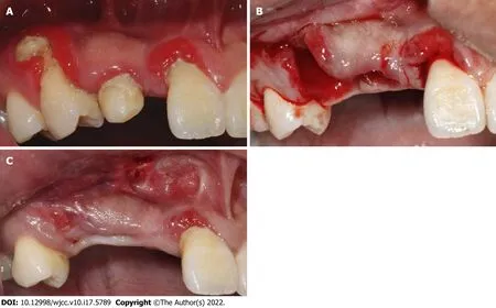
His vital sign was stable,with blood pressure of 120/82 mmHg,heart rate of 75 beats per minute and body temperature of 36.7°C.Oral examination showed tooth 12 deficiency,and deciduous tooth 53 retention with a mobility degree of II,the root of tooth 14 was exposed and the buccal alveolar bone completely absorbed,poor oral hygiene with dental calculus and bleeding on probing(Figure 1A).
Laboratory examinations
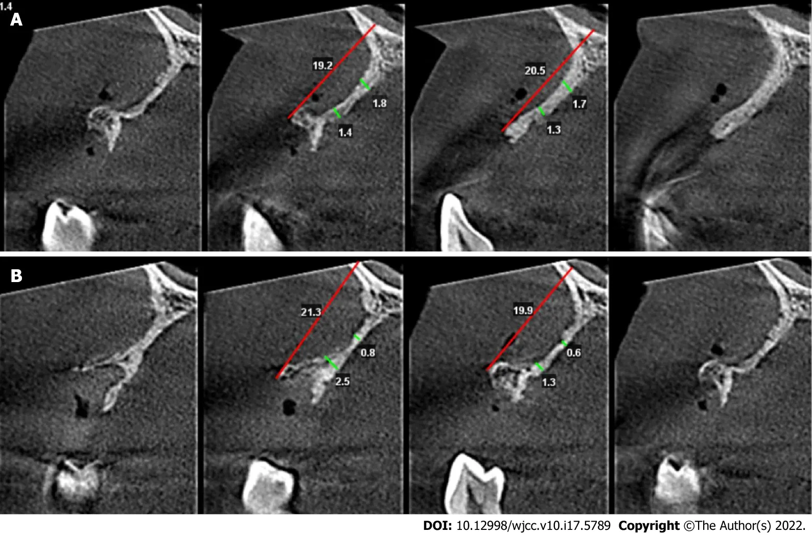
The initial treatment was extraction of teeth 53 and 14(Figure 1B),periodontal scaling and oral hygiene maintenance.Three weeks after teeth extraction and soft tissue healing(Figure 1C),clinical examination indicated that the bone quantity of right maxillary regions was insufficient for implant placement.CBCT scanning revealed the vertical height of alveolar bone was sufficient(19.2-21.3 mm)but the horizontal width of alveolar bone was merely 0.6-2.5 mm at the site 5 mm and 10 mm below the alveolar crest at sites 12(Figure 2A)and 14(Figure 2B).Horizontal bone augmentation and postponed implant placement was planned for this continuous severe horizontal bone defect.Bone augmentation was treated by GBR,with the graft composed of a 1:1 mixture of autogenous bone and ABBM,covered by bilayer absorbable membranes.The graft and membranes were fixed by PDMS combined with four corner pins.This study was approved by the Zhejiang Provincial People’s Hospital Institutional Review Board(No.2021QT267)and the participant gave signed informed consent.
Imaging examinations
Cone beam computed tomography(CBCT)scanning revealed the vertical height of alveolar bone was sufficient(19.2-21.3 mm)but the horizontal width of alveolar crest was merely 0.6-2.5 mm.
FlNAL DlAGNOSlS
VIIWho felt foolish but John, when he awoke, twenty-four hours after, and found himself without purse, without mantle, and without Princess? He tore his hair, he beat his breast, he trampled41 on the bouquet, and tore the scarf of the traitress to atoms
TREATMENT
Blood analysis and other laboratory findings were normal.
33. Cottage was made of bread and roofed with cakes, while the window was made of transparent sugar: Note that gingerbread is not used in the description of the house, only bread. Germany s rich tradition of creating gingerbread houses and other items has caused the house to be described as gingerbread in subsequent rewritings and tellings. To read an excellent history of gingerbread as a food, visit The History of Gingerbread.
Autogenous bone harvest:A vestibular incision followed by two divergent vertical incisions were made below the mucogingival border in the mandibular intercanine region under local anesthesia,and a full-thickness mucoperiostal flap was elevated to expose the bone.Autogenous bone fragments were harvested using an autogenous bone drill(Osstem,Korea)within the safety margin to the tooth apex and the mental foramen,then mixed with particle bone graft(Bio-Oss,Geistlich,Switzerland)at a ratio of 1:1.Prior to soft tissue closure of the donor site,sharp edges were removed and a gelatin sponge was applied to the remaining defect as a hemostatic dressing.The wounds were closed with 4-0 resorbable suture(Coated VICRYL,Ethicon,United States).To minimize postoperative swelling and hematoma,an extraoral pressure dressing was applied to the donor sites and maintained for 3 d.
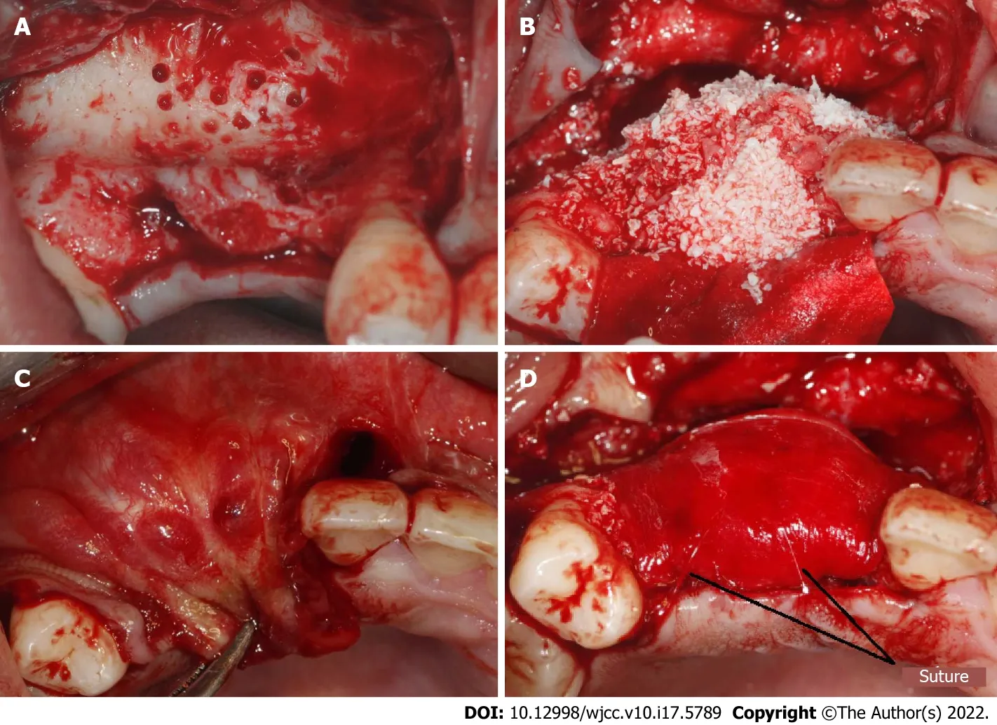
Recipient site preparation:After disinfection and local anesthesia,a mid-crestal incision along with an intrasulcus incision of the adjacent tooth were performed,extending into the distal of teeth 11 and 15,with two divergent vertical incisions made one tooth away from teeth 11 and 15 and a full-thickness flap was reflected.Decortication holes were prepared on the surface of the recipient region(Figure 3A).The mixed particle bone graft was adapted to the recipient sites,with the width more than 10 mm,then entirely covered by bilayer absorbable collagen membrane(Bio-Gide,Geistlich,Switzerland)(Figure 3B).Four titanium pins(Trausim,China)were used to fix the four marginal angles of the collagen membrane;two of which were located on the buccal side and two on the palatal side,avoiding the anatomical structures such as root and maxillary sinus.A periosteal release incision was made 2-3 mm beneath the planned apical position of the graft material and membrane.Incremental incisions of 1 to 3 mm into the periosteum and submucosa were made perpendicular to the base of the inner surface of the flap[21].The flap on the palatal side was partially reflected and the buccal flap advancement was evaluated to determine if deeper incisions into the submucosa were needed to attain more advancement to make sure the soft tissue could be closed without any tension(Figure 3C)[22].A 6-0 absorbable suture(Coated VICRYL,Ethicon,United States)was used to fix the absorbable membranes and bone graft material using PDMS(Figure 3D).
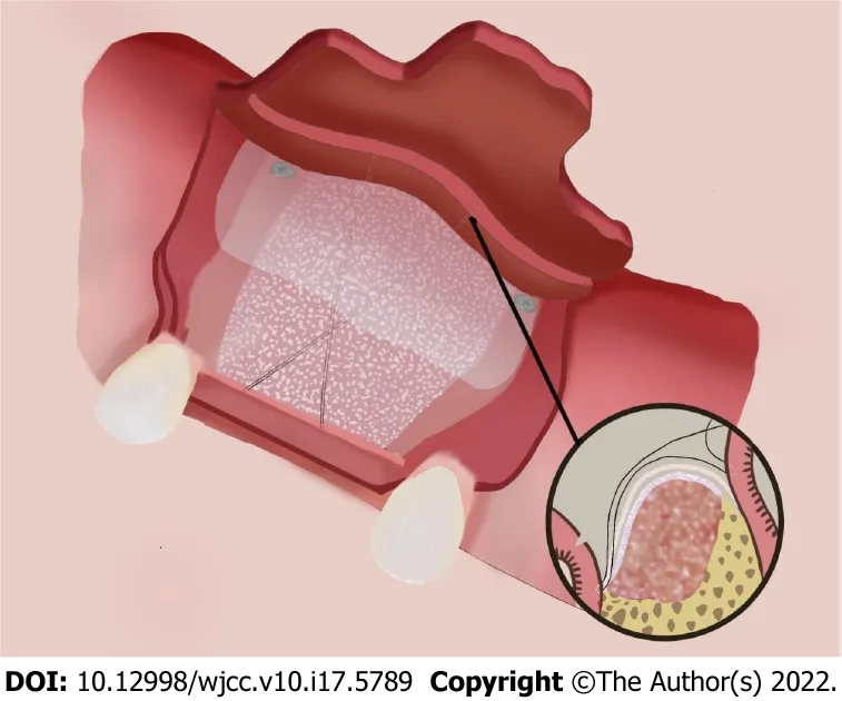
PDMS and titanium pins fixation technique: after accurate placement of the four titanium pins,the suture needle was introduced through the palatine mucosa at a point one third distal to the mesial corner pin at the palatal site.The needle was carried over the membrane and stitched through the periosteum 2-3 mm apical to the periosteal release incision at a point one third mesial to the distal buccal pin.The needle was looped back at the exterior surface of the grafts and passed through the palatine mucosa,tightening the suture,and then a knot was tied at the exterior of the palatine mucosa to stabilize the suture.The same procedure was repeated at a point one third mesial to the distal corner pin at the palatal site to a point one third distal to the mesial buccal corner pin and then knotted.The two sutures formed a figure of eight cross at the midpoint of the graft material on the buccal side(Figure 4).After tightening the suture,we checked if the bone graft and the membrane were completely immobilized and positioned correctly.Two PDMS prevented potential movement and migration of the bone graft and membranes.Finally,the mucoperiosteal flap was released to ensure a tension-free closure and the flaps were sutured by two layers,combining horizontal mattress sutures with single interrupted sutures.Vertical incisions were closed using single interrupted sutures,which were removed 7 d after surgery,while the mattress suture remained in place until 2 wk.The patient was instructed to use antibiotics and 0.2% chlorhexidine mouth rinse twice a day for 7 d to prevent infection.Two implants(Ankylos A11;Friadent,Germany)were placed after 10 mo’s healing without any bone augmentation surgery,and the final prostheses were finished at 3 mo after implant placement.
OUTCOME AND FOLLOW-UP
The cross-sections of the grafted area were measured to evaluate the width of the reconstructed alveolar ridge.CBCT imaging was performed preoperatively,immediately after bone augmentation surgery,and before and after implant placement.The CBCT images were superimposed before bone grafting and after implantation using a software system(3Shape,Denmark);the yellow line represented the alveolar ridge before bone grafting.The result showed that an average augmentation of approximately 10 mm in the alveolar ridge width was achieved at the surgical site after 10 mo’ of healing.Two sites were selected to analyze the volume of bone augmentation,5 and 10 mm below the alveolar crest.Significant bone width increment was achieved by this modified GBR technique.CBCT images showed that at the tooth 12 site,the bone width was increased from 1.83 to 8.83 mm at a point 5 mm below the crest,and from 1.70 to 9.47 mm at a point 10 mm below the crest(Figure 5A).At the tooth 14 site,the bone width was increased from 0.72 to 9.23 mm and from 4.22 to 11.55 mm at points 5 and 10 mm below the crest,respectively(Figure 5B).

DlSCUSSlON
Various techniques and materials have been used for bone augmentation procedures.It is difficult to choose GBR or Onlay bone blocks in cases of continuous severe horizontal alveolar bone defects.In this case,the remaining alveolar ridge width was < 2 mm at most sites and it seemed that onlay bone graft was the first choice for horizontal bone augmentation according to the decision tree of horizontal bone augmentation[5].Moreover,bone block is reported to be more effective than GBR in maintaining the volume at the initial stages of healing,and GBR experiences more changes compared with block grafts[2].However,in the present case,it was difficult to use bone blocks.Firstly,the residual bone of most sites was only 1 mm or less and it was too thin to be used for retention and stabilization of the graft bone bock by titanium screws.Secondly,it was difficult to obtain bone blocks of sufficient size and thickness in the mandibular intercanine region and the external oblique line due to the large area of bone defect,and the patient refused additional surgery outside the oral cavity,such as the fibula or the ilium,and he refused to use any allogenic bone.Finally,GBR might have been a good alternative choice,and an average of approximately 10 mm horizontal bone was gained by GBR to fix the absorbable collagen membranes and particulate grafts with PDMS and four corner pins after 10 mo of healing.
The final diagnosis of the presented case was deficiency of tooth 12,and deciduous tooth 53 retention,severe periodontitis of tooth 14,severe continuous horizontal alveolar bone defects at sites of teeth 12,13 and 14,and chronic gingivitis.
Bone augmentation surgery was performed 3 wk after tooth extraction and the soft tissue was healing at that time.More recently,clinicians have preferred to use non-form-stable collagen membranes to reconstruct severely thin ridges instead of titanium mesh or Poly Tetra Fluoro Ethylene membrane which is a more sensitive technique,with poor angiogenesis,higher rate of soft tissue dehiscence and difficulty in secondary removal[10,23].Moreover,significant scar tissue was found in the area of the bone defect and the soft tissue was a thin gingival biological type,therefore absorbable collagen membrane was selected for GBR.
The patient had no other personal or family history.
For severe horizontal bone defect,the key factor for successful GBR is to fix the graft material and membranes at the desired position,reduce the movement,and maintain the spatial shape[24].The stability of the bone substitute and collagen membrane can be enhanced by the application of fixation pins or by the use of block bone substitute instead of particulate bone substitute[25].The sausage technique using a membrane fixed with titanium pins to stabilize the particle grafts has been reported[18];however,several potential risks have been documented such as damage to the adjacent roots and underlying anatomical vital structures,and the need for an extensive reopening procedure to retrieve the nonabsorbable pins[20].The PVMS technique may be preferable to fix the absorbable collagen membrane and particulate graft materials for single implant sites.However,a limitation to this technique is the tensile strength of the absorbable suture material,and possible migration of the particulate graft material in an apicocoronal direction[13],it is only possible to fix the membrane by means of a linear-guided suture,resulting in possible migration of the particulate graft material in an apicocoronal direction.Thus,for continuous severe horizontal bone defect,the use of pins is still recommended because PVMS may not provide sufficient graft stabilization.So far,the relationship between the new bone regeneration and the various fixation methods used is still unknown,and future research is needed to establish the optimal fixation method for adequate bone regeneration[26].
Wang LH conceived the study design and carried out the study,and drafted the manuscript;Ruan Y and Zhao WY participated in the literature searching,acquisition of data,analysis and interpretation of data;Chen JP supervised writing the manuscript;Yang F designed the treatment plan and performed the surgery;all authors gave final approval of the version to be published.
Titanium pins and sutures can be combined effectively and flexibly.Compared with the sausage technique using pins only,multiple pins are required,which is costly,and it is difficult to operate the pins on the lingual or palatal side,and sometimes it is also difficult to avoid hazardous sites and damage to adjacent roots during insertion.Reducing the number of titanium pins can reduce the risk of complications and the difficulty of surgery.This combined technique is flexible and is very useful in clinical practice.
CONCLUSlON
For continuous severe horizontal bone defect,PDMS combined with four corner pins may provide an alternative to traditional methods to obtain better bone regeneration,and is a feasible technique to maintain the space and stabilize the graft and membranes in severe horizontal bone defect.Nevertheless,well-designed future clinical studies are needed to verify that the technique described here generates comparable and reproducible results.
FOOTNOTES
In the present case,the stability of the graft and membrane seemed unable to be provided only by the PDMS technique,thus,the use of four corner pins was still mandatory.Firstly,the four corner pins can limit the possible movement of the membrane and the particulate graft material in the apicocoronal direction.Secondly,the four corner pins might compensate the gradually decreasing strength of the suture due to degradation and absorption.According to the manufacturer,the tensile strength of the VICRYL suture is approximately 75% of its original strength after 14 d and approximately 25% after 28 d
.All of the original tensile strength is lost by 5 wk after implantation.Absorption of the suture is essentially complete between 56 and 70 d.In contrast,the Bio-Gide membrane showed obvious tissue integration at 2 wk postimplantation,and almost complete degradation at week 4[27].Membrane thickness decreased significantly at week 4,and at week 12,the Bio-Gide was almost absorbed and there was a significant increase in mean bone formation[28].However,it should be kept in mind that the membrane absorption time might not be the most important,and angiogenesis of the membrane plays a crucial role in GBR[29].The prolonged biodegradation of the membranes might be associated with decreased tissue integration,vascularization and foreign body reactions[27].The stability of the graft and membrane,as well as vascularization of the membrane,are crucial to the success of GBR,especially in the early stage of bone regeneration.So far there is no evidence for the time required for membrane fixation,and whether the biodegradation period of the absorbable suture material affects the result of GBR is still unknown.
Written consent was obtained from the patient to participate in the study.
The authors declare that they have no conflicts of interest to disclose.
The first part might be left out, but it gives us a few particulars, and these are useful We were staying in the country at a gentleman s seat, where it happened that the master was absent for a few days
The king s oath means that the daughter will marry beneath her class, a blow to her pride. Von Franz considers this a breaking of the stalemate between father and daughter over her refusal to leave (172).Return to place in story.
The authors have read the CARE Checklist(2016),and the manuscript was prepared and revised according to the CARE Checklist(2016).
The patient had IgA nephropathy and hypertension,which were well controlled after treatment,and the conditions have been stable for > 5 years.
China
“Now I must hasten away to warmer countries,” said the Snow Queen. “I will go and look into the black craters6 of the tops of the burning mountains, Etna and Vesuvius, as they are called,—I shall make them look white, which will be good for them, and for the lemons and the grapes.” And away flew the Snow Queen, leaving little Kay quite alone in the great hall which was so many miles in length; so he sat and looked at his pieces of ice, and was thinking so deeply, and sat so still, that any one might have supposed he was frozen.
Three months later my friends and I gathered at the same restaurant. To life in the Big Apple! they cheered as we tapped our glasses together. My chance of a lifetime! We talked for hours. I told them of my plan to save money by moving out of my beach cottage and renting a room for the few remaining months. Our friend offered, I have a fellow South African friend who is considering renting one of the four bedrooms in his house. His name is Barry. A great guy. He scribbled6 on a napkin() . This is his number. He s a forty-two-year-old confirmed bachelor. Says he s much too busy being a single dad to be a husband.
Lin-Hong Wang 0000-0001-9215-3845;Yan Ruan 0000-0001-9139-9237;Wen-Yan Zhao 0000-0001-8257-2595;Jian-Ping Chen 0000-0002-5607-0601;Fan Yang 0000-0002-1308-4827.
Zhejiang Stomatological Association,vice-chairman;Chinese Stomatological Association,Standing Committee member.
Wu YXJ
Of course I promised I would, for I was too happy to think of what my parents would say, or indeed of anything except Richard was not at our meeting place as he had arranged
A
Wu YXJ
1 Benic GI,H?mmerle CH.Horizontal bone augmentation by means of guided bone regeneration.
2014;66: 13-40[PMID: 25123759 DOI: 10.1111/prd.12039]
2 Elnayef B,Porta C,Suárez-López Del Amo F,Mordini L,Gargallo-Albiol J,Hernández-Alfaro F.The Fate of Lateral Ridge Augmentation:A Systematic Review and Meta-Analysis.
2018;33: 622-635[PMID: 29763500 DOI: 10.11607/jomi.6290]
3 Chiapasco M,Casentini P.Horizontal bone-augmentation procedures in implant dentistry: prosthetically guided regeneration.
2018;77: 213-240[PMID: 29478251 DOI: 10.1111/prd.12219]
4 Aghaloo TL,Moy PK.Which hard tissue augmentation techniques are the most successful in furnishing bony support for implant placement?
2007;22 Suppl: 49-70[PMID: 18437791]
5 Fu JH,Wang HL.Horizontal bone augmentation: the decision tree.
2011;31: 429-436[PMID: 21837309]
6 Lehmijoki M,Holming H,Thorén H,Stoor P.Rehabilitation of the severely atrophied dentoalveolar ridge in the aesthetic region with corticocancellous grafts from the iliac crest and dental implants.
2016;21: e614-e620[PMID: 27475690 DOI: 10.4317/medoral.21146]
7 Scheerlinck LM,Muradin MS,van der Bilt A,Meijer GJ,Koole R,Van Cann EM.Donor site complications in bone grafting: comparison of iliac crest,calvarial,and mandibular ramus bone.
2013;28: 222-227[PMID: 23377069 DOI: 10.11607/jomi.2603]
8 Sbordone C,Toti P,Guidetti F,Califano L,Santoro A,Sbordone L.Volume changes of iliac crest autogenous bone grafts after vertical and horizontal alveolar ridge augmentation of atrophic maxillas and mandibles: a 6-year computerized tomographic follow-up.
2012;70: 2559-2565[PMID: 22959878 DOI: 10.1016/j.joms.2012.07.040]
9 Urban IA,Monje A.Guided Bone Regeneration in Alveolar Bone Reconstruction.
2019;31: 331-338[PMID: 30947850 DOI: 10.1016/j.coms.2019.01.003]
10 Wessing B,Lettner S,Zechner W.Guided Bone Regeneration with Collagen Membranes and Particulate Graft Materials: A Systematic Review and Meta-Analysis.
2018;33: 87-100[PMID: 28938035 DOI: 10.11607/jomi.5461]
11 Wang HL,Boyapati L."PASS" principles for predictable bone regeneration.
2006;15: 8-17[PMID: 16569956 DOI: 10.1097/01.id.0000204762.39826.0f]
12 An YZ,Strauss FJ,Park JY,Shen YQ,Thoma DS,Lee JS.Membrane fixation enhances guided bone regeneration in standardized calvarial defects: A pre-clinical study.
2022;49: 177-187[PMID: 34866208 DOI: 10.1111/jcpe.13583]
13 Urban IA,Lozada JL,Wessing B,Suárez-López del Amo F,Wang HL.Vertical Bone Grafting and Periosteal Vertical Mattress Suture for the Fixation of Resorbable Membranes and Stabilization of Particulate Grafts in Horizontal Guided Bone Regeneration to Achieve More Predictable Results: A Technical Report.
2016;36: 153-159[PMID: 26901293 DOI: 10.11607/prd.2627]
14 Johnson TM,Vargas SM,Wagner JC,Lincicum AR,Stancoven BW,Lancaster DD.The Triangle Suture for Membrane Fixation in Guided Bone Regeneration Procedures: A Report of two Cases.
2022[PMID: 34986274 DOI: 10.1002/cap.10193]
15 Kamat SM,Khandeparker RV,Akkara F,Dhupar V,Mysore A.SauFRa Technique for the Fixation of Resorbable Membranes in Horizontal Guided Bone Regeneration:A Technical Report.
2020;46: 609-613[PMID: 32315438 DOI: 10.1563/aaid-joi-D-19-00265]
16 Kirsch A,Ackermann KL,Hurzeler MB,Durr W,Hutmacher D.Development and clinical application of titanium minipins for fixation of nonresorbable barrier membranes.
1998;29: 368-381[PMID: 9728148]
17 Shalev TH,Kurtzman GM,Shalev AH,Johnson DK,Kersten MEM.Continuous Periosteal Strapping Sutures for Stabilization of Osseous Grafts With Resorbable Membranes for Buccal Ridge Augmentation:A Technique Report.
2017;43: 283-290[PMID: 28628357 DOI: 10.1563/aaid-joi-D-17-00060]
18 Wang HL,Misch C,Neiva RF."Sandwich" bone augmentation technique: rationale and report of pilot cases.
2004;24: 232-245[PMID: 15227771]
19 Siar CH,Toh CG,Romanos G,Ng KH.Subcutaneous reactions and degradation characteristics of collagenous and noncollagenous membranes in a macaque model.
2011;22: 113-120[PMID: 20678135 DOI: 10.1111/j.1600-0501.2010.01970.x]
20 Lorenzoni M,Pertl C,Polansky RA,Jakse N,Wegscheider WA.Evaluation of implants placed with barrier membranes.A restrospective follow-up study up to five years.
2002;13: 274-280[PMID: 12010157 DOI: 10.1034/j.1600-0501.2002.130306.x]
21 Zazou N,Diab N,Bahaa S,El Arab AE,Aziz OA,El Nahass H.Clinical comparison of different flap advancement techniques to periosteal releasing incision in guided bone regeneration:A randomized controlled trial.
2021;23: 107-116[PMID: 33155422 DOI: 10.1111/cid.12960]
22 Romanos GE.Periosteal releasing incision for successful coverage of augmented sites.A technical note.
2010;36: 25-30[PMID: 20218867 DOI: 10.1563/AAID-JOI-D-09-00068]
23 Atef M,Tarek A,Shaheen M,Alarawi RM,Askar N.Horizontal ridge augmentation using native collagen membrane vs titanium mesh in atrophic maxillary ridges: Randomized clinical trial.
2020;22: 156-166[PMID: 32185856 DOI: 10.1111/cid.12892]
24 Carpio L,Loza J,Lynch S,Genco R.Guided bone regeneration around endosseous implants with anorganic bovine bone mineral.A randomized controlled trial comparing bioabsorbable versus non-resorbable barriers.
2000;71: 1743-1749[PMID: 11128923 DOI: 10.1902/jop.2000.71.11.1743]
25 Mir-Mari J,Wui H,Jung RE,H?mmerle CH,Benic GI.Influence of blinded wound closure on the volume stability of different GBR materials: an in vitro cone-beam computed tomographic examination.
2016;27: 258-265[PMID: 25856209 DOI: 10.1111/clr.12590]
26 Cucchi A,Chierico A,Fontana F,Mazzocco F,Cinquegrana C,Belleggia F,Rossetti P,Soardi CM,Todisco M,Luongo R,Signorini L,Ronda M,Pistilli R.Statements and Recommendations for Guided Bone Regeneration: Consensus Report of the Guided Bone Regeneration Symposium Held in Bologna,October 15 to 16,2016.
2019;28: 388-399[PMID: 31344018 DOI: 10.1097/ID.0000000000000909]
27 Rothamel D,Schwarz F,Sager M,Herten M,Sculean A,Becker J.Biodegradation of differently cross-linked collagen membranes: an experimental study in the rat.
2005;16: 369-378[PMID: 15877758 DOI: 10.1111/j.1600-0501.2005.01108.x]
28 H?mmerle CH,Jung RE,Yaman D,Lang NP.Ridge augmentation by applying bioresorbable membranes and deproteinized bovine bone mineral: a report of twelve consecutive cases.
2008;19: 19-25[PMID: 17956571 DOI: 10.1111/j.1600-0501.2007.01407.x]
29 Schwarz F,Rothamel D,Herten M,Wüstefeld M,Sager M,Ferrari D,Becker J.Immunohistochemical characterization of guided bone regeneration at a dehiscence-type defect using different barrier membranes: an experimental study in dogs.
2008;19: 402-415[PMID: 18324961 DOI: 10.1111/j.1600-0501.2007.01486.x]
 World Journal of Clinical Cases2022年17期
- World Journal of Clinical Cases的其它文章
- Repetitive transcranial magnetic stimulation for post-traumatic stress disorder:Lights and shadows
- Response to dacomitinib in advanced non-small-cell lung cancer harboring the rare delE709_T710insD mutation:A case report
- Loss of human epidermal receptor-2 in human epidermal receptor-2+breast cancer after neoadjuvant treatment:A case report
- Tumor-like disorder of the brachial plexus region in a patient with hemophilia:A case report
- High-frame-rate contrast-enhanced ultrasound findings of liver metastasis of duodenal gastrointestinal stromal tumor:A case report and literature review
- Gitelman syndrome:A case report
