High-frame-rate contrast-enhanced ultrasound findings of liver metastasis of duodenal gastrointestinal stromal tumor:A case report and literature review
lNTRODUCTlON
Gastrointestinal stromal tumors(GISTs)are the most common mesenchymal tumors originating from the digestive tract,and they account for 1%-3% of gastrointestinal tumors.Their clinical manifestations have not been established,and no specific tumor markers have been reported;approximately 15%-50% of GISTs are found due to liver metastasis[1].Duodenal GISTs are rare,accounting for 12%-18% of small intestine GISTs and 1%-4% of all GISTs,and only 6 cases of liver metastasis of duodenal GISTs have been reported[2-7].Usually,computed tomography(CT)scan,ultrasound endoscopy,and digestive tract contrast can help in the evaluation of the size,local immersion,and metastasis and location of the GIST[8].Herein,we report a case of metastatic duodenal GIST that was initially accurately diagnosed by high frame rate contrast-enhanced ultrasound(H-CEUS).This report is intended to introduce a new imaging technology,which could not only provide important information in diagnosing rare disease liver metastasis of duodenal GIST but may also contribute to the diagnosis of many other hepatic lesions.
CASE PRESENTATlON
Chief complaints
A 53-year-old Chinese man was admitted to our hospital with a progressively enlarged liver lesion for 2 years.
History of present illness
The lesion in the liver had been found during a routine re-examination after surgery two years earlier,and no significant progression was observed during subsequent annual examinations until three months earlier.The maximum diameter of the lesion had grown from 1.3 cm to 3.5 cm within a year,accompanied by mild abdominal discomfort.The patient reported to our hospital for further diagnosis.
History of past illness
The patient had a 30-year history of fatty liver but no history of hepatitis and liver cirrhosis and had undergone duodenal stromal tumor resection twice in our hospital in March 2012 and September 2016.He had no significant symptoms but slightly abdominal discomfort occasionally.His dietary was regular,but the sleep was not that well.
Personal and family history
The patient had a 30-year history of alcohol consumption.His father had a gastrointestinal-related disease,but the details are unknown.
Physical examination
If you want a slingshot, I hope your father teaches you how to make one instead of buying one. I hope you learn to dig in the dirt and read books, and when you learn to use computers, you also learn how to add and subtract in your head.
Laboratory examinations
The results of the laboratory tests were negative,except an ALT level of 75 U/L.The tumor markers,AFP,CEA,CA19-9,had normal concentrations.The blood tests and fecal,coagulation function,and
antibody examinations showed normal results.
Imaging examinations
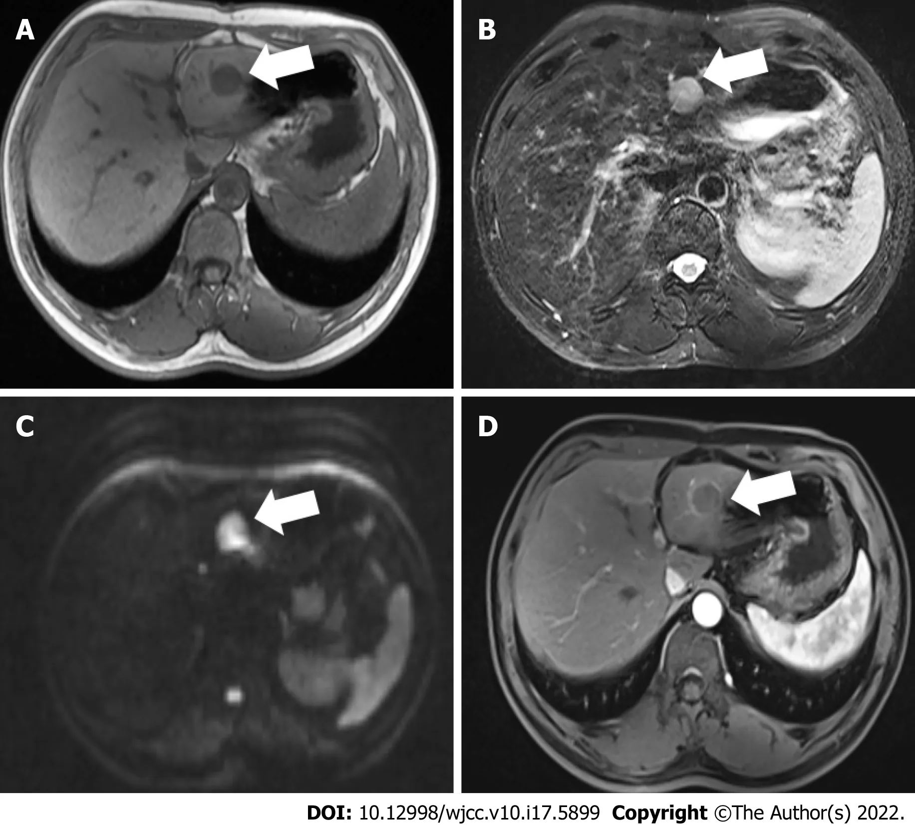
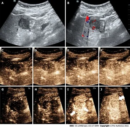
Three months earlier,magnetic resonance imaging(MRI)showed a 2.3 cm occupying lesion in the left external liver lobe(Figure 1).The patient underwent an abdominal ultrasound examination using the Resona9 ultrasound system(Mindray Medical International,China)equipped with an SC6-1U(1-6 MHz)transducer.Conventional ultrasound(US)showed an uneven hypo-echo lesion with a peripheral hypoechoic halo located in the left lobe of the liver.The lesion had an approximate size of 3.5 × 2.2 × 2.4 cm
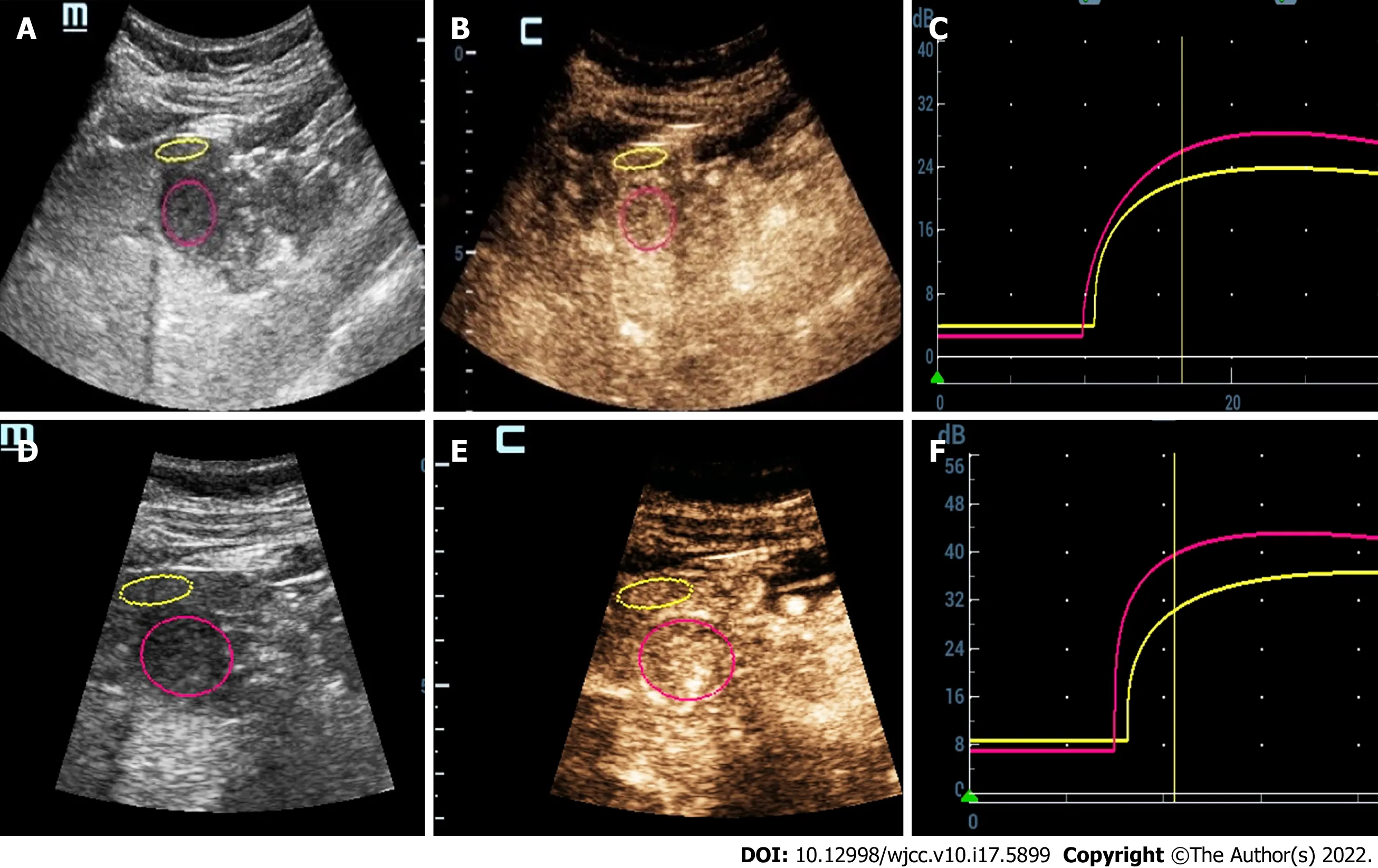
,a round shape,and slightly clear margins.Color Doppler flow imaging(CDFI)showed the dotlinear blood flow signal within the lesion(Figure 2A and B).Given the history of duodenal GIST,the patient’s doctor suggested further CEUS diagnosis and obtained patient's consent.The depth,gain,and focus were thoroughly adjusted for optimal display according to the operator's habits.After a bolus injection of 1.5 mL of Sonovue(Bracco,Italy)suspension,an ultrasound contrast agent,with 5 mL physiological saline(Italy,Bracco),the timer was activated.The target lesion and surrounding liver parenchyma were continuously observed for 5 min.Based on the accepted guidelines,the arterial,portal,and late phases were defined after 10-30 s,30-120 s,and 121-360 s of the contrast agent injection,respectively.On CEUS,the solid nodule appeared heterogeneous,and there was hyper-enhancement during the arterial phase without a significant concentric perfusion and rim-like enhancement(Figure 2C-F).Quantitative analysis showed a peak intensity difference between the lesion and liver parenchyma(ΔPI)of 3.58 dB(Figure 3A-C).The enhancement of the lesion was washed out rapidly and gradually;a heterogeneous enhancement and hypo-enhancement were observed during the portal and late phases.During the late phase,the contrast agent was barely perfused.These features were suggestive of malignancy.To observe the process and direction of the contrast agent more precisely and better assess the liver lesion,we carried out a second H-CEUS after the patient rested for 1 h.During the arterial phase,a solid lesion that was enhanced from the periphery to the center was observed;a heterogeneous hyper-enhancement at peak was also observed.We also found two irregular branch-like vascular columns around the lesion.Rim enhancement was more significant this time(Figure 3G-J).The video showing the H-CEUS in arterial phase is displayed(Video 1).Quantitative analysis showed a peak intensity difference between the lesion and liver parenchyma(ΔPI)of 6.63 dB(Figure 3D-F).Similar to the CEUS findings,the contrast agent was washed out rapidly during the portal and late phases,and,finally,no enhancement was observed.The arterial phase of the lesion was suggestive of a malignancy,and metastasis was suspected based on the history.
The entire abdomen was soft,and there was no pressure pain,rebound pain,and muscle tension.A vertical surgery scar of approximately 15 cm was found.All the other vital signs were stable,and no positive signs were revealed.
Pathological findings and immunohistochemical staining
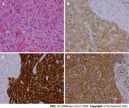
The final pathological findings were as follows:(1)Microscopic: the short and spindle cells were patchy;and(2)Immunohistochemical results: CD117,Dog-1,and Vimentin were positive,and Ki-67 was > 5%(Figure 4).
Gene mutational analysis
The patient currently takes 400 mg imatine per day and has no other symptoms.Although the patient had not quit drinking,he had reduced the frequency and amount of alcohol consumption to a large extent.
FlNAL DlAGNOSlS
The final diagnosis of the presented case was liver metastasis of duodenal GIST.
TREATMENT
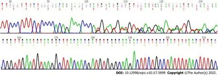
Molecular testing revealed
(c.1655_1699del 15 type),
,c
,c
,
,and
wild type mutations,which were associated with poor prognosis(Figure 5).
I think about you, I thought, and blushed a bit. To him I said in a dull stupid voice, I don t think very much really; I think about getting new clothes or going on my holidays or what we ll have for lunch.
OUTCOME AND FOLLOW-UP
The patient remained on imatine therapy,and was followed up.
DlSCUSSlON
Duodenal GIST is rare,accounting for 12%-18% of small intestine GISTs and 1%-4% of all GISTs[9-11].The clinical features of duodenal GIST include mild gastrointestinal bleeding,abdominal pain,and an abdominal mass[12].The diagnosis of duodenal GIST is usually based on histopathological and imaging findings.Usually,ultrasound endoscopy,CT scan,MRI,and digestive tract contrast can help in evaluating the size,local immersion,metastasis,and location of the GIST[13-15].
Metastatic liver cancer(MLC)is one of the most common metastatic tumors,and is usually associated with a lower survival rate[16,17].The most common type of liver metastasis derives from colorectal cancer.GISTs only account for 1%-3% of gastrointestinal tumors.Due to the higher malignancy and poor prognosis of MLCs,early detection and therapy are of great significance.
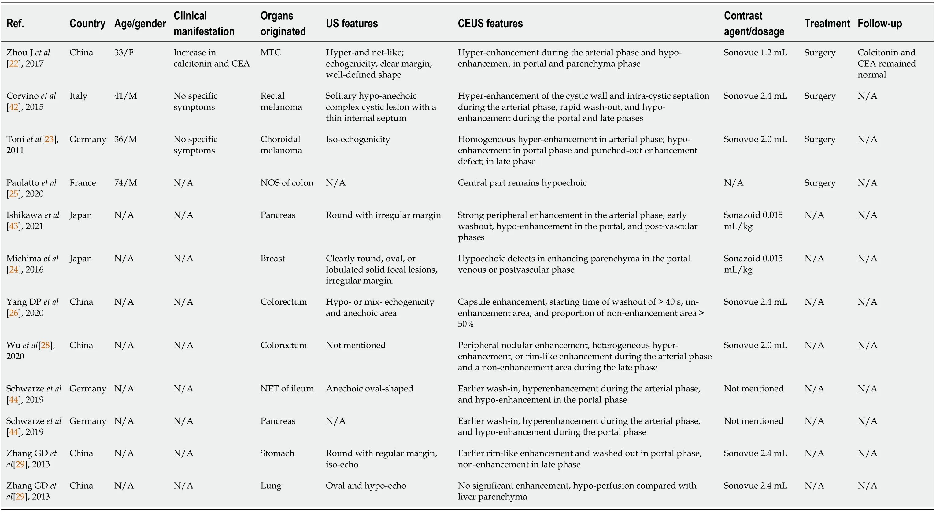
CT and MRI are the preferred imaging modalities for diagnosing MLC.Liver metastasis often shows heterogeneous hypodense lesions with progressive concentric enhancement in CT scans,with a sensitivity of up to 97%[18],while the MRI technique can detect smaller lesions[18,19].These two imaging modalities are not used in real-time evaluation,and their scanning durations are longer.Their radiation and high cost should be taken into consideration for neonates and pregnant women.CEUS has been widely used to detect focal liver lesions(FLLs)in recent years[20,21],but there have only been 10 reports of liver metastatic lesions so far(Table 1).Herein,we collected information regarding the age,gender,clinical manifestation,MLC’s originated organs,treatment,and follow-up from four cases and six clinical research studies.As shown in Table 1,MLCs mainly originated from gastrointestinal organs,except for three patients in whom the primary malignant tumors were medullary thyroid cancer[22],choroidal melanoma[23]and breast cancer[24].MLCs usually have no specific symptoms.The US and CEUS features are depicted in Table 1.We found that MLCs often present as oval or round with irregular margins,and their echo characteristics are not specific.For MLCs from colorectal and ileal lesions,there is a central part with no echo[25,26].They may consist of a hemorrhage and a necrotic area.It was reported that approximately 50% of all GISTs show cystic or necrotic areas[27].CEUS is more sensitive in detecting avascular areas than US when part of the isoechoic or hypoechoic area was necrotic.Regarding the CEUS findings,the majority of these cases presented with homogeneous or heterogeneous hyper-enhancement during the arterial phase,mostly earlier than liver parenchyma.The contrast agents washed out very rapidly during the portal phase and presented hypo-enhancement until the end of the late phase.Wu
[28]showed that metastatic lesion presented non-enhancement during the late phase.Zhang
[29]found that lung primary lesion’s MLC showed no significant enhancement in CEUS,which was contrary to other findings.We deduced that it was probably associated with the pathological type,but more supportive literature was required to make further conclusions.For this present case,CEUS showed a solid nodule that appeared heterogeneous and hyper-enhanced during the arterial phase;these were consistent with malignant liver lesion perfusionpatterns previously reported[20,30].As we all know,hyper-enhancement of atypical hemangiomas during the arterial phase or non-enhancement during the portal and late phases can lead to a misleading diagnosis.Intrahepatic cholangiocellular carcinomas behave like metastases,washing out rapidly and appearing as defects during the late phase[31].Consequently,further imaging characteristics are needed for a precise diagnosis.
The contrast agent was a pure-blood pool tracer for showing microcirculation perfusion of solid organs[21,32].It can detect smaller(> 40 μm)blood vessels better than CDFI(> 100 μm)and is widely used in FLLs[33,34].While it is partly affected by the frame rate,the frame frequency of CEUS is within 9-15 Hz,which is probably not adequate for detecting quick wash-in progression and the vascular architecture during the early arterial phase.H-CEUS provides a frame rate of tens of thousands of images per second to compensate for the reduced focusing of the acoustic beam and enhance the signalto-noise ratio.H-CEUS can show microcirculation differences and wash-in patterns during the early arterial phase and vascular morphogenesis.In this case of H-CEUS technology,the enhancement appeared at the peripheral part of the lesion,and there was concentric perfusion.This may be mainly due to the increase in frame rate,which facilitated a better display of the first enhancement area,and the site where the change in enhancement over time demonstrated the direction of contrast perfusion.Nevertheless,when the image frame rate is low,it is impossible to accurately display the contrast enhancement area,as the enhancement appears almost simultaneously in various parts of the lesion.Instead,it is presented as an entire perfusion on CEUS.Besides,vascular morphology is one of the important features for identifying and diagnosing the nature of FLL,and irregular vascular morphology is usually the main manifestation of malignant FLL[35,36].The vascular morphology can be demonstrated by recording the path of contrast agent microbubbles,as the bubbles cannot enter the tissue gap through the vessel wall,and can only continuously move in the vessel[37-40].The arterial phase is important for observing the vascular morphology,and rapid flow of arterial blood leads to the rapid movement of contrast agents.Therefore,only when the frame rate of contrast imaging reaches a certain threshold.In our case,we observed irregular branch-like vascular columns,which suggested the presence of malignancy;resulting in the final diagnosis of“l(fā)iver metastasis of duodenal GIST.”Compared with the CEUS technology,H-CEUS is more suitable for accurately detecting the movement of contrast agents to ascertain the vascular morphology of liver lesions.
The prognosis and treatment of liver metastasis of duodenal GIST have not been established due to the limited number of reported cases[2,3].Based on our literature review,we found that distant metastases can occur years after the surgical excision of the primary duodenal lesion.Both patients reported in the two cases experienced bleeding after rupture of the liver metastatic lesion[2,3,6,41].Given that liver metastases of duodenal GISTs are negatively correlated with disease prognosis,early detection is important.Consequently,our report is useful,as it is the first to highlight the value and utility of H-CEUS for diagnosis of patients with liver metastases of duodenal GISTs.This case reminds clinicians that the H-CEUS technology can facilitate the diagnosis of liver metastatic cancers,resulting in improved diagnosis,treatment,and outcomes.
CONCLUSlON
Duodenal GISTs are relatively rare,but they usually have a poor prognosis and are associated with high incidences of metastases.The H-CEUS findings were as follows: a solid lesion enhanced from the periphery to the center with two irregular branch-like vessel columns during the early arterial phase with peak heterogeneous hyper-enhancement and rim enhancement.The contrast agent was washed out rapidly during the portal phase and showed no enhancement during the late phase.To the best of our knowledge,this report is the first to describe the H-CEUS patterns of this disease.It also highlights the challenges associated with the CEUS findings of metastatic hepatic cancers.
We thank Dr.Jin Y,Department of Gastroenterology,Shengjing Hospital of China Medical University for her pathological diagnosis.
It was now a year since the prince had returned home, for his father felt that it was time that his son should learn how to rule the kingdom which would one day be his
ACKNOWLEDGEMENTS
So now there was only the youngest at home, and when the other two never came he also begged for a ship that he might go in search of his lost brothers
FOOTNOTES
Chen JH and Huang Y reviewed the literature and contributed to manuscript drafting;Huang Y performed the high frame rate contrast-enhanced ultrasound and biopsy operation;all authors issued final approval for the version to be submitted.
the Guide Project for Key Research and Development Foundation of Liaoning Province,No.2019JH8/10300008;the 345 Talent Project;and the Liaoning Baiqianwan Talents Program.
Informed written consent was obtained from the patient for publication of this report and any accompanying images.
I believe honesty is one of the greatest gifts there is. I know they call it a lot of fancy names these days, like integrity and forthrightness1. But it doesn t make any difference what they call it; it s still what makes a man a good citizen. This is my code, and I try to live by.
Yes, thank you so much, I sniffed12. Before long, I was allowed in to see Mike. He was hooked13 up to monitors, IVs, and was wearing an oxygen mask, but I was so happy to be with him again. You re going to be fine, I said, stroking his arm. I hoped. I looked to the doctor at his bedside.
The authors declare that they have no conflicts of interest.
When did he come? Was he in the crowd? Wait a bit; we are coming to him! On the third day a little figure came without horse or carriage and walked jauntily27 up to the palace
The authors have read the CARE Checklist(2016),and the manuscript was prepared and revised according to the CARE Checklist(2016).
This article is an open-access article that was selected by an in-house editor and fully peer-reviewed by external reviewers.It is distributed in accordance with the Creative Commons Attribution NonCommercial(CC BYNC 4.0)license,which permits others to distribute,remix,adapt,build upon this work non-commercially,and license their derivative works on different terms,provided the original work is properly cited and the use is noncommercial.See: https://creativecommons.org/Licenses/by-nc/4.0/
China
Julius Heuscher believes the white bird or dove symbolizes the need to never forget home after one has left it (Heuscher 1974).Return to place in story.
Jia-Hui Chen 0000-0001-7616-7163;Ying Huang 0000-0002-3361-7184.
Wang LL
A
Wang LL
1 Gaitanidis A,Alevizakos M,Tsaroucha A,Simopoulos C,Pitiakoudis M.Incidence and predictors of synchronous liver metastases in patients with gastrointestinal stromal tumors(GISTs).
2018;216: 492-497[PMID: 29690997 DOI: 10.1016/j.amjsurg.2018.04.011]
2 Mori T,Kataoka M,Kaiho T,Yanagisawa S,Nishimura M,Kobayashi S,Okaniwa A,Ashizawa Y,Oka Y,Ohya M.[A Case of Liver Metastasis 28 Years after Resection for Gastrointestinal Stromal Tumor of Duodenum].
2020;47: 1810-1812[PMID: 33468837]
3 Koike K,Kubota H,Takayanagi Y,Ikeda T,Ito T.[A case of gastrointestinal stromal tumor of the duodenum with ruptured liver metastasis during administration of imatinib].
2020;117: 914-918[PMID: 33041303 DOI: 10.11405/nisshoshi.117.914]
4 Kishi Y,Takaki S,Fukuhara T,Mori N,Okanobu H,Tsuji K,Maeda T,Fujiwara M,Nagai K,Furukawa Y.[A case of liver metastasis 11 years after resection of a gastrointestinal stromal tumor of the duodenum].
2019;116: 1030-1038[PMID: 31827043 DOI: 10.11405/nisshoshi.116.1030]
5 Stratopoulos C,Soonawalla Z,Piris J,Friend PJ.Hepatopancreatoduodenectomy for metastatic duodenal gastrointestinal stromal tumor.
2006;5: 147-150[PMID: 16481303]
6 Sakata M,Kaneyoshi T,Fushimi T,Watanabe J.Rare cause of cystic liver lesions: Liver metastasis of gastrointestinal stromal tumors.
2021;5: 408-409[PMID: 33732891 DOI: 10.1002/jgh3.12487]
7 Inoue M,Shishida M,Watanabe A,Kajikawa R,Kajiwara R,Sawada H,Ohmori I,Miyamoto K,Ikeda M,Toyota K,Sadamoto S,Takahashi T.A liver metastasis 7 years after resection of a low-risk duodenal gastrointestinal stromal tumor.
2021;14: 1464-1469[PMID: 34117599 DOI: 10.1007/s12328-021-01464-w]
8 McGuirk M,Gachabayov M,Gogna S,Da Dong X.Robotic duodenal(D3)resection with Roux-en-Y duodenojejunostomy reconstruction for large GIST tumor: Step by step with video.
2021;36: 130[PMID: 33370658 DOI: 10.1016/j.suronc.2020.12.006]
9 Cassier PA,Ducimetière F,Lurkin A,Ranchère-Vince D,Scoazec JY,Bringuier PP,Decouvelaere AV,Méeus P,Cellier D,Blay JY,Ray-Coquard I.A prospective epidemiological study of new incident GISTs during two consecutive years in Rh?ne Alpes region: incidence and molecular distribution of GIST in a European region.
2010;103: 165-170[PMID: 20588273 DOI: 10.1038/sj.bjc.6605743]
10 Rubin BP,Heinrich MC,Corless CL.Gastrointestinal stromal tumour.
2007;369: 1731-1741[PMID: 17512858 DOI: 10.1016/s0140-6736(07)60780-6]
11 Sandvik OM,S?reide K,Kval?y JT,Gudlaugsson E,S?reide JA.Epidemiology of gastrointestinal stromal tumours: single-institution experience and clinical presentation over three decades.
2011;35: 515-520[PMID: 21489899 DOI: 10.1016/j.canep.2011.03.002]
12 Qiu H,Zhang P,Feng X,Chen T,Sun X,Yu J,Chen Z,Li Y,Tao K,Li G,Zhou Z.Changes of diagnosis and treatment for gastrointestinal stromal tumors during a 18-year period in four medical centers of China.
2016;19: 1265-1270[PMID: 27928797]
13 Karaca C,Turner BG,Cizginer S,Forcione D,Brugge W.Accuracy of EUS in the evaluation of small gastric subepithelial lesions.
2010;71: 722-727[PMID: 20171632 DOI: 10.1016/j.gie.2009.10.019]
14 Park CH,Kim EH,Jung DH,Chung H,Park JC,Shin SK,Lee YC,Kim H,Lee SK.Impact of periodic endoscopy on incidentally diagnosed gastric gastrointestinal stromal tumors: findings in surgically resected and confirmed lesions.
2015;22: 2933-2939[PMID: 25808096 DOI: 10.1245/s10434-015-4517-0]
15 Zhou Z,Lu J,Morelli JN,Hu D,Li Z,Xiao P,Hu X,Shen Y.Utility of noncontrast MRI in the detection and risk grading of gastrointestinal stromal tumor: a comparison with contrast-enhanced CT.
2021;11: 2453-2464[PMID: 34079715 DOI: 10.21037/qims-20-578]
16 Obayashi J,Kawaguchi K,Manabe S,Nagae H,Wakisaka M,Koike J,Takagi M,Kitagawa H.Prognostic factors indicating survival with native liver after Kasai procedure for biliary atresia.
2017;33: 1047-1052[PMID: 28852838 DOI: 10.1007/s00383-017-4135-y]
17 Hara Y,Ogata Y,Shirouzu K.Early tumor growth in metastatic organs influenced by the microenvironment is an important factor which provides organ specificity of colon cancer metastasis.
2000;19: 497-504[PMID: 11277329]
18 Tsili AC,Alexiou G,Naka C,Argyropoulou MI.Imaging of colorectal cancer liver metastases using contrast-enhanced US,multidetector CT,MRI,and FDG PET/Ct:A meta-analysis.
2021;62: 302-312[PMID: 32506935 DOI: 10.1177/0284185120925481]
19 Niekel MC,Bipat S,Stoker J.Diagnostic imaging of colorectal liver metastases with CT,MR imaging,FDG PET,and/or FDG PET/Ct:A meta-analysis of prospective studies including patients who have not previously undergone treatment.
2010;257: 674-684[PMID: 20829538 DOI: 10.1148/radiol.10100729]
20 Wilson SR,Feinstein SB.Introduction: 4th Guidelines and Good Clinical Practice Recommendations for Contrast Enhanced Ultrasound(CEUS)in the Liver-Update 2020 WFUMB in Cooperation with EFSUMB,AFSUMB,AIUM and FLAUS.
2020;46: 3483-3484[PMID: 32888748 DOI: 10.1016/j.ultrasmedbio.2020.08.015]
21 Claudon M,Dietrich CF,Choi BI,Cosgrove DO,Kudo M,Nols?e CP,Piscaglia F,Wilson SR,Barr RG,Chammas MC,Chaubal NG,Chen MH,Clevert DA,Correas JM,Ding H,Forsberg F,Fowlkes JB,Gibson RN,Goldberg BB,Lassau N,Leen EL,Mattrey RF,Moriyasu F,Solbiati L,Weskott HP,Xu HX;World Federation for Ultrasound in Medicine;European Federation of Societies for Ultrasound.Guidelines and good clinical practice recommendations for Contrast Enhanced Ultrasound(CEUS)in the liver - update 2012: A WFUMB-EFSUMB initiative in cooperation with representatives of AFSUMB,AIUM,ASUM,FLAUS and ICUS.
2013;39: 187-210[PMID: 23137926 DOI: 10.1016/j.ultrasmedbio.2012.09.002]
22 Zhou J,Luo Y,Ma BY,Ling WW,Zhu XL.Contrast-enhanced ultrasound diagnosis of hepatic metastasis of concurrent medullary-papillary thyroid carcinoma:A case report.
2017;96: e9065[PMID: 29390301 DOI: 10.1097/md.0000000000009065]
23 De Toni EN,Gallmeier E,Auernhammer CJ,Clevert DA.Contrast-enhanced ultrasound for surveillance of choroidal carcinoma patients: features of liver metastasis arising several years after treatment of the primary tumor.
2011;4: 336-342[PMID: 21769292 DOI: 10.1159/000329453]
24 Mishima M,Toh U,Iwakuma N,Takenaka M,Furukawa M,Akagi Y.Evaluation of contrast Sonazoid-enhanced ultrasonography for the detection of hepatic metastases in breast cancer.
2016;23: 231-241[PMID: 25143060 DOI: 10.1007/s12282-014-0560-0]
25 Paulatto L,Dioguardi Burgio M,Sartoris R,Beaufrère A,Cauchy F,Paradis V,Vilgrain V,Ronot M.Colorectal liver metastases: radiopathological correlation.
2020;11: 99[PMID: 32844319 DOI: 10.1186/s13244-020-00904-4]
26 Yang D,Zhuang B,Wang W,Xie X.Differential diagnosis of liver metastases of gastrointestinal stromal tumors from colorectal cancer based on combined tumor biomarker with features of conventional ultrasound and contrast-enhanced ultrasound.
2020;45: 2717-2725[PMID: 32458028 DOI: 10.1007/s00261-020-02592-6]
27 Baheti AD,Shinagare AB,O'Neill AC,Krajewski KM,Hornick JL,George S,Ramaiya NH,Tirumani SH.MDCT and clinicopathological features of small bowel gastrointestinal stromal tumours in 102 patients: a single institute experience.
2015;88: 20150085[PMID: 26111069 DOI: 10.1259/bjr.20150085]
28 Wu XF,Bai XM,Yang W,Sun Y,Wang H,Wu W,Chen MH,Yan K.Differentiation of atypical hepatic hemangioma from liver metastases: Diagnostic performance of a novel type of color contrast enhanced ultrasound.
2020;26: 960-972[PMID: 32206006 DOI: 10.3748/wjg.v26.i9.960]
29 Zhang GD,Zou SD,Wang H,He J,Huang D.[Contrast-enhanced ultrasound in diagnosing 55 cases of various types of liver metastatic carcinoma].
2013;21: 2849-2850[DOI: 10.11569/wcjd.v21.i27.2849]
30 Chammas MC,Bordini AL.Contrast-enhanced ultrasonography for the evaluation of malignant focal liver lesions.
2022;41: 4-24[PMID: 34724777 DOI: 10.14366/usg.21001]
31 Xu HX,Lu MD,Liu GJ,Xie XY,Xu ZF,Zheng YL,Liang JY.Imaging of peripheral cholangiocarcinoma with lowmechanical index contrast-enhanced sonography and SonoVue: initial experience.
2006;25: 23-33[PMID: 16371552 DOI: 10.7863/jum.2006.25.1.23]
32 Sidhu PS,Cantisani V,Dietrich CF,Gilja OH,Saftoiu A,Bartels E,Bertolotto M,Calliada F,Clevert DA,Cosgrove D,Deganello A,D'Onofrio M,Drudi FM,Freeman S,Harvey C,Jenssen C,Jung EM,Klauser AS,Lassau N,Meloni MF,Leen E,Nicolau C,Nolsoe C,Piscaglia F,Prada F,Prosch H,Radzina M,Savelli L,Weskott HP,Wijkstra H.The EFSUMB Guidelines and Recommendations for the Clinical Practice of Contrast-Enhanced Ultrasound(CEUS)in Non-Hepatic Applications: Update 2017(Long Version).
2018;39: e2-e44[PMID: 29510439 DOI: 10.1055/a-0586-1107]
33 Jiang Y,Zhang M,Zhu Y,Zhu D.Diagnostic role of contrast-enhanced ultrasonography
conventional B-mode ultrasonography in cirrhotic patients with early hepatocellular carcinoma:A retrospective study.
2021;12: 2403-2411[PMID: 34790401 DOI: 10.21037/jgo-21-611]
34 Guo HL,Lu XZ,Hu HT,Ruan SM,Zheng X,Xie XY,Lu MD,Kuang M,Shen SL,Chen LD,Wang W.Contrast-Enhanced Ultrasound-Based Nomogram: A Potential Predictor of Individually Postoperative Early Recurrence for Patients With Combined Hepatocellular-Cholangiocarcinoma.
2021[PMID: 34751450 DOI: 10.1002/jum.15869]
35 Ferraioli G,Meloni MF.Contrast-enhanced ultrasonography of the liver using SonoVue.
2018;37: 25-35 [PMID: 28830058 DOI: 10.14366/usg.17037]
36 Zarzour JG,Porter KK,Tchelepi H,Robbin ML.Contrast-enhanced ultrasound of benign liver lesions.
2018;43: 848-860[PMID: 29167944 DOI: 10.1007/s00261-017-1402-2]
37 Kang XN,Zhang XY,Bai J,Wang ZY,Yin WJ,Li L.Analysis of B-ultrasound and contrast-enhanced ultrasound characteristics of different hepatic neuroendocrine neoplasm.
2019;11: 436-448[PMID: 31139313 DOI: 10.4251/wjgo.v11.i5.436]
38 Jang HJ,Kim TK,Burns PN,Wilson SR.CEUS: An essential component in a multimodality approach to small nodules in patients at high-risk for hepatocellular carcinoma.
2015;84: 1623-1635[PMID: 26092406 DOI: 10.1016/j.ejrad.2015.05.020]
39 Kitao A,Zen Y,Matsui O,Gabata T,Nakanuma Y.Hepatocarcinogenesis: multistep changes of drainage vessels at CT during arterial portography and hepatic arteriography--radiologic-pathologic correlation.
2009;252: 605-614[PMID: 19703890 DOI: 10.1148/radiol.2522081414]
40 Fukukura Y,Nakashima O,Kusaba A,Kage M,Kojiro M.Angioarchitecture and blood circulation in focal nodular hyperplasia of the liver.
1998;29: 470-475[PMID: 9764996 DOI: 10.1016/s0168-8278(98)80067-6]
41 Sakakura C,Kumano T,Mizuta Y,Yamaoka N,Sagara Y,Hagiwara A,Otsuji E.Successful treatment of huge peritoneal metastasis from duodenal gastrointestinal stromal tumor resistant for imatinib mesylate.
2007;34: 2144-2146[PMID: 18219926]
42 Corvino A,Catalano O,Corvino F,Petrillo A.Rectal melanoma presenting as a solitary complex cystic liver lesion: role of contrast-specific low-MI real-time ultrasound imaging.
2016;19: 135-139[PMID: 27298643 DOI: 10.1007/s40477-015-0182-1]
43 Ishikawa T,Ohno E,Mizutani Y,Iida T,Koya T,Sasaki Y,Ogawa H,Kinoshita F,Hirooka Y,Kawashima H.Comparison of contrast-enhanced transabdominal ultrasonography following endoscopic ultrasonography with GD-EOBDTPA-enhanced MRI for the sequential diagnosis of liver metastasis in patients with pancreatic cancer.
2021[PMID: 34878726 DOI: 10.1002/jhbp.1097]
44 Schwarze V,Marschner C,Grosu S,Rübenthaler J,Kn?sel T,Clevert DA.[Modern sonographic imaging of abdominal neuroendocrine tumors].
2019;59: 1002-1009[PMID: 31440790 DOI: 10.1007/s00117-019-00586-0]
 World Journal of Clinical Cases2022年17期
- World Journal of Clinical Cases的其它文章
- Repetitive transcranial magnetic stimulation for post-traumatic stress disorder:Lights and shadows
- Response to dacomitinib in advanced non-small-cell lung cancer harboring the rare delE709_T710insD mutation:A case report
- Loss of human epidermal receptor-2 in human epidermal receptor-2+breast cancer after neoadjuvant treatment:A case report
- Tumor-like disorder of the brachial plexus region in a patient with hemophilia:A case report
- Gitelman syndrome:A case report
- Primary renal small cell carcinoma:A case report
