Chondromyxoid fibroma of the cervical spine:A case report
lNTRODUCTlON
Chondromyxoid fibroma(CMF)is a rare benign tumour of cartilaginous origin.It accounts for less than 0.5% of bone tumours and less than 2% of benign bone tumours[1].CMF was first reported in 1948 by Jaffe and Lichtenstien[2],and it was defined by the World Health Organization as "a benign tumour characterized by lobules of spindle-shaped or stellate cells with abundant myxoid or chondroid intercellular material separated by zones of more cellular tissue rich in spindle-shaped or round cells with varying number of multinucleated giant cells of different sizes”.The morbidity rate is higher in male patients than female patients,and the male-to-female ratio is approximately 1.28:1[3].Karyotype analysis of CMF tumour cells has demonstrated that all the cells contain clonal rearrangements of chromosome 6 and 6q13,which are not related to other types of bone and soft tissue tumours.Inv(6)(p25q13)was seen only in CMF.The persistent occurrence of 6q13 rearrangements suggests a specific oncogenic mechanism in CMFs that most likely involves the activation of oncogenes.CMF of the cervical spine is rare,but cases have been reported in various parts of the human body[4].One case of CMF of the C7 vertebral body has been treated surgically.The operative procedure was successful,and the postoperative follow-up was satisfactory.
The year I moved to Alaska, I lived with my husband s family while he stayed in Montana and worked. I had never been around a huge family before, and he was the oldest of ten children, most of them married with kids of their own. They all lived within a forty-mile radius1 and used any excuse for a family gathering2.
CASE PRESENTATlON
Chief complaints
A 24-year-old young woman was admitted to the hospital because of "neck and shoulder pain for more than one month,aggravated with activity limitation for one day".
History of present illness
One month earlier,neck and shoulder pain began without obvious inducement,alternating on both sides,which was aggravated after fatigue and slightly relieved after rest.There was no sign of radiation pain,numbness or weakness of the upper limbs.One day before admission,the distending pain in the neck and shoulder was aggravated again,accompanied by limitation of neck movement.
History of past illness
The patient had been healthy in the past.
Personal and family history
Controversy about the imaging characteristics of CMF also exist.However,the literature suggests a multidisciplinary approach for distinguishing CMF from chondrosarcoma and concludes that CMF has some characteristic radiological characteristics in typical sites[9].Tumours showing oval osteolytic lesions generally occur in the long or flat bones of young people.CMFs are isointense on T1WI and have moderate-to-high signal intensity or contrast peripheral enhancement on T2STIR/T2FS.Traditional "bite" features,low signal intensity margins on all MRI sequences,and the absence of or minimal bone marrow or soft tissue oedema might also be useful features.
Physical examination
Informed written consent was obtained from the patient for publication of this report and any accompanying images.
Laboratory examinations
After admission,no obvious abnormalities were found in relevant laboratory examinations,female breast colour Doppler ultrasound,genitourinary system colour Doppler ultrasound or lung computed tomography(CT).A Tuberculosis(TB)test was also negative.
Imaging examinations
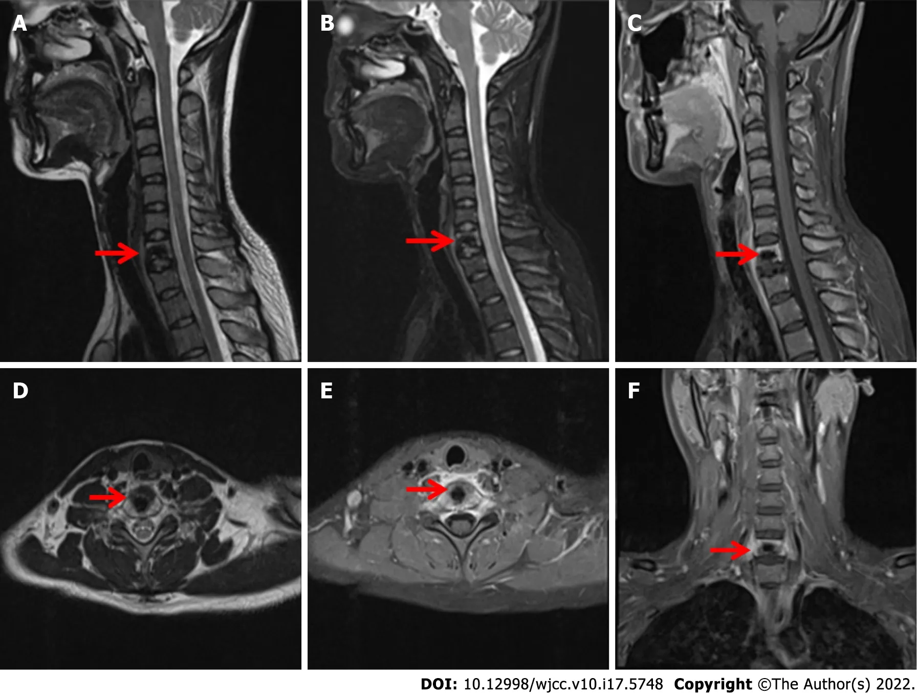
Cervical CT(Figure 1)showed that the bone structure of each cervical vertebral body was complete.The C7 vertebral body and C7/T1 intervertebral disc showed irregular high-density shadows with clear boundaries,and other bone density shadows showed no obvious abnormalities.Magnetic resonance imaging(MRI)(Figure 2)of the cervical vertebrae showed patchy abnormal signals at C7,low signals on T1-weighted images(T1WI),high and low mixed signals in T2WI and fat suppression images,swelling of adjacent soft tissue and inhomogeneous enhancement after enhancement.No abnormal signal was found in the C3/4-C6/7 intervertebral discs.The signal of the cervical spinal cord was normal.

FlNAL DlAGNOSlS
The patient was diagnosed with cervical CMF.
TREATMENT
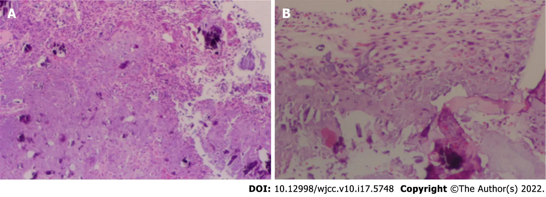
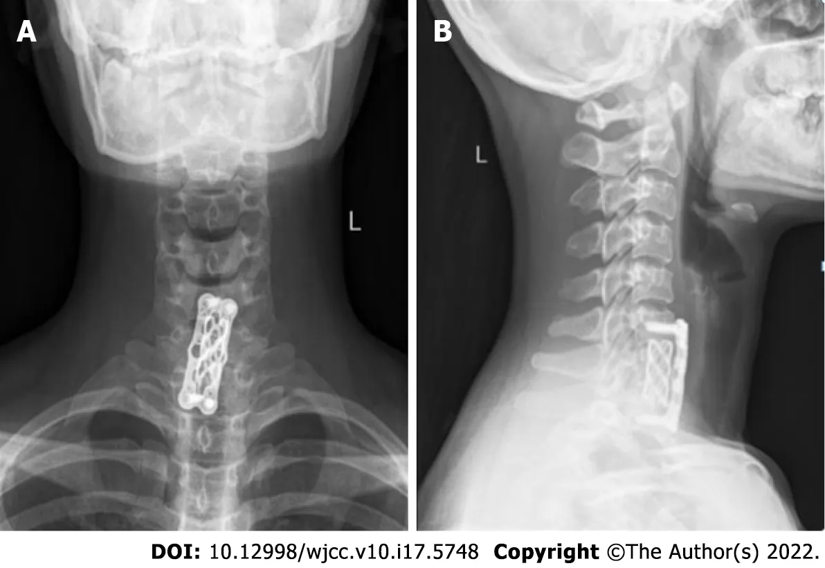
Under general anaesthesia with the patient in the supine position,We performed an anterior cervical surgery on the patient.C-arm fluoroscopy was used to locate the intervertebral space with a locating needle,and a distractor was placed in the centre of the front of the C6/7 and C7/T1 vertebral bodies to expand them.And subtotal resection of the C7 vertebral body was performed with a bone rongeur.A large amount of white chyle focus tissue in the vertebral body had eroded most of the bone in the vertebral body,and specimens were obtained for pathological examination(Figure 3).The cervical canal and nerve root canal were decompressed,and the adhesion of the spinal cord and bilateral nerve roots was released.The length from the lower edge of the C6 vertebral body to the upper edge of the T1 vertebral body was measured.A titanium cage of appropriate length was selected,and autogenous iliac bone was implanted into the titanium cage between C6 and T1.A hole was drilled in front of the C6 and T1 vertebral bodies,and titanium plate screws were installed.Before the end of the operation,C-arm fluoroscopy showed that the positions of the titanium plate and screw remained acceptable.
The man was amazed, and said that this would not only cost her her life, but would also bring upon him a greater misfortune than the one he was already under
OUTCOME AND FOLLOW-UP
An epidemiological survey of primary spinal tumours in Chinese patients showed that the proportion of benign tumours is larger than that of malignant tumours,and the proportion of male patients is larger than that of female patients.Benign tumours are more likely to occur at the age of 21 to 40 years old,and malignant tumours are more likely to occur at the age of 41 to 60 years old.The distribution of primary spinal tumours in the cervical,thoracic,lumbar and sacral segments is roughly the same,with most benign tumours occurring in cervical vertebrae and most malignant tumours appearing in sacral vertebrae.The age of morbidity and the location of the tumour in our case were consistent with the characteristics of primary spinal tumours in Chinese patients.Simple curettage,without filling the defect,leads to approximately 20% to 80% tumour recurrence[16].This recurrence might result from incomplete tumour excision and leave pseudopod processes extending from the main tumour to the cavernous body(sutures)after simple curettage.Curettage plus bone grafting or cement fixation has a much lower recurrence rate of approximately 7%[17].To prevent tumour recurrence,we performed total resection of the vertebral body and intervertebral disc,and internal fixation was performed to simultaneously maintain the stability of the entire spine.
DlSCUSSlON
Retrospective study of this case revealed no obvious abnormalities in the body from the preoperative screening of patients,with the exception of the cervical spine.Laboratory tests showed that relevant tumour markers and inflammatory markers were not significantly increased.The diagnosis was confirmed according to the comprehensive judgement of imaging examinations and intraoperative pathology.The diagnosis of CMF is difficult because of its overlapping features with other bony lesions.Pathology reveals that CMF has mucinous and fibrous components[5].The tumour cells were primarily arranged in lobules characterised by sparse cells in the centre,dense cells in the periphery,and more cells(primarily spindle cells,multinucleated giant cells and chondroblasts)between the lobules.Tumour cells were arranged in a fusiform or stellate pattern in a myxoid stroma.Calcification occurred in more than one-third of these cases but rarely became prominent.Hyaline cartilage and chondroblastoma-like areas were not uncommon.Approximately 18% of the tumours showed odd nuclei,and trabecular perforations were rare.Immunohistochemically(IHC),the tumour was positive for S100,which suggests that it was a chondroid tumour.Diseases that may be differentiated from CMF include low grade chondrosarcoma,enchondroma,chondromyxoid fifibroma-like osteosarcoma,chondroblastoma,and giant cell tumor of bone[6].
The differential diagnosis between CMF and chondrosarcoma is crucial.CMF also presents as a lowgrade malignancy with histological nuclear atypia similar to chondrosarcoma[7].The treatment and prognosis of CMF and chondrosarcoma vary greatly.Approximately 22% of CMFs are misdiagnosed as chondrosarcoma,which is a common malignant tumour in many bone tumour diseases.Chondrosarcoma morbidity peaks in the sixth and seventh decades of life,whereas CMF occurs in the second and third decades of life.Chondrosarcomas have specific demographic and radiographic features.CMF is often misdiagnosed as chondroblastoma.However,chondroid matrix,chondrocyte-like and multinucleated giant cell hyaline cartilage are predominantly absent in chondroblastoma,whereas CMF has myxochondroid matrix lobules with spindle and stem-like cells.Similarly,enchondroma has pathologically well-defined lobules with "bony encasement" and lacks atypia and myxoid areas,which is significantly different from the pathological appearance of CMF.Patients with CMF may be misdiagnosed with giant cell tumor(GCT)in the early stage,but the age of GCT patients is relatively older than that of CMF patients,and the cellular and imaging features of GCT are different from those of CMF.GCT usually originates from the metaphysis and is accompanied by epiphyseal extension.Multinucleated giant cells with the same background nuclei as interstitial cells are the main defining histological feature of GCT,whereas CMF shows only a few giant cells located close to the periphery.GCT shows neither myxoid nor cartilaginous components.
Another disease that can be confused with CMF is osteosarcoma.There are rare low-grade osteosarcomas.Chondromyxoid fibromatoid osteosarcoma(CMF-OS)was first reported in 1989,which presented with the histological and biological behaviours of low malignancy.Microscopically,it had a CMF-like appearance that consisted of loosely packed cellular gates in a highly mucoid stroma background and lobulated by fibrovessels.The most salient feature in differentiating CMF from CMFOS should be careful study of whether the bone is neoplastic,especially in lesions with malignant radiological features.IHC staining of vimentin-positive and S100-negative cells might be helpful in distinguishing CMF from CMF-OS.Zhong
[8]summarised the differences between CMF-OS and CMF using age of morbidity,tumour location,imaging examination,medical history,IHC and genetic characteristics from a series of case reviews.Finally,based on the above differences,combined with the imaging and pathological features of our case,we concluded that the nature of the patient's tumor was cervical CMF.
The patient,who had a daughter,with no family history of heredity.
That was it. That was all he said. See what happens? We are going to get up in the air, and see what happens? Couldn t we have another plan, one that s been worked out just a little better?
CMF is a benign tumor that may be associated with active growth of cartilage at the predilection site.At present,active surgical treatment,such as resection or curettage,is recommended for such tumors.Regardless of which treatment is adopted,the tumor should be totally removed as far as possible,because the residual tumor will lead to recurrence of the lesion.To prevent spinal instability after tumor resection,internal fixation should be performed at the same time to maintain spinal stability.It has been reported in the literature that there remains a certain proportion of recurrence after total resection of the lesion,and the longest recurrence time is 30 years;therefore,long-term follow-up is recommended.In this case,the lesion was located in the cervical vertebra,the lesion was completely resected,and autogenous bone was transplanted at the same time.There was no tumor recurrence after more than two years of follow-up,but close follow-up should be continued.
After the operation,the patient's symptoms were significantly alleviated,and the patient could walk independently.The neck was fixed with a common neck brace for 6 wk.At the 2-year follow-up,the patient's symptoms had not recurred.
CONCLUSlON
CMF was reported in upper and lower cervical vertebrae,and the tumour invades all parts of the vertebral body,transverse foramen and posterior soft tissue.The tumour had only caused obvious disc erosion in our case.We resected all of the tumours of the vertebral body and intervertebral disc
surgery and performed iliac bone implantation and anterior fusion.The occipital bone and atlas maintain 50% of the flexion and extension range of motion of the cervical spine,and the atlas and axis maintain 50% of the rotation range of motion of the vertebral body.The age of the patient was fully considered in the preoperative strategy.Anterior fusion of the two segments of the lower cervical spine was performed.Therefore,it would not have a significant impact on the patient's quality of life in the future.The internal fixation position of the patient was good,and no discomfort,such as neck movement or dysphagia,was shown(Figure 4).
FOOTNOTES
Li C,Li S and Hu W contributed to study conception and design;Li C and Hu W collected and analyzed the clinical data and wrote the manuscript;Li C,Li S and Hu W submitted and revised the manuscript;and All authors read and approved the final version of manuscript.
Physical examination showed tenderness and percussion pain of the cervical spinous process.No radiating nerve pain was detected,and other related pathological signs were negative.
The authors declare that there is no conflict of interest.
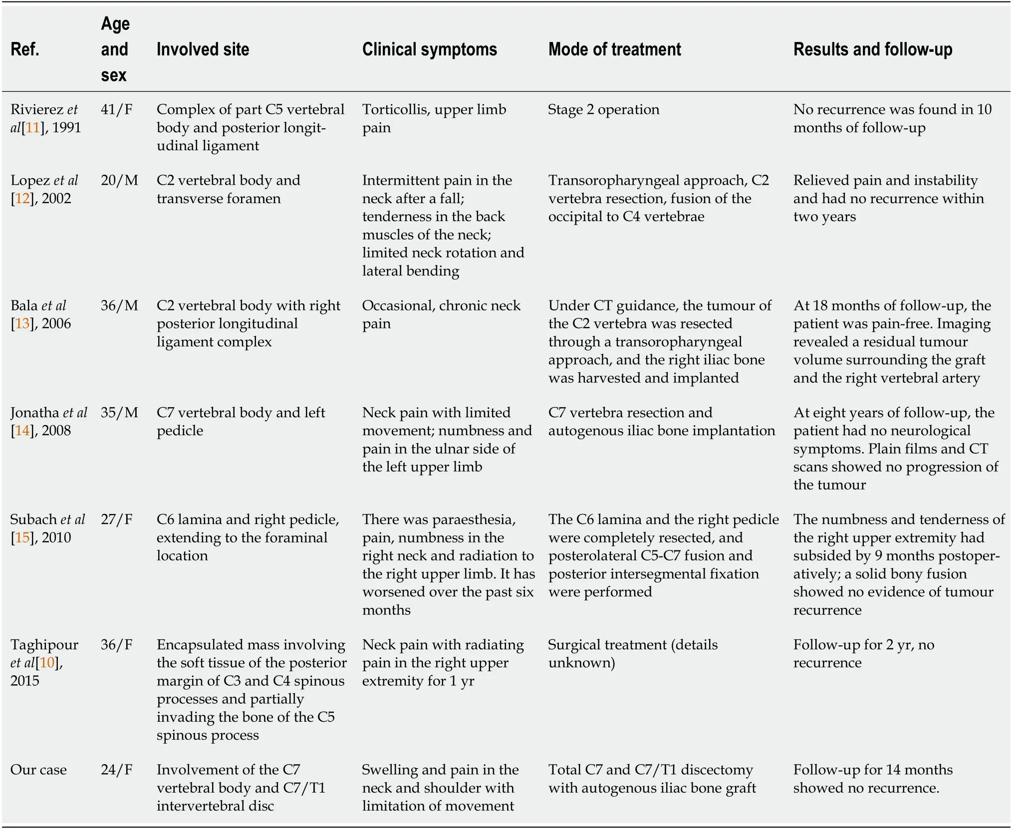
CMF is generally a slow-growing asymptomatic lesion.Most CMFs are located in the metaphyseal region of the long bones,approximately one-third of which formed in the tibia,foot tubular bones,distal femur,and pelvis.Although CMF has a predilection site,most of the reported cases have been atypical.Therefore,we cannot avoid CMF in the differential diagnosis of bone tumours in various parts of the human body.The clinical manifestations vary according to the size,location and extent of the lesion.The common symptoms of CMF in cervical spine are neck and shoulder pain,limitation of movement,numbness and radiating pain of upper limbs after nerve root damage.Although CMF is a relatively rare benign tumour,vertebral CMF only accounts for 8% of all CMFs,and cervical CMF is even rarer,with fewer than 12 cases reported[10].The features of vertebral CMF include the erosion of cortical bone,even beyond the extent of the periosteum into the surrounding soft tissue or spinal canal,and this feature indicates more serious destruction,which may indicate the malignant transformation of tumours.All cervical CMFs reported in the English literature over a 30-year period from 1991 to 2021(Table 1)were retrieved and are summarized below.
If it s as you say, you may take as much rampion away with you as you like, but on one condition only -- that you give me the child11 your wife will shortly bring into the world. All shall go well with it, and I will look after it like a mother. 12
The authors have read the CARE Checklist(2016),and the manuscript was prepared and revised according to the CARE Checklist(2016).
This article is an open-access article that was selected by an in-house editor and fully peer-reviewed by external reviewers.It is distributed in accordance with the Creative Commons Attribution NonCommercial(CC BYNC 4.0)license,which permits others to distribute,remix,adapt,build upon this work non-commercially,and license their derivative works on different terms,provided the original work is properly cited and the use is noncommercial.See: https://creativecommons.org/Licenses/by-nc/4.0/
China
She would not answer her telephone the next morning but in his mails that afternoon came an envelope that he knew had come from Amy. The handwriting was not beautiful, but it was without question hers. Inside was only a card on which she had written:
One day when the Queen was at church, and the two children sat and played with their father, he gazed again full of grief on the stone statue, and sighing, wailed43: Oh, if I could only restore you to life, my most trusty John! Suddenly the stone began to speak, and said: Yes, you can restore me to life again if you are prepared to sacrifice what you hold most dear
Cheng Li 0000-0002-1173-9148;Sen Li 0000-0003-2837-4808;Wei Hu 0000-0002-8110-9969.
I looked up, surprised. She seemed so calm and gentle. Seemingly11 out of the blue, someone had found me and offered help. I wiped the tears from my cheek.
He was very careful not to leave enough space for the dragon to jump out, but unluckily there was just room for his great mouth, and with one snap the king vanished down his wide red jaws16
Ma YJ
A
Ma YJ
1 Soni R,Kapoor C,Shah M,Turakhiya J,Golwala P.Chondromyxoid Fibroma:A Rare Case Report and Review of Literature.
2016;8: e803[PMID: 27833828 DOI: 10.7759/cureus.803]
2 JAFFE HL,LICHTENSTEIN L.Chondromyxoid fibroma of bone;a distinctive benign tumor likely to be mistaken especially for chondrosarcoma.
1948;45: 541-551[PMID: 18891025]
3 Douis H,Saifuddin A.The imaging of cartilaginous bone tumours.II.Chondrosarcoma.
2013;42: 611-626[PMID: 23053201 DOI: 10.1007/s00256-012-1521-3]
4 Yasuda T,Nishio J,Sumegi J,Kapels KM,Althof PA,Sawyer JR,Reith JD,Bridge JA.Aberrations of 6q13 mapped to the COL12A1 locus in chondromyxoid fibroma.
2009;22: 1499-1506[PMID: 19648885 DOI: 10.1038/modpathol.2009.101]
5 HemanthaKumar G,Sathish M.Diagnosis and Literature Review of Chondromyxoid Fibroma - A Pathological Puzzle.
2019;9: 101-105[PMID: 32405500 DOI: 10.13107/jocr.2019.v09.i04.1500]
6 Wangsiricharoen S,Wakely PE Jr,Siddiqui MT,Ali SZ.Cytopathology of chondromyxoid fibroma:A case series and review of the literature.
2021;10: 366-381[PMID: 33958292 DOI: 10.1016/j.jasc.2021.04.001]
7 Vasudeva N,Shyam Kumar C,Ayyappa Naidu CR.Chondromyxoid Fibroma of Distal Phalanx of the Great Toe: A Rare Clinical Entity.
2020;12: e7133[PMID: 32257678 DOI: 10.7759/cureus.7133]
8 Zhong J,Si L,Geng J,Xing Y,Hu Y,Jiao Q,Zhang H,Yao W.Chondromyxoid fibroma-like osteosarcoma:A case series and literature review.
2020;21: 53[PMID: 31996205 DOI: 10.1186/s12891-020-3063-5]
9 Cappelle S,Pans S,Sciot R.Imaging features of chondromyxoid fibroma: report of 15 cases and literature review.
2016;89: 20160088[PMID: 27226218 DOI: 10.1259/bjr.20160088]
10 Taghipour Zahir S,Sefidrokh Sharahjin N,Sadlu Parizi F,Rahmani K.Chondromyxoid Fibroma of Two Cervical Vertebrae with Involvement of Surrounding Soft Tissue: Radiologic Diagnostic Dilemma.
2015;12: e19273 [PMID: 26587204 DOI: 10.5812/iranjradiol.19273]
11 Rivierez M,Richard S,Pradat P,Devred C.[Chondromyxoid fibroma of the cervical spine.Apropos of a case treated by partial vertebrectomy].
1991;37: 264-268[PMID: 1922638]
12 Lopez-Ben R,Siegal GP,Hadley MN.Chondromyxoid fibroma of the cervical spine: case report.
2002;50: 409-411[PMID: 11844279 DOI: 10.1097/00006123-200202000-00034]
13 Bala A,Robbins P,Knuckey N,Wong G,Lee G.Spinal chondromyxoid fibroma of C2.
2006;13: 140-146[PMID: 16410218 DOI: 10.1016/j.jocn.2005.03.022]
14 Jonathan A,Rajshekhar V,Chacko G.Chondromyxoid fibroma of the seventh cervical vertebra.
2008;56: 84-87[PMID: 18310848 DOI: 10.4103/0028-3886.39323]
15 Subach BR,Copay AG,Martin MM,Schuler TC,Romero-Gutierrez M.An unusual occurrence of chondromyxoid fibroma with secondary aneurysmal bone cyst in the cervical spine.
2010;10: e5-e9[PMID: 20036621 DOI: 10.1016/j.spinee.2009.11.016]
16 Zillmer DA,Dorfman HD.Chondromyxoid fibroma of bone: thirty-six cases with clinicopathologic correlation.
1989;20: 952-964[PMID: 2793160 DOI: 10.1016/0046-8177(89)90267-0]
17 Basak B,Haragan A,Shackcloth M,Thekkudan J.Chondromyxoid Fibroma of the Rib: A Rare Benign Tumor With Potential for Local Recurrence.
2021;13: e19172[PMID: 34873515 DOI: 10.7759/cureus.19172]
 World Journal of Clinical Cases2022年17期
- World Journal of Clinical Cases的其它文章
- Repetitive transcranial magnetic stimulation for post-traumatic stress disorder:Lights and shadows
- Response to dacomitinib in advanced non-small-cell lung cancer harboring the rare delE709_T710insD mutation:A case report
- Loss of human epidermal receptor-2 in human epidermal receptor-2+breast cancer after neoadjuvant treatment:A case report
- Tumor-like disorder of the brachial plexus region in a patient with hemophilia:A case report
- High-frame-rate contrast-enhanced ultrasound findings of liver metastasis of duodenal gastrointestinal stromal tumor:A case report and literature review
- Gitelman syndrome:A case report
