Glutamine deprivation impairs function of infiltrating CD8+ T cells in hepatocellular carcinoma by inducing mitochondrial damage and apoptosis
INTRODUCTION
CD8
T cells are important effector immune cells in the tumor microenvironment that primarily kill tumor cells by secreting granzyme B(GZMB)and perforin(PRF1)[1].Owing to factors that include chronic stimulation by tumor antigens,the physicochemical state of the tumor microenvironment is imbalanced,including low pH,hypoxia,and low nutrition availability[2,3],which trigger T cell dysfunction and ultimately result in cell depletion[4,5].Exhausted T(Tex)cells are characterized by loss of effector functions,elevated and sustained expression of inhibitory receptors(IRs),and a distinct metabolic profile[6,7].Recently,the mechanisms of CD8
T cell exhaustion have become a hot research topic,and there is increased interest on how changes in metabolomics correlate with changes in immune cell functions.
Tumor cells consume high levels of energy sources such as glucose[8],resulting in nutrient depravation in the tumor microenvironment.This deprives the energy sources of immune cells,altering the phenotype and function of affected immune cells[9].Glutamine(Gln)is the most abundant free amino acid in serum[10].Gln is not only involved in the occurrence,development,and metastasis of tumors[11,12],but also regulates the growth and function of immune cells[13].Gln has been reported to regulate the phenotype of CD4
cells,and increasing Gln levels can skew regulatory CD4
T cells toward more inflammatory subtypes[14,15].However,whether Gln also regulates CD8
T cells and the mechanism underlying this regulation have not been reported.
10. Bewitched by a wicked witch: No version of the story explains why a witch cast the spell on the prince. However, it is interesting that a witch is mentioned in passing as the culprit. Witches are often responsible for casting transformation spells in other tales, so the witch is essentially38 a stereotype39 used in this story.Return to place in story.
Mitochondria are important intracellular organelles that provide energy and biosynthetic substrates for cell survival through oxidative phosphorylation[16].Mitochondria are also highly nutrient-sensitive.When cells are deficient in nutrients,their mitochondria undergo depolarization and appear damaged.Damaged mitochondria can release pro-apoptotic proteins such as cytochrome c and other substances to induce the formation of apoptotic complexes that activate caspase 9,which causes apoptosis.Recent research has demonstrated mitochondrial damage in tumor tissues that is closely related to remodeling of the tumor microenvironment.It has also been shown in the literature that after glucose deprivation,CD8
T cells in the tumor microenvironment produce reactive oxygen species(ROS),which subsequently trigger mitochondrial damage[17].Whether Gln similarly regulates the function of mitochondria in CD8
T cells is still unclear.
Then again the wise woman stood before her, and said, Little Two-eyes, what are you crying for? Have I not reason to cry? she answered, the goat, which when I said the little rhyme, spread the table so beautifully, my mother has killed, and now I must suffer hunger and want again
Mitochondria are the primary source of ROS production,and JC-1 is a dye used to reflect mitochondrial integrity.To examine whether hepatoma cells have a regulatory effect on CD8
T cell mitochondria,CD8
T cells were co-cultured with Huh-7 cells.Flow cytometry analysis revealed that CD8
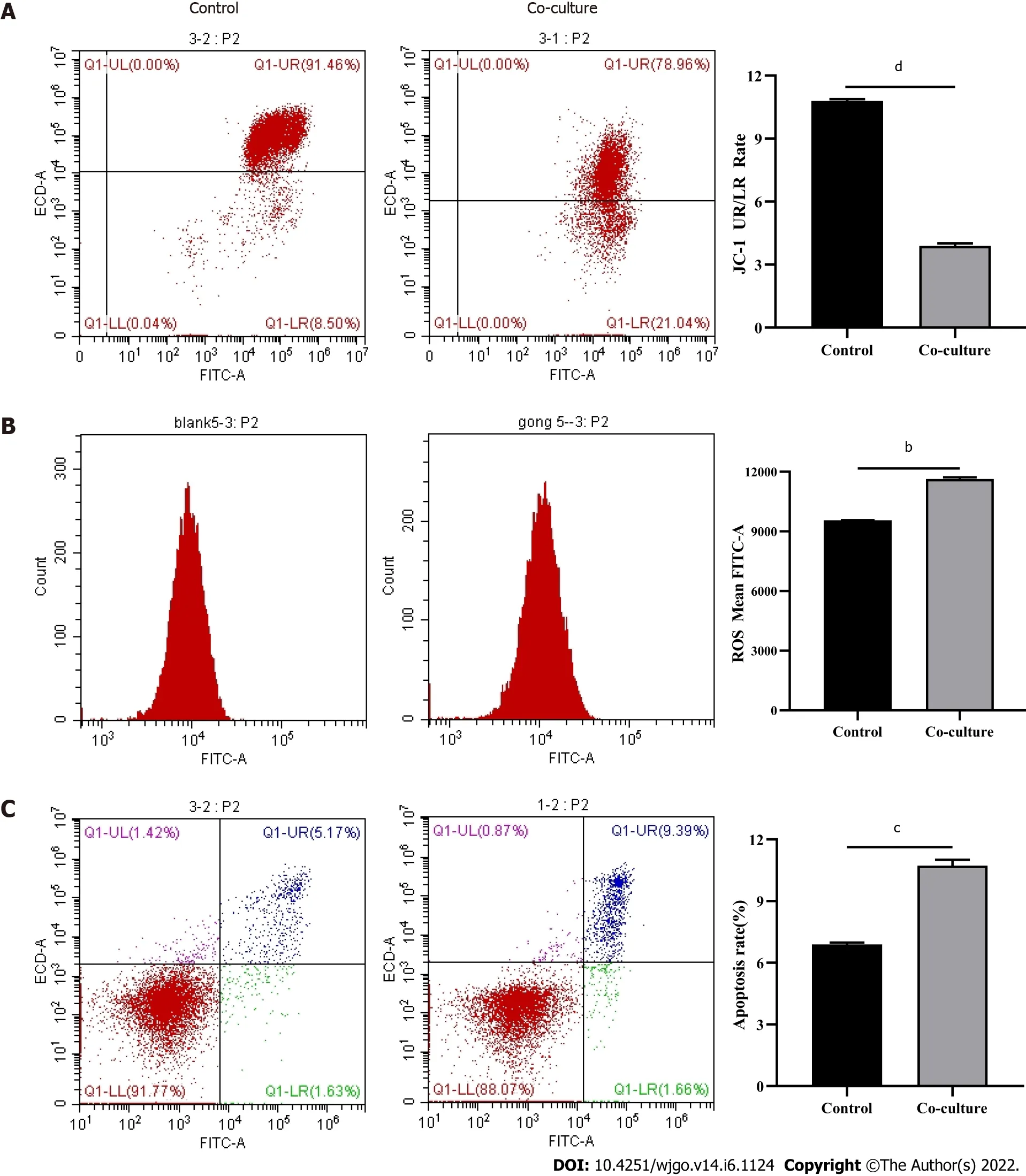
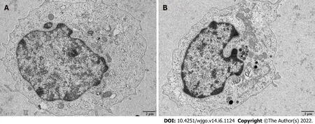
T cells from the co-culture group had decreased JC-1 staining(Figure 5A)and increased levels of ROS(Figure 5B)and apoptosis(Figure 5C)compared with the control group.Next,mitochondrial morphology was further studied by TEM to examine the ultrastructure of mitochondria.Mitochondria of CD8
T cells from the control group were identified with well-defined integral two-layer membranes and regular cristae(Figure 6A).In contrast,mitochondria of CD8
T cells in the co-culture group showed significant swelling(Figure 6B).In summary,co-culture with hepatoma cells induced damage to the mitochondria of CD8
T cells and caused apoptosis.
MATERIALS AND METHODS
Immunohistochemical staining
Immunohistochemical staining was performed on surgically resected tissues from patients with clinicopathologically confirmed hepatocellular carcinoma(HCC)and adjacent non-cancerous tissues.The study was approved by the Medical Ethics Committee of North China University of Science and Technology(No.2018109).
Immunofluorescence
Immunofluorescence staining of paraffin-embedded human liver tissue sections was performed using anti-PD-1/CD279(66220-1-Ig,Proteintech,Rosemont,IL,United States)and anti-CD8α(PB9249,Boster Bio,Pleasanton,CA,United States)antibodies.Antigen retrieval was performed with EDTA buffer(AR0023,Boster Bio).Tissue sections were blocked with 10% goat serum(AR1009,Boster Bio),and then incubated with rabbit anti-CD8α antibody(1:200 dilution)and mouse anti-PD-1 antibody(1:4000 dilution)at 4 °C overnight.Secondary antibodies including DyLight594 fluorescein goat anti-mouse(BA1141,Boster Bio)and fluorescein DyLight488 goat anti-rabbit(BA1127,Boster Bio)IgGs were then added and incubated for 45 min at 37 °C.DAPI staining solution(AR1176,Boster Bio)was used for counterstaining at room temperature for 3 min,and then washed with PBS(pH 7.2-7.6)(AR0030,Boster Bio).Slides were mounted using anti-fluorescent quench mounting medium(AR1109,Boster Bio).Finally,whole-slide scanning(APERIOVERSA8,Leica,Wetzlar,Germany)and image acquisition(BX51,Olympus,Tokyo,Japan)were performed.
Cell sorting
First,mRNA was extracted from sorted CD8
T cells using the MiniBEST Universal RNA Extraction Kit(Cat.#9767;TaKaRa,Dalian,China)according to the manufacturer’s protocol.Extracted mRNA was reverse-transcribed into cDNA after mixing mRNA template with the Primescript RT Reagent Kit with gDNA Eraser(AK3920;TaKaRa)for quantitative polymerase chain reaction(qPCR).The qPCR reactions were run on a Light Cycler 480SYBRGreenIMaster(04887352001;Roche,Basel,Switzerland).The primer sequences are as follows:
(forward: 5′-GGTGCGGTGGCTTCCTGAT-3′ and reverse: 5′-ACTGCTGGGTCGGCTCCTGT-3′);
(forward: 5′-TGCCGCTTCTACAGTTTCCA-3′ and reverse: 5′-CCACCTCGTTGTCCGTGAG-3′);and
(forward: 5′-TCAAGAAGGTGGTGAAGCAGG-3′ and reverse: 5′-TCAAAGGTGGAGGAGTGGGT-3′).The qPCR steps included initial denaturation at 95 °C for 10 min,and 40 cycles of denaturation at 95 °C for 10 s,annealing at 60 °C for 15 s,and extension at 72 °C for 20 s.All qPCR reactions were performed on a Bio-Rad real-time quantitative fluorescence PCR instrument(CFX Connect,Bio-Rad,Hercules,CA,United States).Relative gene expression was calculated with respect to the internal standard(
).Expression levels were normalized against
using the 2
method.
Cell culture and activation
The hepatoma cell line HuH-7(RRID: CVCL_0336)was confirmed
short tandem repeat markers by Procell Life Science &Technology Co.,Ltd.(Hyderabad,India).The results showed that the DNA typing of this cell line matched 100% with other typing in the CRC cell bank,and no human cell crosscontamination was found.HuH-7 cells were cultured in Dulbecco’s modified eagle medium(DMEM)(Sigma-Aldrich,St.Louis,MO,United States),supplemented with 10% fetal bovine serum(FBS)(Gibco,Waltham,MA,United States)and 1% penicillin/streptomycin(Hyclone).CD8
T cells were cultured
using Roswell Park Memorial Institute(RPMI)1640 medium(SH30809.01B;Hyclone),supplemented with 10% FBS(Gibco)and 1% penicillin/streptomycin(Hyclone).CD8
T cells were stimulated with 10 μg/mL anti-human CD3 antibody(kx10-3A;Beijing Kexin Biological,Beijing,China)and 2.5 μg/mL anti-human CD28 antibody(kx10-28A;Beijing Kexin Biological)in medium containing 100 U/mL IL-2(PeproTech,Rocky Hill,NJ,United States)for 2 d.Both cell types were maintained at 37 °C in an atmosphere containing 50 mL/L CO
.
Cell co-culture
HuH-7 and CD8
T cells were co-cultured in transwell inserts for 3 d,and the initial number of both cell types used was equal.
RNA sequencing of CD8+ T cells
Total RNA was extracted from CD8
T cells,and total RNA sequencing(RNA-Seq)libraries were obtained by a three-step method:(1)RNA library construction and on-board sequencing were performed.Sequencing data were aligned to the reference genome of the project species to obtain comprehensive transcript information and to perform gene expression quantification;(2)The transcriptome sequencing project was completed on the Illumina sequencing platform(San Diego,CA,United States),and the IlluminaPE library(approximately 300 bp)was constructed for sequencing;and(3)The obtained sequencing data were analyzed by bioinformatics after performing quality control.
42. Red eyes, and cannot see far: Red eyes are an image associated with sorrow and with demonic fury. Eyesight is associated with mental perception, indicating that the witch s poor eyesight means poor reasoning ability, which allows Hansel and Gretel to best her (Olderr 1986). The Grimms are setting up the circumstances for Hansel and Gretel s escape from the witch.Return to place in story.
Flow cytometry analysis
Glutamine deprivation impairs the function of infiltrating CD8
T cells in hepatocellular carcinoma through the mitochondrial damage and apoptotic pathways.
Quantitative real-time polymerase chain reaction
First,100 mL of peripheral blood was collected from healthy volunteers,from which peripheral blood mononuclear cells(PBMCs)were obtained by diluting peripheral blood with an equal volume of PBS followed by centrifugation(800
,20 min).The pellet was then washed with 5 × volume of PBS and centrifuged(200
,5 min).Cells were resuspended in x-vivo15(SH30809.01B,Hyclone,Logan,UT,United States).PBMCs were stained with anti-human CD8 antibody(Biolegend,San Diego,CA,United States).Cell sorting was performed using a MofloXDP sorting flow cytometer(Beckman Coulter,Brea,CA,United States).The post-classification purity of all included samples was > 85%.
Transmission electron microscopy
Cells in each group were digested to prepare cell suspensions at a density of 1 × 10
cells/mL.After discarding the supernatant,the cells were fixed with 3% glutaraldehyde fixative for 2 h and 1% osmic acid for 1 h.After dehydration with a graded ethanol series and acetone,the cells were embedded in epoxy resin,ultrathin sectioned,and double stained with uranium and lead.The ultrastructural changes of mitochondria in each group of cells were observed by transmission electron microscopy(TEM).
Statistical analysis
All analyses were performed with SPSS 21.0 software(IBM Corp.,Armonk,NY,United States)and GraphPad Prism 8.0.2(GraphPad Software,Inc.,San Diego,CA,United States).Measurement data were first examined using the Kolmogorov-Smirnov test to check whether the measurement data of each group had a normal distribution.Results are expressed as the mean ± SE for measurement data with a normal distribution.Comparisons between two groups were performed using the two-tailed Student’s
test.One-way analysis of variance was used for multiple group comparisons,and the LSD-
test was used for pairwise comparisons.Significance was accepted at
< 0.05.
RESULTS
CD8+ T cell infiltration in liver tissues
There were a large number of infiltrating PD-1
CD8
T cells in liver cancer tissues.Next,we co-cultured CD8
T cells and Huh-7 cells to explore the regulatory effect of hepatoma cells on CD8
T cells.Flow cytometry results revealed increased PD-1 expression and decreased secretion of perforin(PRF1)and granzyme B(GZMB)by CD8
T cells in the co-culture group.Meanwhile,JC-1 staining was decreased and the levels of reactive oxygen species and apoptosis were increased in CD8
T cells of the co-culture group;additionally,the mitochondria of these cells were swollen.When CD8
T cells were treated with the mitochondrial protective and damaging agents,their function was restored and inhibited,respectively,through the mitochondrial damage and apoptotic pathways.Subsequently,complete medium without glutamine was used to culture cells.As expected,CD8
T cells showed functional downregulation,mitochondrial damage,and apoptosis.
Hepatoma cells induce decreased CD8+ T cell function
After observing a large number of infiltrating PD-1
CD8
T cells in HCC tissues,we next co-cultured CD8
T cells with Huh-7 cells to evaluate the functional alterations of CD8
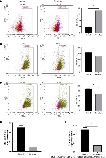
T cells.Flow cytometry and quantitative real-time PCR were used to examine the function of CD8
T cells,which indicated that compared with the control group,CD8
T cells in the co-culture group expressed higher levels of PD-1(Figure 3A)and lower levels of GZMB and PRF1(Figure 3B-E).Increased expression of IRs and reduced ability to secrete effector molecules suggested functional inhibition of CD8
T cells;thus,hepatoma cells impaired the function of CD8
T cells.
Hepatoma cells induce differential gene expression in CD8+ T cells
Immunohistochemical staining and immunofluorescence were performed on surgically resected liver tissues from patients.Differentially expressed genes in infiltrating CD8
T cells in hepatocellular carcinoma were detected using RNA sequencing.Activated CD8
T cells were co-cultured with Huh-7 cells for 3 d.The function and mitochondrial status of CD8
T cells were analyzed by flow cytometry,quantitative polymerase chain reaction,and transmission electron microscopy.Next,CD8
T cells were treated with the mitochondrial protective and damaging agents.Functional alterations in CD8
T cells were detected by flow cytometry.Then,complete medium without glutamine was used to culture cells,and their functional changes and mitochondrial status were detected.
Hepatoma cells induce mitochondrial damage and apoptosis in CD8+ T cells
Therefore,this study investigated whether Gln regulates CD8
T cell function through the mitochondrial damage and apoptotic pathways to clarify the relationship between Gln metabolism and CD8
T cell depletion,which will lay a foundation for developing new anti-tumor treatments.
Mitochondrial damage induced by hepatoma cells impairs CD8+ T cell function
Next,we explored the relationship between mitochondrial damage and T cell function in infiltrating CD8
T cells in HCC.First,we co-cultured CD8
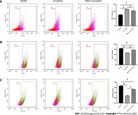
T cells with Huh-7 cells,with mitochondrial division inhibitor 1(Mdivi-1)added to the culture system,which has been shown to protect from mitochondrial damage.Flow cytometry was used to examine the function of Mdivi-1-treated CD8
T cells in co-culture.The results showed reduced expression of PD-1(Figure 7A)and increased expression of PRF1(Figure 7B)and GZMB(Figure 7C)in CD8
T cells from the Mdivi-1 group compared with CD8
T cells from the co-culture alone group,demonstrating that the function of co-cultured CD8
T cells was not decreased after protecting from mitochondrial damage.Next,we cultured CD8
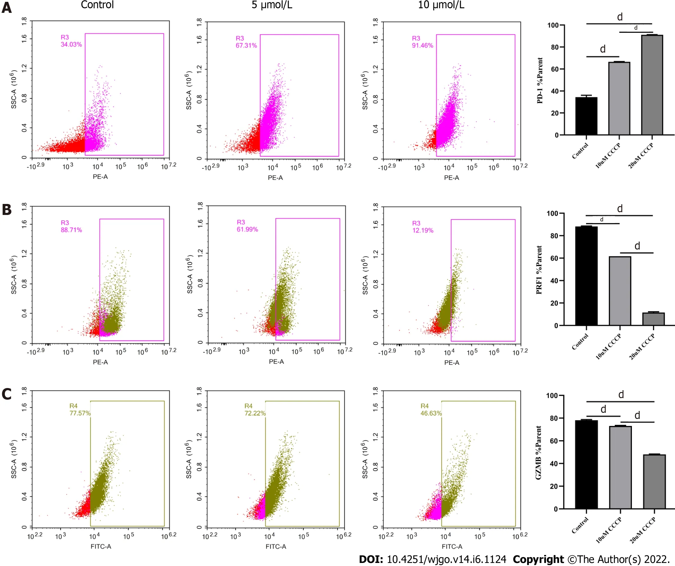
T cells with different concentrations of the oxidative phosphorylation uncoupler carbonyl cyanide-3-chlorophenylhydrazone(CCCP)and examined their function.As shown from flow cytometry analysis,PD-1 expression gradually increased(Figure 8A),and there was a gradual decrease in the secretion of PRF1(Figure 8B)and GZMB(Figure 8C)by CD8
T cells with increasing CCCP concentrations.These results illustrated that mitochondrial damage led to the inhibition of CD8
T cell function.In summary,co-culture with hepatoma cells impaired the function of CD8
T cells through the mitochondrial damage pathway.

Gln deprivation induces mitochondrial damage,apoptosis,and functional impairment of CD8+ T cells
As a primary energy source,Gln affects both the development of tumor cells and the functions of immune cells.Therefore,we assessed whether Gln metabolism affects the function of CD8
T cells.We co-cultured CD8
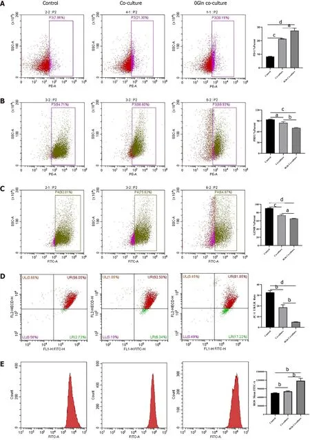
T cells with Huh-7 cells and supplemented the co-culture system with RPMI 1640 with normal concentrations of Gln or without Gln.The findings demonstrated that CD8
T cells in the coculture system without Gln expressed lower PD-1(Figure 9A)and higher PRF1(Figure 9B)and GZMB(Figure 9C)levels compared with the co-culture alone group;additionally,JC-1 staining was decreased(Figure 9D),and there were increased levels of ROS(Figure 9E)and apoptosis(Figure 9F).Next,CD8
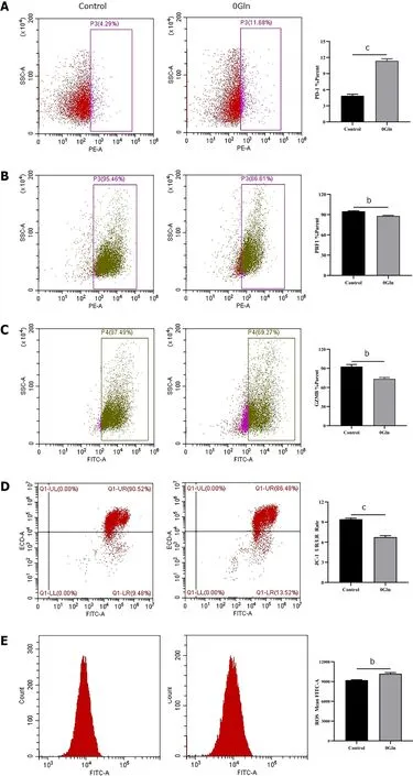
T cells were cultured alone in RPMI 1640 with the normal Gln concentration or without Gln to further validate the effect of Gln on CD8
T cells.The results showed that CD8
T cells in the group lacking Gln had increased PD-1 expression(Figure 10A)and decreased secretion of PRF1(Figure 10B)and GZMB(Figure 10C)compared with those in the control group.Similarly,JC-1 staining was decreased(Figure 10D),and the levels of ROS(Figure 10E)and apoptosis(Figure 10F)were increased.Together,these data showed that hepatoma cells competed with CD8
T cells for Gln.Gln deficiency inhibited the function of CD8
T cells and induced mitochondrial damage and apoptosis.

DISCUSSION
In the tumor immune microenvironment,CD8
T cells are the main immune effector cells and play a crucial role in the host immune environment.A large number of CD8
T cells infiltrate tumor tissues in patients.However,due to chronic stimulation by tumor antigens,most of the T cells are functionally impaired and have differentiated into CD8
T ex cells,which mediate tumor immune escape[18-20].Therefore,understanding the mechanism of decreased CD8
T cell function is essential for tumor immunity.Tex cells are characterized by the loss of effector functions and elevated and sustained expression of IRs[6,21,22].In this study,the expression of PD-1 was increased and the secretion of cellular effector molecules such as GZMB and PRF1 was decreased in CD8
T cells from the co-culture group.Our observations illustrated that the function of infiltrating CD8
T cells was suppressed by HCC cells.Therefore,we next explored the specific mechanisms
which CD8
T cell function was inhibited.
Mitochondria are central to cellular metabolism and play key roles in the functional regulation of immune cells such as CD8
T cells[23].When mitochondria are slightly impaired,the damage is neutralized by fusion with healthy mitochondria.However,when mitochondrial damage exceeds the range buffered by fusion,the cell promotes mitochondrial division and damage[24].Severely injured mitochondria usually show increased ROS production and decreased membrane potential[25],which decreases cellular function.As shown by our data,the mitochondria of CD8
T cells co-cultured with HCC cells exhibited swelling,increased ROS production,and decreased membrane potential,and the cells underwent apoptosis,implying that HCC cells induce mitochondrial damage and apoptosis in CD8
T cells.Next,we examined the relationship between mitochondrial damage and CD8
T cell function by using Mdivi-1 and CCCP to regulate mitochondrial status.Mdivi-1 is a quinazolinone derivative that penetrates cell membrane and attenuates mitochondrial damage by inhibiting mitochondrial division[26].CCCP can interrupt oxidative phosphorylation,which impairs mitochondrial function and prompts mitochondria to produce high levels of ROS[27].Together,these data show that mitochondrial damage can impairs the function of CD8
T cells,
,hepatoma cells can impairs the function of CD8
T cells through the mitochondrial damage and apoptotic pathways.
Studies have shown that tumor cells inhibit anti-tumor immunity by competing for essential nutrients and reducing the metabolic adaptability of tumor-infiltrating immune cells,which in turn decreases the function of immune cells[28].The nutrients required for cellular metabolism include glucose,amino acids,and fatty acids.Both tumor cells and activated T cells show a significant Gln requirement[29].CD4
T cells lacking Gln have diminished proliferative capacity and decreased cytokine secretion.We found that CD8
T cells lacking Gln have a reduced ability to secrete effector molecules and increased expression of inhibitory receptors such as PD-1,suggesting that Gln deprivation inhibits the function of CD8
T cells.In states of energy deficiency,mitochondrial function is abnormal,which further activates pro-apoptotic downstream regulators that induce apoptosis[30].A previous study suggested that upon glucose deprivation,cells largely mobilize oxidative phosphorylation to maintain energy homeostasis,causing mitochondria to produce high levels of ATP and ROS[31].Our data showed that in the absence of Gln,the mitochondria of CD8
T cells underwent morphological changes with reduced mitochondrial membrane potential,generated high levels of ROS,and induced apoptosis.However,the above results were obtained only from
experiments,and more details need to be further studied.
The evening before, the youth went down to the rocks and into the copse, collecting all the drift wood the sea had washed up or the gale15 had blown down, and he piled it up in a great stack outside the door, so that he might not have to fetch any all the next day
36 “You are not afraid of the sea, my dumb child,” said he, as they stood on the deck of the noble ship which was to carry them to the country of the neighboring king
CONCLUSION
We found that Gln deprivation impairs the function of CD8
T cells through the mitochondrial damage and apoptotic pathways.
ARTICLE HIGHLIGHTS
Research background
The functions of infiltrating CD8
T cells are often impaired due to tumor cells causing nutrient deprivation in the tumor microenvironment.Thus,the mechanisms of CD8
T cell dysfunction have become a hot research topic,and there is increased interest on how changes in metabolomics correlate with CD8
T cell dysfunction.
She told him then that she was a princess whom the witch had stolen, and had changed to this shape, but with his kiss she had got her human form again; and if he would be faithful to her, and take her to wife, she could free them both from the witch s power
Research motivation
To explore the effect of glutamine metabolism on the function of tissue-infiltrating CD8
T cells,so as to provide a new strategy for reversing the exhausted CD8
T cells in hepatocellular carcinoma.
Research objectives
This study aimed to investigate whether and how glutamine metabolism affects the function of infiltrating CD8
T cells in hepatocellular carcinoma.
Research methods
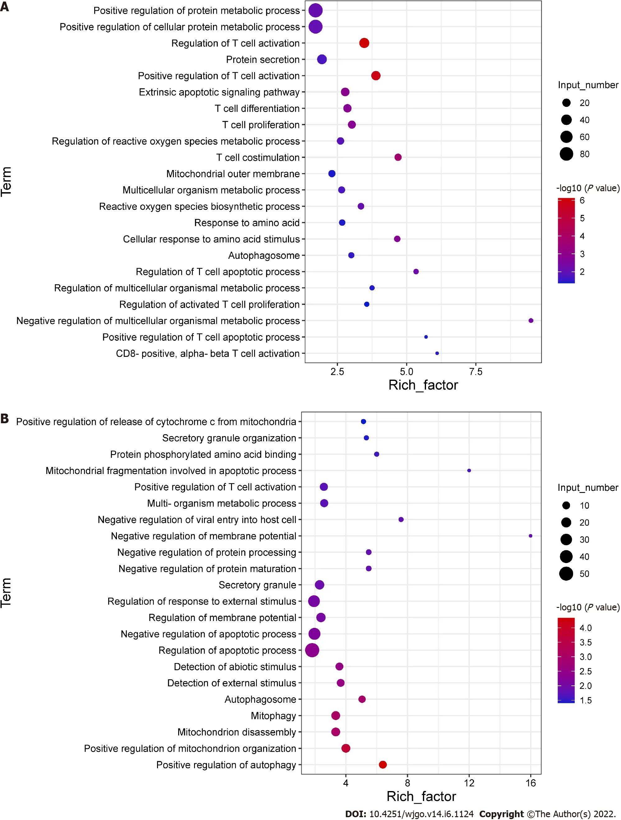
To explore the potential mechanisms of the functional impairment of T cells,we performed RNA-seq of CD8
T cells co-cultured with Huh-7 cells.We found that genes related to mitochondrial damage(such as mitochondrion disassembly)and apoptosis(such as regulation of apoptotic pathways)were upregulated(Figure 4A).The expression of genes related to cell metabolism(such as multi-organism metabolic processes)and T cell function(such as T cell proliferation)was downregulated(Figure 4B).These data illustrated that CD8
T cells co-cultured with hepatoma cells underwent alterations associated with cellular metabolism,mitochondrial damage,and apoptosis.
Dynise Balcavage, 42, an associate creative director at an advertising27 agency and vegan who lives in Philadelphia, said she has been happily married to her omnivorous husband, John Gatti, 53, for seven years
Research results
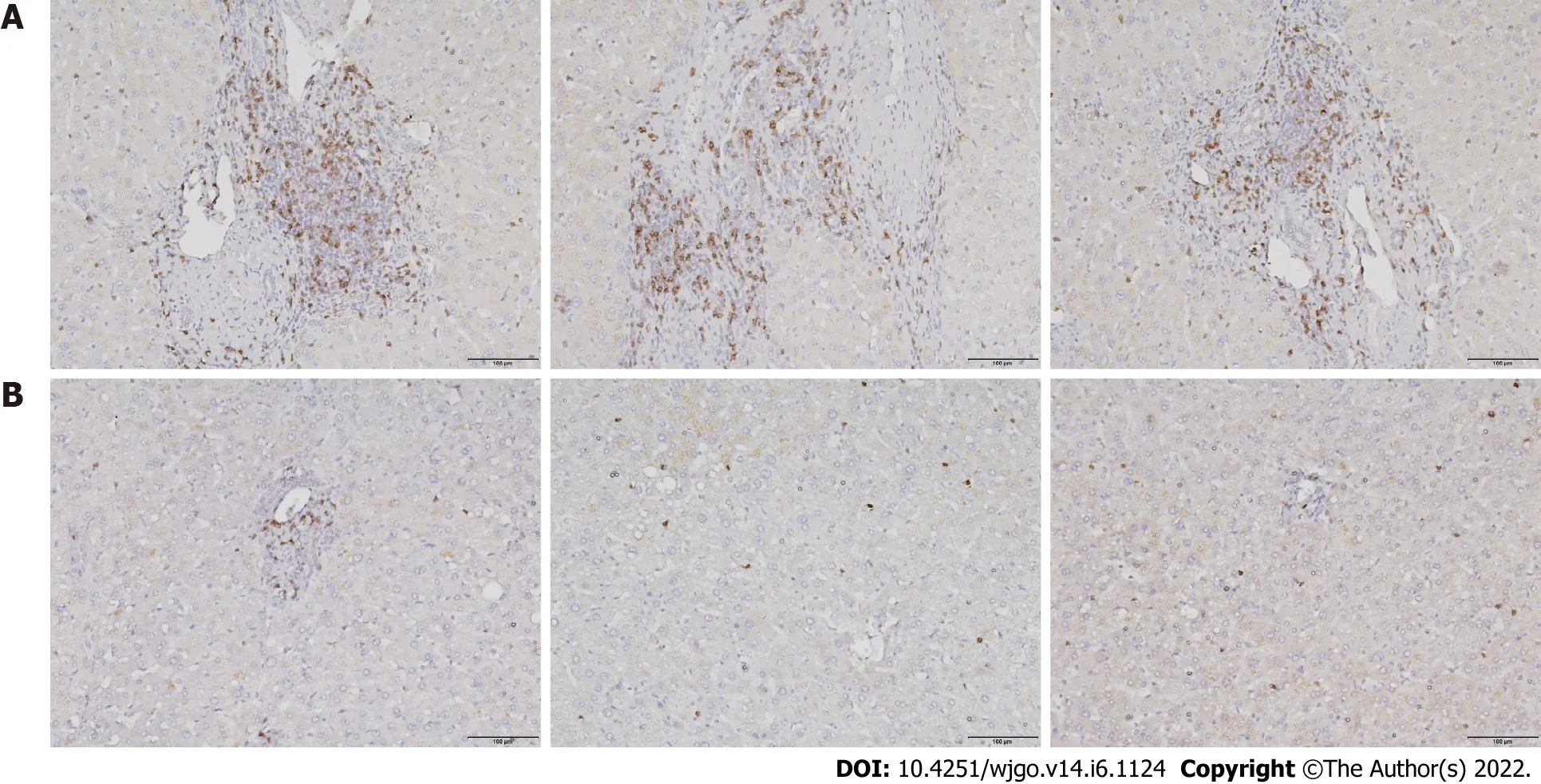
To investigate T cell infiltration in the tumor microenvironment,we performed immunohistochemical staining of resected liver tissues.The results showed significantly more CD8
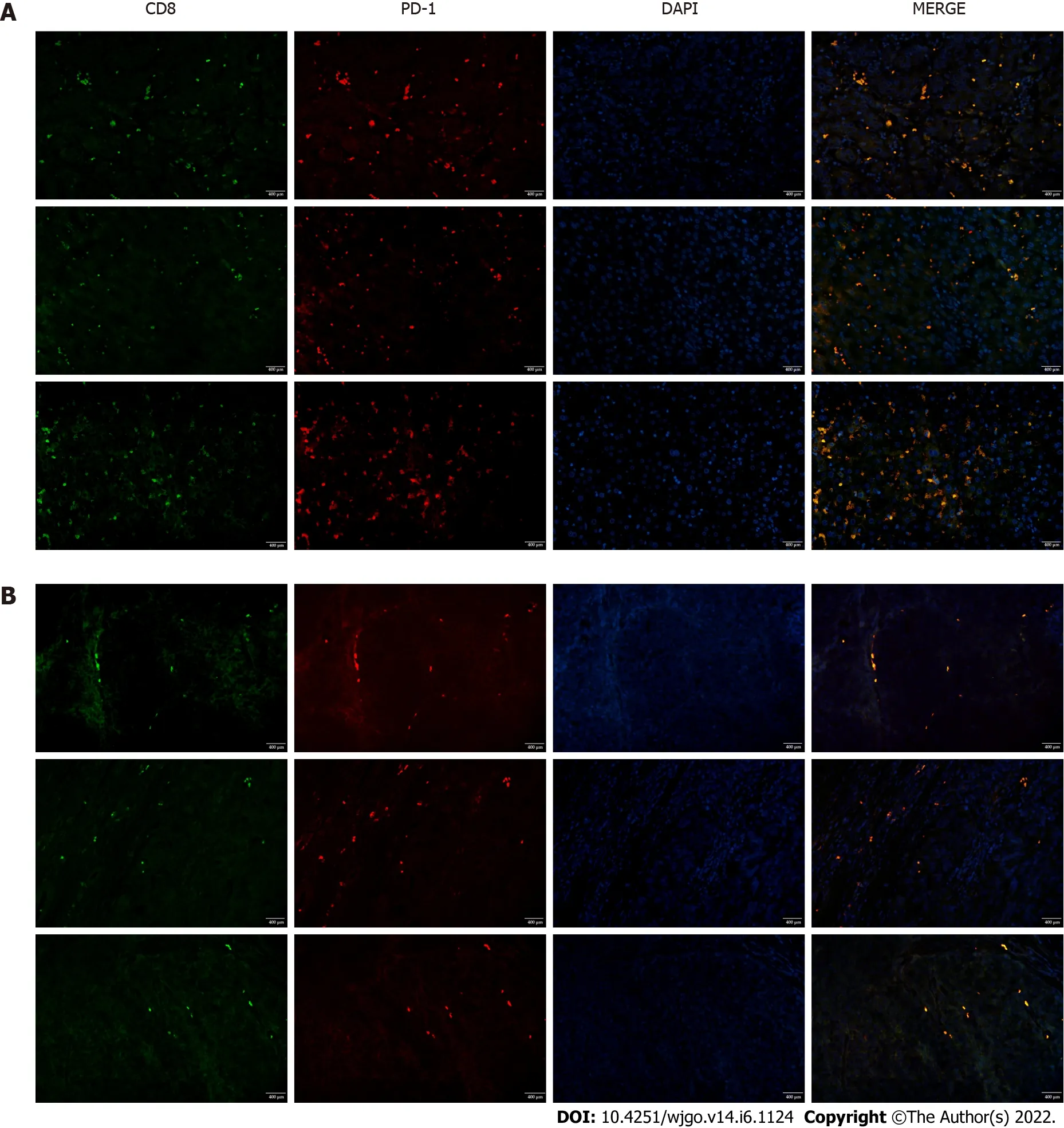
T cells in HCC tissues than in paracancerous tissues(Figure 1).Next,we used immunofluorescence staining to detect PD-1 expression on CD8
T cells.Compared with paracancerous tissues,PD-1 was more abundantly expressed on the surface of CD8
T cells in HCC tissues(Figure 2).
Research conclusions
CD8
T cells isolated from peripheral blood were cultured in groups and stained for intracellular markers.Analytical flow cytometry(NovoCyte,ACEA Biosciences Inc.,San Diego,CA,United States)and a flow analyzer(CytoFLEXS,Beckman Coulter)were used for the analysis.CD8
T cells were stained with an Annexin V-FITC Apoptosis Detection Kit(A211-02;Vazyme,Nanjing,China)to detect apoptosis.CD8
T cells were stained with JC-1 fluorescent dye(J8030;Solarbio,Beijing,China)and a ROS Detection Kit(CA1410;Solarbio)to detect mitochondrial damage and ROS levels,respectively.Cells were fixed and permeabilized with Fixation/Permeabilization solution(554714;BD Biosciences,Franklin Lakes,NJ,United States),and then CD8
T cells were stained with anti-GZMB(515403;Biolegend)and anti-PRF1(154305;Biolegend)antibodies to detect their activation status.Finally,CD8
T cells were stained with PE-conjugated anti-human CD279(PD-1)antibody(329905;Biolegend)to detect their functional changes.
36 “You are not afraid of the sea, my dumb child,” said he, as they stood on the deck of the noble ship which was to carry them to the country of the neighboring king
Research perspectives
From this study,we confirmed the potential mechanisms of CD8
T cell dysfunction induced by glutamine deprivation,which would provide a novel strategy for reversing the exhaustion of CD8
T cells in hepatocellular carcinoma.
FOOTNOTES
Wang W participated in the writing and editing of the manuscript;Guo MN and Li N participated in the data analysis;Pang DQ participated in the clinical specimen collection;Wu JH provided the experimental idea;all the authors approved for the final version of the manuscript.
High-End Talent Funding Project in Hebei Province,No.A202003005;Hebei Provincial Health Commission Office,No.G2019074;and Natural Science Foundation of Hebei Province,No.H2019209355.
The study was approved by the Medical Ethics Committee of North China University of Science and Technology(No.2018109).
All the authors report no relevant conflicts of interest for this article.
27. Barred the door: Before the common use of door knobs and intricate locks, doors were often secured by placing large pieces of wood or metal, usually in the shape of a bar, across the door. These bars were often heavy and difficult for a small child to lift, especially with the stealth needed in this situation.Return to place in story.
No additional data are available.
This article is an open-access article that was selected by an in-house editor and fully peer-reviewed by external reviewers.It is distributed in accordance with the Creative Commons Attribution NonCommercial(CC BYNC 4.0)license,which permits others to distribute,remix,adapt,build upon this work non-commercially,and license their derivative works on different terms,provided the original work is properly cited and the use is noncommercial.See: https://creativecommons.org/Licenses/by-nc/4.0/
As he could do nothing to escape his visit, the only thing that remained was to seem as little afraid as possible; so when the Beast appeared and asked roughly if he had supped well, the merchant answered humbly34 that he had, thanks to his host s kindness
China
Wei Wang 0000-0002-7863-8309;Meng-Nan Guo 0000-0003-0694-7919;Ning Li 0000-0002-4972-5841;De-Quan Pang 0000-0001-7992-3649;Jing-Hua Wu 0000-0002-2829-3496.
So he sat there full of grief and misery9, eating every day only a tiny bit of bread, and drinking only a mouthful of ovine, and he watched death creeping nearer and nearer to him
Gao CC
Wang TQ
Yuan YY
1 Xie Y,Xie F,Zhang L,Zhou X,Huang J,Wang F,Jin J,Zeng L,Zhou F.Targeted Anti-Tumor Immunotherapy Using Tumor Infiltrating Cells.
2021;8: e2101672[PMID: 34658167 DOI: 10.1002/advs.202101672]
2 Wei R,Liu S,Zhang S,Min L,Zhu S.Cellular and Extracellular Components in Tumor Microenvironment and Their Application in Early Diagnosis of Cancers.
2020;2020: 6283796[PMID: 32377504 DOI: 10.1155/2020/6283796]
3 Natua S,Dhamdhere SG,Mutnuru SA,Shukla S.Interplay within tumor microenvironment orchestrates neoplastic RNA metabolism and transcriptome diversity.
2022;13: e1676[PMID: 34109748 DOI: 10.1002/wrna.1676]
4 Ma J,Zheng B,Goswami S,Meng L,Zhang D,Cao C,Li T,Zhu F,Ma L,Zhang Z,Zhang S,Duan M,Chen Q,Gao Q,Zhang X.PD1
CD8
T cells correlate with exhausted signature and poor clinical outcome in hepatocellular carcinoma.
2019;7: 331[PMID: 31783783 DOI: 10.1186/s40425-019-0814-7]
5 Wang X,Lu XJ,Sun B.The pros and cons of dying tumour cells in adaptive immune responses.
2017;17: 591[PMID: 28757605 DOI: 10.1038/nri.2017.87]
6 Doering TA,Crawford A,Angelosanto JM,Paley MA,Ziegler CG,Wherry EJ.Network analysis reveals centrally connected genes and pathways involved in CD8
T cell exhaustion
memory.
2012;37: 1130-1144[PMID: 23159438 DOI: 10.1016/j.immuni.2012.08.021]
7 Schietinger A,Greenberg PD.Tolerance and exhaustion: defining mechanisms of T cell dysfunction.
2014;35: 51-60[PMID: 24210163 DOI: 10.1016/j.it.2013.10.001]
8 Chang CH,Qiu J,O'Sullivan D,Buck MD,Noguchi T,Curtis JD,Chen Q,Gindin M,Gubin MM,van der Windt GJ,Tonc E,Schreiber RD,Pearce EJ,Pearce EL.Metabolic Competition in the Tumor Microenvironment Is a Driver of Cancer Progression.
2015;162: 1229-1241[PMID: 26321679 DOI: 10.1016/j.cell.2015.08.016]
9 Hu M,Chen X,Ma L,Ma Y,Li Y,Song H,Xu J,Zhou L,Li X,Jiang Y,Kong B,Huang P.AMPK Inhibition Suppresses the Malignant Phenotype of Pancreatic Cancer Cells in Part by Attenuating Aerobic Glycolysis.
2019;10: 1870-1878[PMID: 31205544 DOI: 10.7150/jca.28299]
10 Altman BJ,Stine ZE,Dang CV.From Krebs to clinic: glutamine metabolism to cancer therapy.
2016;16: 749[PMID: 28704361 DOI: 10.1038/nrc.2016.114]
11 Xiang L,Mou J,Shao B,Wei Y,Liang H,Takano N,Semenza GL,Xie G.Glutaminase 1 expression in colorectal cancer cells is induced by hypoxia and required for tumor growth,invasion,and metastatic colonization.
2019;10: 40[PMID: 30674873 DOI: 10.1038/s41419-018-1291-5]
12 Ahn CS,Metallo CM.Mitochondria as biosynthetic factories for cancer proliferation.
2015;3: 1[PMID: 25621173 DOI: 10.1186/s40170-015-0128-2]
13 Cruzat V,Macedo Rogero M,Noel Keane K,Curi R,Newsholme P.Glutamine: Metabolism and Immune Function,Supplementation and Clinical Translation.
2018;10[PMID: 30360490 DOI: 10.3390/nu10111564]
14 Gerriets VA,Kishton RJ,Nichols AG,Macintyre AN,Inoue M,Ilkayeva O,Winter PS,Liu X,Priyadharshini B,Slawinska ME,Haeberli L,Huck C,Turka LA,Wood KC,Hale LP,Smith PA,Schneider MA,MacIver NJ,Locasale JW,Newgard CB,Shinohara ML,Rathmell JC.Metabolic programming and PDHK1 control CD4+ T cell subsets and inflammation.
2015;125: 194-207[PMID: 25437876 DOI: 10.1172/JCI76012]
15 Klysz D,Tai X,Robert PA,Craveiro M,Cretenet G,Oburoglu L,Mongellaz C,Floess S,Fritz V,Matias MI,Yong C,Surh N,Marie JC,Huehn J,Zimmermann V,Kinet S,Dardalhon V,Taylor N.Glutamine-dependent α-ketoglutarate production regulates the balance between T helper 1 cell and regulatory T cell generation.
2015;8: ra97[PMID: 26420908 DOI: 10.1126/scisignal.aab2610]
16 Liu Y,Birsoy K.Asparagine,a Key Metabolite in Cellular Response to Mitochondrial Dysfunction.
2021;7: 479-481[PMID: 33896762 DOI: 10.1016/j.trecan.2021.04.001]
17 Yu YR,Imrichova H,Wang H,Chao T,Xiao Z,Gao M,Rincon-Restrepo M,Franco F,Genolet R,Cheng WC,Jandus C,Coukos G,Jiang YF,Locasale JW,Zippelius A,Liu PS,Tang L,Bock C,Vannini N,Ho PC.Disturbed mitochondrial dynamics in CD8
TILs reinforce T cell exhaustion.
2020;21: 1540-1551[PMID: 33020660 DOI: 10.1038/s41590-020-0793-3]
18 Chew V,Lai L,Pan L,Lim CJ,Li J,Ong R,Chua C,Leong JY,Lim KH,Toh HC,Lee SY,Chan CY,Goh BKP,Chung A,Chow PKH,Albani S.Delineation of an immunosuppressive gradient in hepatocellular carcinoma using highdimensional proteomic and transcriptomic analyses.
2017;114: E5900-E5909[PMID: 28674001 DOI: 10.1073/pnas.1706559114]
19 Zheng C,Zheng L,Yoo JK,Guo H,Zhang Y,Guo X,Kang B,Hu R,Huang JY,Zhang Q,Liu Z,Dong M,Hu X,Ouyang W,Peng J,Zhang Z.Landscape of Infiltrating T Cells in Liver Cancer Revealed by Single-Cell Sequencing.
2017;169: 1342-1356.e16[PMID: 28622514 DOI: 10.1016/j.cell.2017.05.035]
20 Zhou G,Sprengers D,Boor PPC,Doukas M,Schutz H,Mancham S,Pedroza-Gonzalez A,Polak WG,de Jonge J,Gaspersz M,Dong H,Thielemans K,Pan Q,IJzermans JNM,Bruno MJ,Kwekkeboom J.Antibodies Against Immune Checkpoint Molecules Restore Functions of Tumor-Infiltrating T Cells in Hepatocellular Carcinomas.
2017;153: 1107-1119.e10[PMID: 28648905 DOI: 10.1053/j.gastro.2017.06.017]
21 Khan O,Giles JR,McDonald S,Manne S,Ngiow SF,Patel KP,Werner MT,Huang AC,Alexander KA,Wu JE,Attanasio J,Yan P,George SM,Bengsch B,Staupe RP,Donahue G,Xu W,Amaravadi RK,Xu X,Karakousis GC,Mitchell TC,Schuchter LM,Kaye J,Berger SL,Wherry EJ.TOX transcriptionally and epigenetically programs CD8
T cell exhaustion.
2019;571: 211-218[PMID: 31207603 DOI: 10.1038/s41586-019-1325-x]
22 Li W,Cheng H,Li G,Zhang L.Mitochondrial Damage and the Road to Exhaustion.
2020;32: 905-907[PMID: 33264601 DOI: 10.1016/j.cmet.2020.11.004]
23 Deng H,Ma J,Liu Y,He P,Dong W.Combining α-Hederin with cisplatin increases the apoptosis of gastric cancer
and
mitochondrial related apoptosis pathway.
2019;120: 109477[PMID: 31562979 DOI: 10.1016/j.biopha.2019.109477]
24 Padman BS,Nguyen TN,Lazarou M.Autophagosome formation and cargo sequestration in the absence of LC3/GABARAPs.
2017;13: 772-774[PMID: 28165849 DOI: 10.1080/15548627.2017.1281492]
25 Tusskorn O,Khunluck T,Prawan A,Senggunprai L,Kukongviriyapan V.Mitochondrial division inhibitor-1 potentiates cisplatin-induced apoptosis
the mitochondrial death pathway in cholangiocarcinoma cells.
2019;111: 109-118[PMID: 30579250 DOI: 10.1016/j.biopha.2018.12.051]
26 Si L,Fu J,Liu W,Hayashi T,Mizuno K,Hattori S,Fujisaki H,Onodera S,Ikejima T.Silibinin-induced mitochondria fission leads to mitophagy,which attenuates silibinin-induced apoptosis in MCF-7 and MDA-MB-231 cells.
2020;685: 108284[PMID: 32014401 DOI: 10.1016/j.abb.2020.108284]
27 Chen YF,Liu H,Luo XJ,Zhao Z,Zou ZY,Li J,Lin XJ,Liang Y.The roles of reactive oxygen species(ROS)and autophagy in the survival and death of leukemia cells.
2017;112: 21-30[PMID: 28325262 DOI: 10.1016/j.critrevonc.2017.02.004]
28 Li X,Wenes M,Romero P,Huang SC,Fendt SM,Ho PC.Navigating metabolic pathways to enhance antitumour immunity and immunotherapy.
2019;16: 425-441[PMID: 30914826 DOI: 10.1038/s41571-019-0203-7]
29 Issaq SH,Mendoza A,Fox SD,Helman LJ.Glutamine synthetase is necessary for sarcoma adaptation to glutamine deprivation and tumor growth.
2019;8: 20[PMID: 30808861 DOI: 10.1038/s41389-019-0129-z]
30 Künzi L,Holt GE.Cigarette smoke activates the parthanatos pathway of cell death in human bronchial epithelial cells.
2019;5: 127[PMID: 31396404 DOI: 10.1038/s41420-019-0205-3]
31 Song SB,Hwang ES.High Levels of ROS Impair Lysosomal Acidity and Autophagy Flux in Glucose-Deprived Fibroblasts by Activating ATM and Erk Pathways.
2020;10[PMID: 32414146 DOI: 10.3390/biom10050761]
 World Journal of Gastrointestinal Oncology2022年6期
- World Journal of Gastrointestinal Oncology的其它文章
- Insight on BRAFV600E mutated colorectal cancer immune microenvironment
- Hepatocellular carcinoma and immunotherapy:Beyond immune checkpoint inhibitors
- Biofeedback therapy combined with Baduanjin on quality of life and gastrointestinal hormone level in patients with colorectal cancer
- Does chronic kidney disease affect the complications and prognosis of patients after primary colorectal cancer surgery?
- Characterizing the patient experience during neoadjuvant therapy for pancreatic ductal adenocarcinoma:A qualitative study
- Clinicopathological differences,risk factors and prognostic scores for western patients with intestinal and diffuse-type gastric cancer
