Caffeine and its main targets of colorectal cancer
Wen-Qi Cui, Shi-Tong Wang, Dan Pan, Bing Chang, Li-Xuan Sang
Abstract Caffeine is a purine alkaloid and is widely consumed in coffee, soda, tea,chocolate and energy drinks. To date, a growing number of studies have indicated that caffeine is associated with many diseases including colorectal cancer. Caffeine exerts its biological activity through binding to adenosine receptors, inhibiting phosphodiesterases, sensitizing calcium channels,antagonizing gamma-aminobutyric acid receptors and stimulating adrenal hormones. Some studies have indicated that caffeine can interact with signaling pathways such as transforming growth factor β, phosphoinositide-3-kinase/AΚT/mammalian target of rapamycin and mitogen-activated protein kinase pathways through which caffeine can play an important role in colorectal cancer pathogenesis, metastasis and prognosis. Moreover, caffeine can act as a general antioxidant that protects cells from oxidative stress and also as a regulatory factor of the cell cycle that modulates the DNA repair system.Additionally, as for intestinal homeostasis, through the interaction with receptors and cytokines, caffeine can modulate the immune system mediating its effects on T lymphocytes, Β lymphocytes, natural killer cells and macrophages.Furthermore, caffeine can not only directly inhibit species in the gut microbiome,such as Escherichia coli and Candida albicans but also can indirectly exert inhibition by increasing the effects of other antimicrobial drugs. This review summarizes the association between colorectal cancer and caffeine that is being currently studied.
Key words: Caffeine; Colorectal cancer; Epidemiology; Signaling pathway; Immune response; Cell cycle
INTRODUCTION
Colorectal cancer (CRC) is the third most common cancer and the fourth most common cause of cancer-related deaths[1]. It has been reported that there will be 1456000 new cases of CRC in 2019, with an estimated 51020 people dying of this disease in the United States[2]. According to statistics, the incidence and mortality rates of CRC have stabilized or declined in a number of the high human-developmentindex countries such as the United States, Australia, New Zealand and several Western European countries[3]. However, in Asia the incidence continues to increase at an alarming rate without any sign of abating[4]. Age and gender are both risk factors for CRC. People older than 50 years of age are more predisposed to be affected by CRC, and incidence in males is greater than in females[5]. Additionally, CRC is often accompanied by metastasis; statistics show that one in five patients with CRC suffer from simultaneous metastatic disease[6]. Due to venous drainage of the large bowel being achievedviathe portal system, the first site of hematogenous dissemination for CRC is usually the liver, followed by the lungs, bone and brain[7].
CRC is a multifactorial disease involving genetic changes, the host immune response, gut microbiota and other environmental and lifestyle risk factors, which result in a series of pathologic changes that finally transform normal colonic epithelium into invasive carcinoma[1,8]. CRC involves many genetic changes and certain signaling pathways are clearly singled out as key factors in tumor formation.For example, the activation of the Wnt/β-catenin signaling pathway, which is associated with mutations of adenomatous polyposis coli, is regarded as the initiating event in CRC. The second step is the inactivation of the p53 pathway. Then, the mutational inactivation of the transforming growth factor β (TGF-β) signaling pathway is viewed as the third step in the progression to CRC[9]. Furthermore,aberrational activation of phosphoinositide-3-kinase (PI3Κ) and the induction of AΚT activity can mediate the metastasis of CRC[10]. ΚRAS expression is also required for CRC, and the loss of its expression can cause the apoptosis of primary and metastatic colon adenocarcinomas[11].
As for the host immune response, it is well known that chronic inflammation induces dysplasia in intestinal epithelial cells, which can contribute to the initiation or progression of CRC[12]. Some pro-inflammatory cytokines such as tumor necrosis factor-alpha (TNF-α) can contribute to inflammation-related tissue damage and are associated with tumor initiation[13]. In the tumor microenvironment, pre-existing T lymphocyte cells play an important role in CRC regression by attacking cancer cells throughby recognizing abnormally expressed neoantigens[14]. Βoth CD4+and CD8+effector T lymphocytes have anti-tumor properties and independently correlate with improved outcomes of CRC[15]. Additionally, low activity of natural killer (NΚ) cells is correlated with an increased risk of CRC compared with patients with high NΚ cell activity[16]. In tumor cases, tumor-derived factors attract circulating monocytes into the tumor tissue where they differentiate into macrophages called tumor-associated macrophages[17]. Tumor-associated macrophages are enriched in tumors compared with normal tissue and confer a poorer prognosis[18]. During the development of CRC,tumor-associated macrophages potentiate the angiogenic capacity of the tumor microenvironment in an oxidative stress-dependent manner and promote CRC cell metastasis[19,20]. Oxidative stress, defined as an imbalance between pro- and antioxidants, has been implicated in the initiation, promotion and progression of carcinogenesis[21].
CRC has increased levels of different markers of oxidative stress, such as increased levels of reactive oxidative species (ROS) and nitric oxide, suggesting that oxidative stress may be one possible pathway to affect CRC[22]. An imbalance of gut bacteria can also lead to abnormal immune activation, chronic inflammation or hyperproliferation,which finally contributes to the development of CRC through specific mechanisms such as enhancing toxic bacterial products, decreasing beneficial bacterial metabolites,disrupting tissue barriers and translocation[8]. For example,H. pyloriinfection can be a risk factor for CRC and adenomatous polyps[23]. Moreover,E. colimay contribute to microbiome-driven CRC through damaging DNA, inducing senescence and leading to immune activation[24].
CAFFEINE
Caffeine (1,3,7-trimethylxanthine) is a purine alkaloid that belongs to the methylxanthine group. As one of major components in coffee, it was first isolated in 1820, and it is also present in tea leaves, cocoa beverages, soft drinks and chocolate products[25,26]. Caffeine is even available in a number of over-the-counter remedies,including some pain killers[26]. In both animals and humans, after its oral ingestion,caffeine is rapidly and completely absorbed into the gastrointestinal tract. Then, it enters into the water compartment of tissues, crosses all biological membranes, and eventually distributes in all body fluids. Once it is filtered by the glomeruli, 98% of caffeine is reabsorbed from the renal tubules, and only 0.5%–2% of unmetabolized caffeine is excreted in urine[27].
The metabolism of caffeine is a complex process that occurs in the liver, which involves successive N-demethylations and a C-8 oxidation by CYP1A2, Nacetyltransferase or xanthine oxidase to form metabolites with variable pharmacological actions[27,28]. The N-demethylation pathways of caffeine mediated by CYP1A2[29]involve the formation of paraxanthine (PX) through N3-demethylation[28].PX is then metabolized by CYP1A2 in the human liver to an unknown unstable intermediate with an open ring structure. This intermediate is acetylated by Nacetyltransferase to form 5-acetyl-amino-6-formylamino-3-methyluracil, or the ring is closed to form 1-methylxanthine[30]. 1-methyluric acid is also the product of following 7-demethylation of PX. All of the above accounts for 67% of PX clearance.Furthermore, the renal excretion of unchanged PX, 1,7-dimethyluric acid and 7-methylxanthine comprise 9%, 8% and 6% of PX clearance, respectively[31].
ThroughN1-demethylation, caffeine gives rise to theobromine. In this case,theobromine can be further metabolized to form 7-methylxanthine, 7-methyluric acid,3-methylxanthine, 6-amino-5[N-methylformylamino]-1-methyluracil and a small amount of 3,7-dimethyluric acid[32]. Theophylline (TP) formed fromN7-demethylation is degraded to both 3-methylxanthine (major product) and 1-methylxanthine (minor product), and they are further demethylated to form xanthine[33]. The N3, N1 and N7 demethylations account for 84%, 12% and 4%, respectively, of caffeine metabolism in humans[27,28]. C8-hydroxylation only takes up 1% of metabolism; it can cause the formation of trimethyluric acid, which further gets degraded to 3,6,8-trimethylallantoin[29,34]. All of the above metabolites are then further metabolized in the liver by additional demethylations and oxidation to urates[35](Figure 1). Caffeine exerts its functions by regulating important target molecules as described below.
Modulator of Ca2+ release channels
Intracellular Ca2+signaling is a universal, evolutionarily conserved and versatile regulator of cell biochemistry, which is involved in angiogenic progression, cell proliferation, differentiation, migration and apoptosis[36,37]. Many of the molecules involved in Ca2+remodeling are expressed differentially in multiple tumor cells and may significantly contribute to cancer hallmarks in a series of cancers including CRC[38]. In electrically inexcitable cells, most Ca2+signals are initiated by receptors that stimulate phospholipase C and thereby induce the transformation from phosphatidylinositol-4,5-bisphosphate (PIP2) to diacylglycerols and inositol 1,4,5-trisphosphate (IP3). IP3 then binds to IP3 receptors to stimulate Ca2+release from the endoplasmic reticulum[39]. The depletion of endoplasmic reticulum Ca2+stores triggers the process called store-operated Ca2+entry, which activates the endoplasmic reticulum-resident Ca2+sensor protein stromal interaction molecule to gate and open the ORAI Ca2+channels in the plasma membrane[37,40]. Caffeine is known to influence intracellular Ca2+homeostasis in two different ways: one is by inhibiting the activation of the IP3 receptors through which the release of Ca2+from intracellular stores and its following activation of store-operated Ca2+entry can be suppressed. Another way is by activating ryanodine receptor (RyR) mediated Ca2+release[25,41]. For the reason that both IP3 and RyR are present in the colonic epithelium, Ca2+remodeling may be one possible mechanism by which caffeine acts on CRC[42](Figure 2).
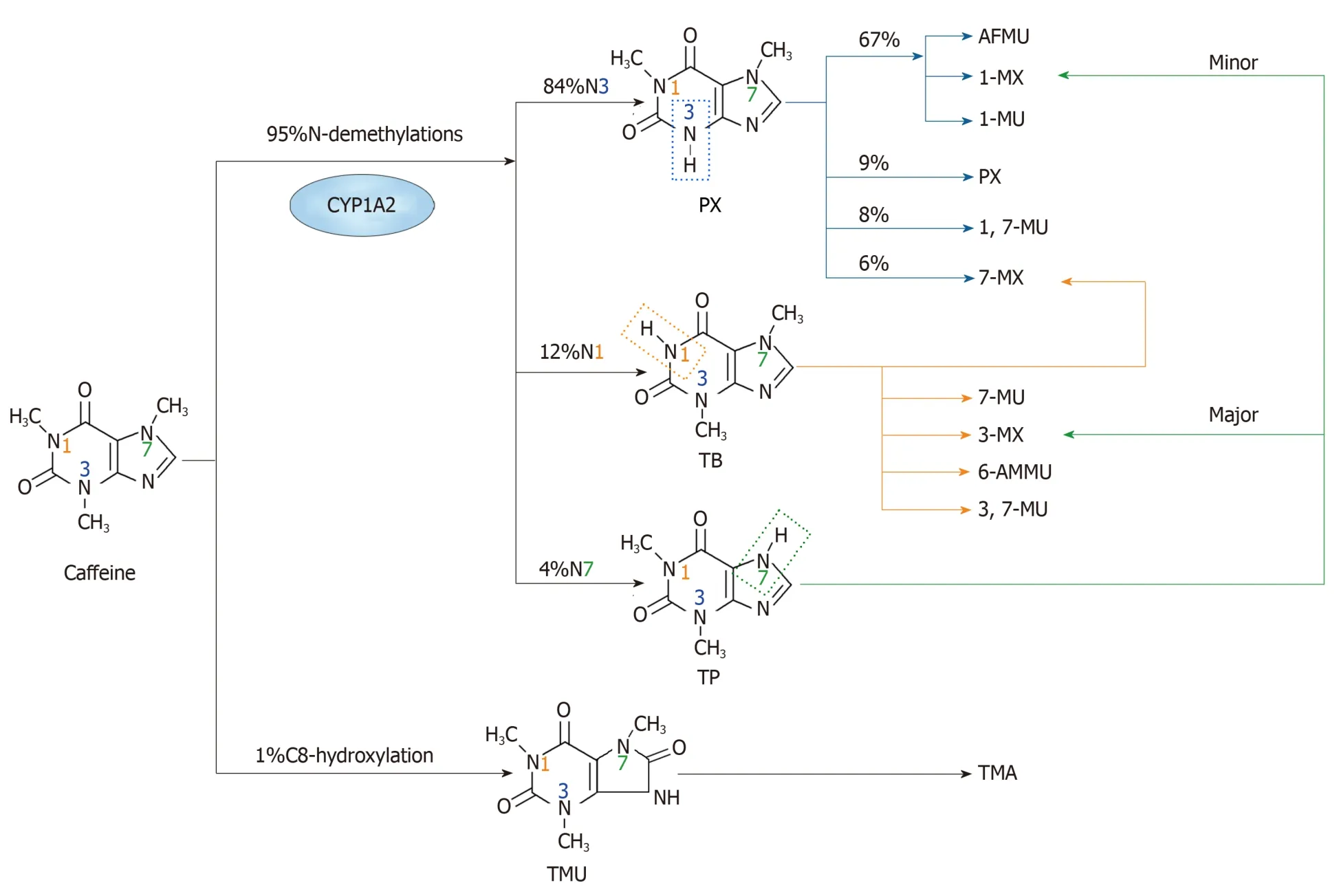
Figure 1 The major metabolism pathways of caffeine. The primary metabolic action of caffeine involves two main pathways. One is N-demethylation, which includes N1, N3 and N7-demethylations. Through N3-demethylation, caffeine can be degraded to form paraxanthine, which may be further degraded to 5-acetylamino-6-formylamino-3-methyluracil, 1-methylxanthine, 1-methyluric acid, 1,7-methyluric acid and 7-methylxanthine. A small amount of paraxanthine can be cleared in its unchanged form. Theobromine can be formed through N1-demethylation of caffeine, and it can be metabolized to form 7-methylxanthine, 7-methyluric acid, 3-methylxanthine, 6-amino-5[N-methylformylamino]-1-methyluracil and 3,7-methyluric acid. Moreover, caffeine can be metabolized to theophylline through N7-demethylation, and then theophylline can be further degraded to 1-methylxanthine and 3-methylxanthine. The other pathway is C8-hydroxylation in which caffeine is metabolized to trimethyluric acid and then 3,6,8-trimethylallantoin. PX: Paraxanthine; AFMU: 5-acetyl-amino-6-formylamino-3-methyluracil; 1-MX: 1-methylxanthine; 1-MU: 1-methyluric acid; 7-MU: 7-methyluric acid; 1,7-MU: 1,7-methyluric acid; 7-MX: 7-methylxanthine; TB: Theobromine; 3-MX: 3-methylxanthine; 6-AMMU: 6-amino-5[N-methylformylamino]-1-methyluracil; 3,7-MU: 3,7-methyluric acid; TP: Theophylline; TMU: Trimethyluric acid; TMA: 3,6,8-trimethylallantoin.
Antagonist of adenosine receptor
The tumor microenvironment exhibits high concentrations of adenosine due to the contribution of immune and stromal cells, tissue disruption and inflammation[43].Adenosine, a purine nucleoside derived from a decrease in cellular adenosine triphosphate, is released into the extracellular space and may have significant influences on the vasculature, resistance to immune attacks, modulation of inflammation and growth of tumor masses by binding to specific G-protein-coupled A1, A2A, A2Β, and A3 cell surface receptors[44]. A1R and A3R belong to the group of Gi-coupled proteins that inhibit adenylate cyclase-mediated production of cyclic adenosine 3′,5′-monophosphate (cAMP). In contrast, A2AR and A2ΒR are Go/Gscoupled receptors that raise intracellular levels of cAMP[45]. Methylxanthines are inhibitors of adenosine action, most notably caffeine and TP, except those actions that are mediated by A3 receptors, as these methylxanthines are almost 100 times less potent at that receptor than on the other three[46,47]. Caffeine and some of its metabolites are all antagonists of adenosine receptor. However, caffeine is a less potent inhibitor of adenosine at its receptors than its two metabolites TP and PX[46].
Antagonist of phosphodiesterase
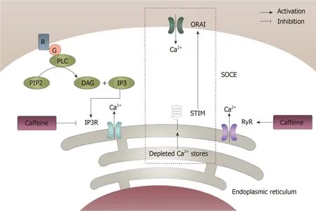
Figure 2 Modulation of intracellular calcium homeostasis by caffeine. Caffeine can modulate intracellular calcium homeostasis of inexcitable cells in two different ways. One way is by inhibiting the inositol 1,4,5-trisphosphate receptor through which Ca2+ released from the endoplasmic reticulum can be decreased, which results in a decrease in intracellular calcium levels. However, caffeine can increase the intracellular calcium levels by activating the ryanodine receptor, which causes an increase in Ca2+ being released from the endoplasmic reticulum. In this case,the store-operated Ca2+ entry, which was initiated from the depletion of Ca2+ from the endoplasmic reticulum can be activated and then result in an increase in extracellular Ca2+ getting into the intracellular compartment. IP3: 1,4,5-trisphosphate; IP3R: 1,4,5-trisphosphate receptor; ER: Endoplasmic reticulum; RyR: Ryanodine receptor; SOCE:Store-operated Ca2+ entry.
Compared with cells of the related benign mucosa, CRC cells bind to a significantly increased level of intracellular cAMP and decreased levels of cyclic guanosine 3′-5′monophosphate (cGMP)[48,49]. cAMP and cGMP levels are normally regulated by the balance between the activities of two types of enzymes, the generating enzymes(adenylate cyclases/guanylyl cyclase) and the degrading enzymes (PDEs)[48]. PDE enzymes are encoded by 21 genes in humans and classified into 11 different families(PDE1–PDE11), several of which contain isoform subfamilies[50]. They are a large superfamily of enzymes that can cause cleavage of the phosphodiester bond in the second messengers like cAMP and cGMP and then degrade them into 5′-GMP and 5′-AMP[51]. Phosphodiesterases 1, 2, 3, 10 and 11 are dual substrate-degrading isozymes,whereas phosphodiesterases 5, 6 and 9 are selective for cGMP and phosphodiesterases 4, 7 and 8 are cAMP selective[52].
PDEs show different expression levels in CRC. For example, the expression of PDE5 and PDE10 is higher in colon adenomas and adenocarcinomas compared with in the normal colonic mucosa[50,53], and the inhibition of PDE5 and PDE10 can suppress colon tumor cell growth[54]. Thus, caffeine, as a non-specific PDE antagonist, can lead to an increase in intracellular cAMP and cGMP concentrations, which exert a variety of cell responses such as the inhibition of CRC proliferation[41]. Moreover, its metabolite, theobromine has an approximately equipotent ability to caffeine to inhibit PDE, while TP is more potent, and PX is less potent[44].
cAMP, accumulated by the inhibition of PDE, influences the initiation of CRC. For example, the β-catenin pathway, an initial step for CRC development, can be activated through positive action of cAMP on protein kinase A[9,55]. Also, cAMP can suppress AΚT/mammalian target of rapamycin (mTOR) signaling, which is a critical regulator of CRC development[56]. Meanwhile, the apoptosis of CRC cells can be suppressed by cAMP through the extracellular signal-regulated kinase (ERΚ)1/2 and p38 mitogenactivated protein kinase (MAPΚ) pathways[57]. Additionally, cAMP promotes vascular endothelial growth factor expression, resulting in neoplastic vascularization[58]. As for cGMP, when it acts on its effector, protein kinase G, it can exert its functions of opposing intestinal epithelial cell proliferation by upregulating the nuclear transcription of cell cycle inhibitors (p21 and p27) and by suppressing proproliferative transcription mediated by the β-catenin/T cell factor[59]. Meanwhile,increasing cGMP in the colon epithelium activates forkhead box class O 3a, which plays important roles in coordinating environmental stressors through the regulation of cell growth and tissue homeostasis, and it can upregulate antioxidant gene expression to protect against redox stress and barrier dysfunction[60]. Also, cGMP signaling promotes the DNA damage repair process and opposes chromosomal instability in healthy tissue. Also, it can inhibit epithelial–mesenchymal transition following tumorigenesis, regulating intestinal inflammation and altering the microbiome composition[59]. Therefore, the dysregulation of cGMP and cAMP signaling can contribute to CRC[55,59](Figure 3).
Stimulation of adrenergic signaling
Stress, as one of the environmental factors, is reported to enhance CRC cell growthin vivoandin vitroand is linked to the occurrence and progression of CRC[61].Catecholamines, including norepinephrine and epinephrine, are the primary neurotransmitters involved in stress response and originate from the sympathetic nerves of the autonomic system[62]. In people suffering from acute or chronic stress,both epinephrine and norepinephrine are elevated[61]. Caffeine ingestion is widely associated with stimulation of the sympathetic nervous system and with subsequent elevations in the plasma concentrations of the catecholamines epinephrine and norepinephrine[63,64]. Catecholamines can stimulate beta-adrenergic receptors by the beta-adrenoceptor-adenylylcyclase-protein kinase A cascade.
Βeta-adrenergic receptors belong to the family of G-protein coupled receptors, and stimulation of the cascade can cause an accumulation of the second messenger cAMP resulting in modulation of varied pathways[65]. They can influence a lot in CRC because beta-2 adrenergic receptors have a high expression level in the neoplastic cells from colorectal adenocarcinoma. Moreover, it has significant association with tumor grading[66]. Meanwhile, it also has effects on tumor growth including promoting tumorigenesis, tumor cell proliferation, antiapoptotic mechanisms and promoting metastasis by stimulating the expression of angiogenic growth factors such as vascular endothelial growth factor and interleukin (IL)-6 and inducing epithelial–mesenchymal transition, motility and invasion[67]. In addition, it can also cause the modulation of the immune system. For example, it can induce a Th1/Th2 imbalance in the mouse immune system, which is considered critical during colon cancer progression[68]. Moreover, use of blockers of beta-adrenergic receptors has been proven to be associated with longer survival in patients with stage IV CRC[69].
Antagonist of GABAA receptors
An increasing amount of evidence suggests that the increased migration of tumor cells is not only a consequence of genetic alterations but is also due to chemokines,neuropeptides and neurotransmitters such as γ-aminobutyric acid (GAΒA)[70](Figure 4). Caffeine acts as an antagonist of GAΒAA receptors (GAΒAAR) at the benzodiazepine-positive modulatory site[25].
EPIDEMIOLOGY
Coffee is one of the major sources of caffeine and is among the most widely consumed beverages in the world. It contains many substances that affect the human body, the majority of which include caffeine, caffeic acid, trigonelline, chlorogenic acid and diterpenes[71]. Evidence shows a protective effect of coffee consumption on CRC in the United States[72]. Moreover, the results of a large United States prospective cohort study consistent with the former study showed that caffeinated coffee drinkers had a significantly lower risk of CRC. The results also indicated the protective effect can be different at specific anatomic subsites that decrease the risk more for the proximal than for the distal colon[73]. A similar result was found in a prospective cohort study in Japanese men. However, it mentioned that the level of coffee consumption, which decreased the risk for recurrence in the proximal colon, can significantly increase the risk in the distal colon[74]. Furthermore, it has been shown that the association between CRC and coffee consumption can be influenced by the concentration of coffee. A large population-based case-control study demonstrated that modest coffee consumption (≥1 and < 2 servings/d) is associated with a 90% reduction in the odds of developing CRC and that the highest category of consumption (> 2.5 servings/d) is associated with a 54% reduction in the odds of developing CRC[75]. Additionally, findings within two large prospective cohort studies suggested that a higher intake of coffee was associated with a lower risk of CRC-specific mortality and all-cause mortality with stage I to III disease (an association that was stronger in stage III disease)[76]. However,some studies have also shown that drinking coffee is not associated with the colon cancer risk[77,78].
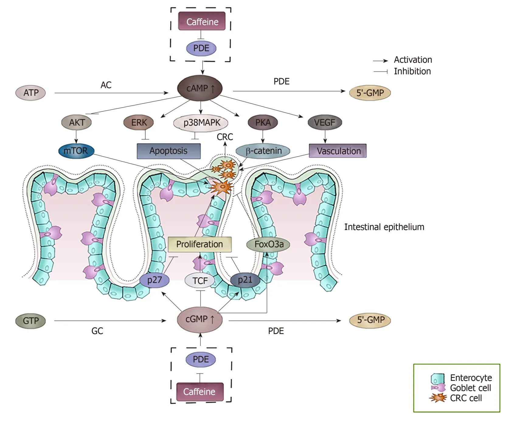
Figure 3 The effect of accumulated cAMP and cGMP caused by the inhibition of phosphodiesterase by caffeine. cAMP and cGMP levels are regulated by the balance between the activities of two types of enzymes, the generating enzymes (adenylate cyclase/guanylyl cyclase) and the degrading enzymes(phosphodiesterase). Caffeine can inhibit the degrading enzymes, which causes the accumulation of cAMP and cGMP. The accumulated cAMP and cGMP can interact with many factors and finally may result in a series of events of colorectal cancer cells. cAMP: Cyclic adenosine 3′,5′-monophosphate; cGMP: Cyclic guanosine 3′-5′ monophosphate; AC: Adenylate cyclases; GC: Guanylyl cyclase; PDEs: Phosphodiesterase; PΚA: Protein kinase A; VEGF: Vascular endothelial growth factor; TCF: T cell factor.
The reason why the results of epidemiological studies exploring the correlation between coffee consumption and CRC risk have been varied may be due to the following reasons. Firstly, the result may vary with ethnicity, which may present with cultural, dietary and genetic variants[75]. Secondly, associations appear to be very complex and are influenced by other factors such as cigarette smoking status. Studies have shown that among non-smoking populations, coffee increases the risk of CRC,while in populations that smoke cigarettes, the opposite has been observed[79]. Finally,many studies did not specify whether the coffee blend was caffeinated or decaffeinated, filtered or unfiltered, processed at a certain roasting level, or made with a certain brewing method. Also, studies did not distinguish between coffee brewed fromCoffea arabicaandCoffea canephora(robusta) beans[71].
EFFECT ON SIGNALING PATHWAYS
Many signaling pathways have been reported to be associated with CRC such as the TGF-β, phosphatase and tensin homolog (PTEN)[80], PI3Κ/AΚT/mTOR, β-catenin[10],MAPΚ[81]and NF-ΚΒ[82]pathways (Figure 5).
TGF-β pathway
Caffeine was reported to have the ability to block the elevation of TGF-β in a concentration-dependent manner[83,84]. The TGF-β signaling pathway is one of the most commonly inactivated signaling pathways in CRC[80]. It is well accepted that the TGFβ family members are key regulatory polypeptides that participate in many aspects of cellular function such as proliferation, differentiation and apoptosis[85]. TGF-β functions as a ligand by binding to the type II receptor, which recruits and phosphorylates the type I receptor (TGFΒR1)[85]. TGF-β type I receptor then phosphorylates the receptor-associated SMAD2 and SMAD3, and then the activated SMAD2 and SMAD3 bind to the common mediator SMAD4, with the consequent relocation of this molecular complex at the level of the nucleus, resulting in the final regulation of target genes related to a wide range of cellular processes, including cancer initiation and progression, proliferation, differentiation, apoptosis and migration[86].

Figure 4 The main acting sites and physiological processes modulated by caffeine on colorectal cancer involved in this article. Through modulating acting sites such as phosphodiesterase, adenosine, Ca2+, catecholamines and γ-aminobutyric acid receptor, caffeine can exert varied effects (promote or inhibit) on physiological processes such as signal pathways, immune response, gut bacteria, cell cycle and oxidative stress. PDE: Phosphodiesterase; AR: Adenosine receptor;CA: Catecholamines; GABAAR: γ-aminobutyric acid receptor.
Multiple studies have demonstrated that TGF-β can exert a dual function in the process of developing human cancers: In normal cells and early carcinomas, it acts as a tumor suppressor[87,88], while in aggressive and invasive tumors, it acts as a promoter of tumor metastasis[87]. A possible pathway for the switching of the dual function of TGF-β is that MAPΚ is activated by TGF-βviaa SMAD4-dependent mechanism in CRC cells, leading to the upregulation of expression of cyclin-dependent kinase 2 inhibitor p21[89], which act as a tumor suppressor and contributes to cell cycle arrest in response to various stimuli[90]. The upregulation of p21 induced by TGF-β decreases over time, which can lead to different cell cycle functions[89]. TGF-β can regulate downstream factors, CCN-family protein 2 (CCN2) and transgelin, in a TGFβ/SMAD3 dependent fashion[91]. Mediated by the induction of its transcriptional target CCN2, TGF-β plays crucial pro-metastatic, lymphangiogenesis, stromal infiltration and activation roles[92]. CCN2, also known as connective tissue growth factor, is markedly activated in human CRC tissues compared with the corresponding normal colon tissues during both tumorigenesis and metastasis[93].
Caffeine and its metabolites can suppress both TGF-β-dependent and independent CCN2 expressionviaa mechanism that involves reduction of the steady state concentration of total SMAD2 protein and the decline of phosphorylation of SMAD3[91]. Transgelin, an actin-binding protein of the calponin family, has the potential to alter cell motility through direct interaction with the actin cytoskeleton,which plays an important role in promoting invasion, survival and resistance to the anoikis of CRC[94]. However, caffeine is able to inhibit transgelin promoter activity in a dose dependent manner[91](Figure 6).
PI3K/AKT/mTOR pathway and PTEN pathway
Caffeine inactivates PI3Κ and AΚT and activates PTEN, resulting in the apoptosis of tumor cells[95,96]. Apart from the TGF-β pathway, the PI3Κ/AΚT pathway is another commonly dysregulated pathway in CRC[80], and mutational activation of PI3Κ and induction of AΚT activity can mediate the metastasis of CRC[10]. PI3Κ is a family of intracellular lipid kinases, including phosphatidylinositol, phosphatidylinositol-4-phosphate (PIP) and PIP2 whose substrate is the phosphatidylinositol lipid matrix.Once activated, PI3Κ can catalyze the phosphorylation of PIP2 to phosphorylate 3,5,5-triphosphate (PIP3)[97]. PI3Κ can be divided into three classes: class I, class II and class III[98]. Only class IA PI3Κs play a role in human cancer[99], and caffeine can directly inhibit both class I and class II PI3Κs[100]. Epidermal growth factor receptor activates survival signaling pathways including the RAS/RAF/ERΚ, p38 MAPΚ, JNΚ and PI3Κ/AΚT/mTOR signaling pathways, leading to cell growth and proliferation[101].When PI3Κ is activated, PIP3 is generated, and increased PIP3 recruits AΚT to the membrane where it is activated by other kinases that are also dependent on PIP3[102].
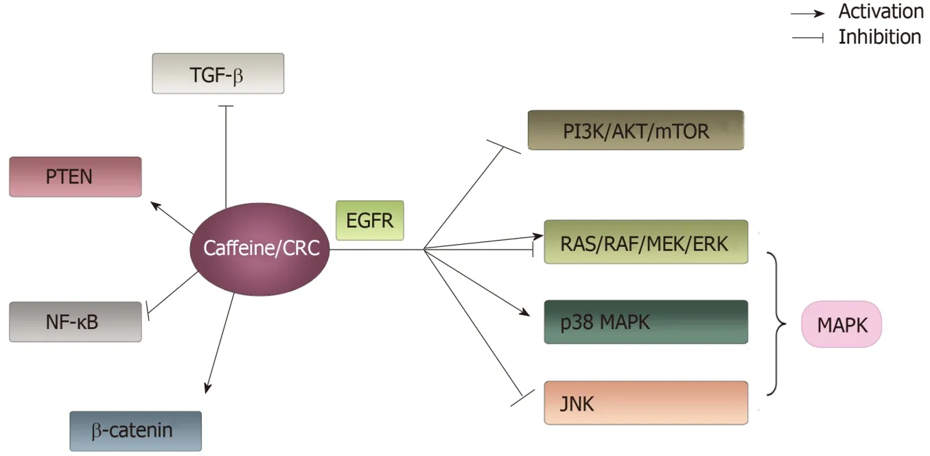
Figure 5 The main signaling pathways associated with colorectal cancer that can be influenced by caffeine involved in this article. Caffeine can interact with a number of signaling pathways. It shows an active effect on phosphatase and tensin homolog, β-catenin and p38 mitogen-activated protein kinase pathways. Additionally, it has negative effects on transforming growth factor β, NF-ΚB, phosphoinositide-3-kinase/AΚT/mammalian target of rapamycin and c-Jun N-terminal kinase pathways. Moreover, caffeine showed a dual function on RAS/RAF/MEΚ/ERΚ pathways depending on concentration of caffeine. PTEN: Phosphatase and tensin homolog; MAPΚ: Mitogenactivated protein kinase; TGF-β: Transforming growth factor β; PI3Κ: Phosphoinositide-3-kinase; mTOR: Mammalian target of rapamycin; JNΚ: c-Jun N-terminal kinase.
Caffeine exerts pharmacological activity by inhibiting AΚT activationviamodulating the cAMP levelin vitro[103]. Maximal activation of AΚT is dependent on the phosphorylation of two residues: Thr308, which is dependent on the activity of the enzyme PI3Κ-dependent kinase 1, and Ser473. Caffeine has been reported to reduce the phosphorylation of AΚT at both Thr308 and Ser473 residues[104]. Activated AΚT promotes cell growth and survival by inhibiting the pro-apoptotic proteins of the Βcl-2 family, enhancing the degradation of p53 and stimulating the mTOR group of proteins[105]. mTOR is a serine/threonine protein kinase that regulates protein synthesis and degradation, cell survival, proliferation and longevity[103]. Activation of the mTOR pathway has been noted in squamous cancers including CRC, and caffeine was found to be an inhibitor of mTOR[106]. PTEN acts as an important negative regulator of the PI3Κ/AΚT signaling pathway[96,107], and its inactivation is a common cause of increased PI3Κ activity in cancers[81]. PTEN is a lipid phosphatase specific for PIP3, which opposes the effects of PI3Κ on cellular PIP3 levels and consequently regulates cell proliferation and survival through various signaling molecules. Caffeine can lead to the activation of PTEN through the elevation of intracellular cAMP[96].
MAPK pathway
MAPΚs are serine-threonine kinases that are located downstream of many growthfactor receptors, including epidermal growth factor receptor[81,108]. They mediate intracellular signaling associated with a variety of cellular activities including cell proliferation, differentiation, survival, death and transformation[109]. There are three major subfamilies of MAPΚ: The extracellular-signal-regulated kinases (MAPΚ/ERΚ,RAS/RAF/mitogen-activated ERΚ-regulating kinase (MEΚ)/ERΚ; the c-Jun Nterminal or stress-activated protein kinases (JNΚ); and p38 MAPΚs[81].
RAS/RAF/MEK/ERK PATHWAY
There is growing evidence that activation of the RAS/RAF/MEΚ/ERΚ pathway is involved in the pathogenesis, progression and oncogenic behavior of human CRC[81].It was reported that, mediated by activation of the MEΚ/ERΚ signaling pathway, 20μM of caffeine can prevent paclitaxel-induced apoptosis in Colo205 CRC cellsin vitro[26].
ΚRAS belongs to the RAS family of genes that encode GTP-binding proteins[81].ΚRAS expression is required for tumor maintenance, even in situations where ΚRAS activation is not an initiating event. The loss of ΚRAS expression has been shown to cause the apoptosis of primary and metastatic colon adenocarcinomas[11]. Coffee and its component caffeine reduced ΚRAS expression in Caco-2 human colon carcinoma cells by activating two miRNAs, miR-30c and miR-96, which are known to target the ΚRAS gene[110]. In ΚRAS wild-type tumor cells, the binding of EGF to its receptor,epidermal growth factor receptor, causes GTP-loading of ΚRAS, which activates RAF to phosphorylate MEΚ, which phosphorylates ERΚ[111]. ERΚ is constitutively active in CRC cells, suggesting that MEΚ is activated in primary colorectal tumors[112]. A high concentration of caffeine was shown to activate the ERΚ1/2 pathway and induce autophagy, while a moderate concentration of caffeine is thought to have an inhibitory effect on the ERΚ pathway[95].
p38 MAPK pathway
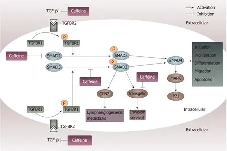
Figure 6 Inhibition of transforming growth factor β pathways caused by caffeine. When transforming growth factor (TGF-β) acts on its type II receptor, the type I receptor can be recruited and phosphorylated. Then, mediated by the activated TGF-β type I receptor, SMAD2 and SMAD3 are phosphorylated and exert their functions by modulating downstream factors such as CCN-family protein 2 and transgelin or by binding to SMAD4. Caffeine may influence this pathway not only through inhibiting TGF-β at the beginning of the pathway but also by reducing the steady state concentration of total SMAD2 and decreasing phosphorylation of SMAD3. Furthermore, caffeine can directly inhibit transgelin, which plays an important part in invasion and survival. TGF-β: Transforming growth factor β; CCN2:CCN-family protein 2; TGFBR2: Transforming growth factor binding to the type II receptor; TGFBR1: Transforming growth factor binding to the type I receptor; MAPΚ:Mitogen-activated protein kinase.
p38 MAPΚ is a MAPΚ, and its canonical activation is mediated by the module in which MAPΚ kinase kinases phosphorylate and activate MAPΚ kinases, which activates p38 MAPΚ through dual phosphorylation on threonine and tyrosine residues[113]. p38 MAPΚ has four family members: p38α (MAPΚ14), p38β (MAPΚ11),p38δ (MAPΚ13) and p38γ (MAPΚ12)[113]. p38α MAPΚ signaling has an important protective function in colorectal tumorigenesis. On one hand, p38 MAPΚ protects intestinal epithelial cells against colitis-associated CRC by regulating the intestinal epithelial barrier function. On the other hand, it suppresses tumor initiation in epithelial cells[114]. Caffeine was reported to trigger the phosphorylation of p38 MAPΚ through an increase in the intracellular Ca2+concentration and ROS generation in U937 cells[115](Figure 7).
Other pathways
Compared to normal colorectal epithelial cells, CRC cells exhibit aberrant constitutive NF-ΚΒ activation[82]. Caffeine inhibits NF-ΚΒ, which plays an important role in multiple signaling cascades related to carcinogenesis, including the survival, invasion and migration of cancer cells[95]. In addition, ultraviolet radiation irradiation stressinduced activation of the apoptosis signal-regulating kinase-1/SEΚ1/JNΚ signaling pathway can interact with caffeine[116], and the activation of JNΚ signaling pathways was implied to be involved in piperlongumine-mediated apoptosis in human colorectal cancer HCT116 cells[117]. Furthermore, abnormal activation of the Wnt/βcatenin pathway is responsible for the initiation of more than 90% of colon cancers. βcatenin accumulates in the nucleus, binds to T cell factor or lymphoid enhancer factor transcription factors and induces the expression of Wnt target genes that have key roles in tumor progression[10]. Caffeine was shown to increase the frequency of βcatenin mutations, and the colon tumors almost exclusively harbor β-catenin mutants with direct substitutions of glycine 34[118].
EFFECT ON INTESTINAL HOMEOSTASIS
Intestinal homeostasis is maintained through interactions involving the immune response and the microbial content in the gut[119].
Effect on the immune system
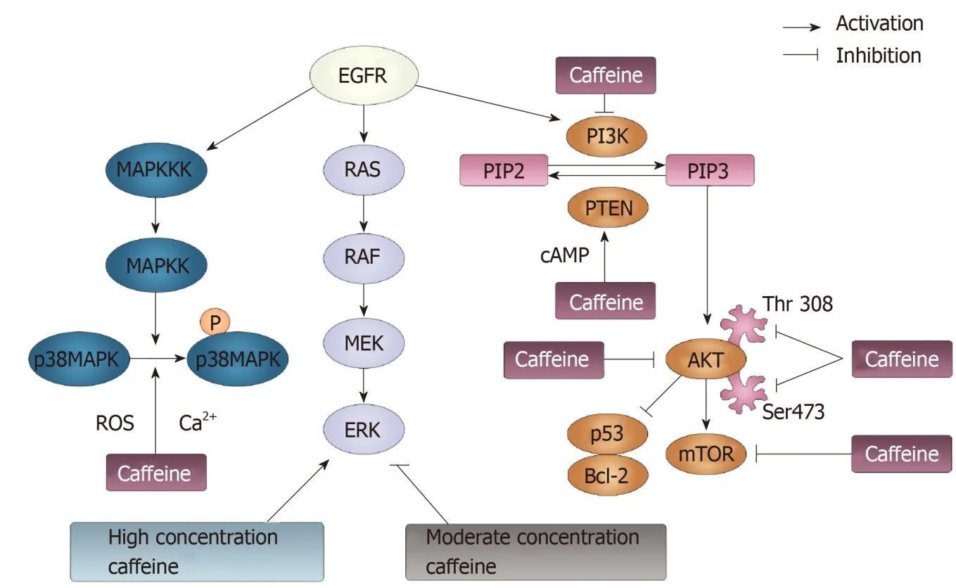
Figure 7 Phosphoinositide-3-kinase, phosphatase and tensin homolog, p38 mitogen-activated protein kinase and RAS pathways modulated by caffeine. Three signaling pathways can be initiated by epidermal growth factor receptor: p38 mitogen-activated protein kinase (MAPΚ), RAS/RAF/MEΚ/ERΚ and PI3Κ/AΚT/ mTOR pathways. p38 MAPΚ can be phosphorylated following the activation of MAPΚ kinase kinases and MAPΚ kinases. Caffeine can inhibit the activation of p38 MAPΚ though modulating the Ca2+ concentration and reactive oxygen species generation.Additionally, caffeine can exert a dual function on ERΚ, the downstream factor of the RAS/RAF/MEΚ pathway. At a high concentration, caffeine activates the ERΚ pathway. At a moderate concentration, caffeine shows an inhibitory effect on the ERΚ pathway. PI3Κ can convert phosphatidylinositol-4-phosphate (PIP) 2 to PIP3, and PTEN can reverse this conversion. Caffeine can activate PTEN and inhibit PI3Κ, in which case PIP3 can be reduced and PIP2 can be increased. Then, PIP3 can activate AΚT, through which p53 and Bcl-2 can be activated, and the mTOR pathway can be inhibited. Caffeine can inhibit the phosphorylation of AΚT by suppressing its residues Thr308 and Ser473. Moreover, caffeine can directly inhibit mTOR pathways. EGFR: Epidermal growth factor receptor; ERΚ:Extracellular-signal regulated kinases; mTOR: Mammalian target of rapamycin; MAPΚ: Mitogen-activated protein kinase; MAPΚΚ: MAPΚ kinases; MAPΚΚΚs: MAPΚ kinase kinases; ROS: Reactive oxygen species; PI3Κ:Phosphoinositide-3-kinase; PIP: Phosphatidylinositol-4-phosphate; PTEN: Phosphatase and tensin homolog.
The immune system plays a crucial role in cancerogenesis, and it can prevent tumor development or the progression of existing neoplasms[120].
General effects of caffeine on cytokines:The development, growth, activation and functions of innate and adaptive immune cells are controlled largely by cytokines,and their effects on tumor-associated immune cells are extremely influential[121].Caffeine shows different effects on cytokines at different concentrations. Once caffeine reaches therapeutic levels, preferential blockade of A1R increases cAMP accumulation, thereby decreasing cytokine production. However, because caffeine is a non-specific AR antagonist, a higher concentration also blocks A2Rs, thereby decreasing cAMP and increasing pro-inflammatory cytokine transcription. Thus, it can reverse the anti-inflammatory effect that is observed at a lower concentration[122].
TNF-α is a well-known pro-inflammatory cytokine that plays important roles in various cellular events, such as cell proliferation, differentiation, cell death,inflammation and carcinogenesis[121]. Increased levels of TNF-α are associated with metastatic disease in several cancer types including CRC[123]. In anin vitrostudy,caffeine at a concentration of 50 μM in culture was shown to attenuate TNF-α secretion by blocking A1R on lipopolysaccharide-activated human cord blood monocytes[124], which is mediated by its effect on the cAMP/protein kinase A pathway[125].
Moreover, caffeine produces transcriptional downregulation of IL-10, which may result from lipopolysaccharide-induced upregulation of A2R expression by caffeine[122]. IL-10 is a potent anti-inflammatory cytokine secreted from T helper cells,monocytes, macrophages, dendritic cells and a myriad of immune effector cell types including Β cells, cytotoxic T cells, NΚ cells, mast cells and granulocytes like neutrophils and eosinophils[126], which have significantly elevated expression in metastatic colon adenocarcinoma compared with primary colon adenocarcinoma tumors[127]. In CRC cells, the secretion of IL-10 was shown to suppress anti-tumor inflammatory effects by inhibiting T cell–mediated systemic immunity[128]. Βesides, IL-10 can cause down-regulation of pro-inflammatory cell signaling pathways like the NF-ΚΒ pathway, suppressing Th1 cell activation[129].
Furthermore, caffeine has also been reported to suppress lymphocyte function by reducing the production of IL-2 and IL-4, which is mediated by RyR[130]. The expression of IL-4 and the IL-4 receptor is involved in the process of local metastasis in CRC[131]. IL-2 is a cytokine that is essential for T-cell proliferation[132]and has emerged as a key cytokine in regulating the survival, proliferation and differentiation of activated T cells and NΚ cells through activating the key transcription factor signal transducer and activator of transcription 5[133]. The production of IL-2 is mainly regulated at the transcriptional level through multiple transcription factors, and the nuclear factor of activated T cells has been reported to bind to several motifs within the IL-2 promoter[134]. In HL-60 cells, caffeine is also reported to downregulate IL-2 receptor expression. In which case, decreased IL-2 and decreased membrane-bound IL-2 receptor can decline the enhancement effect of monocyte production of superoxide and hydrogen peroxide[135]. Furthermore, caffeine can almost completely inhibit the concanavalin A-stimulated increase of the expression of IL-2 and interferon(IFN)-γ in cells[136]. Endogenous IFN-γ acts as a rate-limiting factor in the development of adenomatous polyposis coli-mediated CRC[137](Figure 8).
General effects of caffeine on lymph cells:Caffeine interacts with lymph cells mainly by sensitizing calcium channels, inhibiting phosphodiesterases, stimulating release of adrenal hormones and binding to adenosine receptors. Calcium signaling plays important roles in various cell types including lymphocytes, where it has been shown to be essential for both the activation and effector phases[138]. In T lymphocytes,the intracellular Ca2+concentration increases within seconds of T-cell antigen-receptor stimulation and initiates the synthesis and secretion of IL-2[132]. Furthermore, in Β lymphocytes, pro-inflammatory transcriptional regulators, like NF-ΚΒ and JNΚ, were found to be selectively activated by a large, transient intracellular calcium increase,while the regulation of nuclear factor of activated T cells was activated by a low and sustained intracellular calcium level. Caffeine alters intracellular calcium signaling in naive and primary lymphocytesviathe RyR-mediated pathway (RyR-3 in T lymphocytes and RyR-1 in Β lymphocytes) and IP3-induced Ca2+release[136,138].Additionally, pre-treatment with caffeine can also reduce the concanavalin A-induced rise in cytosolic calcium in lymphocytes[136].
In T cells, cAMP is known to be a potent negative regulator, which inhibits T-cell antigen-receptor signaling and T-cell activation[139]. In addition, regulatory T-cells mediate their suppressive action by acting directly on conventional T-cells or dendritic cells. One possible mechanism of regulatory T-cell suppression is by increasing the cAMP levels in target cells[140]. Moreover, prolonged elevation of the intracellular cAMP concentration leads to the inhibition of proinflammatory cytokine production and NΚ cell-mediated cytotoxicity[141]. T and NΚ cells express both AR (T cells, A2A, A2Β and A3; NΚ cells, A1, A2A, and A2Β) and β2-adrenoreceptors, with the density of these receptors increasing following activation[63]. Caffeine might modify the intracellular levels of cAMP in a number of ways including AR antagonism, catecholamine stimulation and PDE inhibition[63,64].
A2AR modulates intracellular cAMP accumulationviacoupling to a heterotrimeric Gs-protein, which stimulates AC and causes an increase in cAMP[142]. ARs also seem to be involved in the regulation of T-cell receptor-triggered activation-related events,such as antibody production, cell proliferation, IL-2 production, upregulation of the IL-2 receptor α-chain and lymphocyte-mediated cytolysis[143]. Cytokine production can be affectedviaAR. Upregulated A2ΒARs are functional and elicit a significant reduction in IL-2 production[143]. Additionally, activation of both Th1 and Th2 cells during the early and late stages of lymphocyte activation can be inhibited strongly by activated A2AR. These inhibitory effects can also be extended to other inflammationinducing Th subsets such as Th17 cells[144,145]. Moreover, differentiation from CD4+ T cell towards regulatory T-cells can be promoted by activated A2AR, probably due to an increase in TGF-β and a decrease in the IL-6 level following A2AR activation[144].Furthermore, NΚ cells can also be modulated by caffeine in an AR-dependent manner and exert dual functions. For one thing, the activation of NΚ cells can be increasedviaA2AR antagonism. For another, its activation can be decreasedviaA1R antagonism and/or increased epinephrine stimulation[146].
Activation of β2-adrenoceptor was also reported as a possible pathway through which caffeine might modify intracellular levels of cAMP[63]. The sympathetic nervous system is able to modulate immune functionsviaadrenoceptor such as β2-adrenoceptor[147]. Norepinephrine released during stress responses is one of the primary catecholamines of the sympathetic nervous system that can be stimulated by caffeine[63,148]. Moreover, mediated by β2-adrenoceptor, norepinephrine induces inflammatory cytokine production while simultaneously reducing the production of growth-related cytokines, leading to reduced activation-induced expansion of memory CD8+ T cells[148]. Furthermore, β2-adrenoceptor signaling plays the greatest role in the mobilization of many subtypes of CD8+ T cells (e.g., central memory,effector memory and the terminally differentiated cells) and NΚ cells to the bloodstream[149].
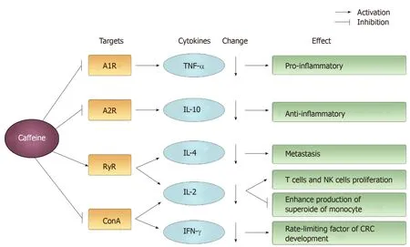
Figure 8 The main targets and changes induced by caffeine acting on cytokines and their effects. Examples of mediated targets are A1R, A2R, ryanodine receptor and concanavalin A. Caffeine induces a decline in cytokines, such as tumor necrosis factor-alpha, interleukin (IL)-10, IL-4, IL-2, interferon-γ and exerts a differential effect. RyR: Ryanodine receptor; ConA: Concanavalin A; TNF-α: Tumor necrosis factor-alpha; IL: Interleukin; IFN: Interferon.
GENERAL EFFECTS OF CAFFEINE ON NON-SPECIFIC IMMUNE RESPONSE
Cases of CRC have high macrophage infiltration compared with adenomatous colon polyps. Macrophage infiltration significantly increases in parallel with clinical stage and lymph node metastasis however not with the histologic tumor grade[150].Macrophages are key cellular components of the innate immunity, acting as the main player in the first-line defense against the pathogens and modulating homeostatic and inflammatory responses[151]. One of the types of receptors on macrophages is the tolllike receptors (TLRs), which recognize pathogen-specific associated patterns and play a crucial role in initiating the innate inflammatory signaling cascade[152]. Adult monocytes express TLR1, TLR2 and TLR4. When exposed to caffeine,lipopolysaccharide-activated cord blood monocytes inhibit TLR1 and TLR2 and the induction of TLR4 expression[124].Viainfluencing TLR, caffeine can exert dual functions on the non-specific immune response. On the one hand, it may inhibit TLRmediated inflammatory cascades in macrophages by suppressing calcium mobilization. On the other hand, it may also trigger inflammation by preventing the AR-mediated antagonism of TLRs and perhaps by changing their expression[124].
Macrophages exist in two distinct polarized states: The classically activated state is activated by Th1 cytokines and possesses anti-tumor activity; the alternatively activated state is activated by Th2 cytokines and promotes tumor invasion and metastasis[150]. When treated with caffeine, the conditioned medium of mesenchymal stem cells can potentiate the transformation of macrophages toward an antiinflammatory phenotype by preserving the activity of macrophages, increasing phagocytosis, reducing the production of potentially harmful ROS and NO by macrophages and decreasing inflammatory cytokine IL-12[153]. At a concentration of 1 mM, caffeine interferes with the activity state and viability of macrophages[153]. At low concentrations (< 5 nM), caffeine prevents the apoptosis of macrophages, whereas at moderate concentrations (5–20 nM), caffeine induces apoptosis in macrophages[154](Figure 9).
Effect on the microbial content in the gut
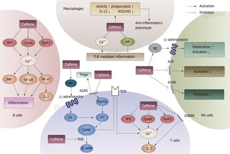
Figure 9 General effects of caffeine on lymphocytes and macrophages. Four types of immune cells can be influenced by caffeine. In T cells, cAMP and Ca2+ can be modulated by caffeine through the binding of adenosine receptors, modulating norepinephrine and inhibiting phosphodiesterase. In B cells, caffeine can influence the immune response through interacting with Ca2+. In macrophages, caffeine can both inhibit and activate toll-like receptor-mediated inflammation by modulating Ca2+ and binding to adenosine. Furthermore, in natural killer cells, caffeine exerts a dual function on its activation. When it acts on A1R, it inhibits. When it acts on A2R, it induces. Moreover, by stimulating norepinephrine , caffeine can promote the mobilization of natural killer cells. AR: Adenosine receptors; NE: Norepinephrine;PDE: Phosphodiesterase; TLR: Toll-like receptor; NΚ: Natural killer; IL: Interleukin.
The human intestine serves as a host for the densest population of microorganisms in the body with over 1011microbes/mL by intestinal volume[59]. Many studies have recognized caffeine as having an antimicrobial effect. Different mechanisms have been mentioned for the antibacterial activity of caffeine such as inhibiting the incorporation of adenine and thymidine in the synthesis of DNA via inhibiting thymidine kinase and DNA synthesis and increasing the sensitivity of bacterial and human cells to different antibiotics[155,156].
Under normal conditions, E. coli, as part of the intestinal flora, coexists harmoniously with its host, which promotes normal intestinal homeostasis and rarely causes disease. However, some pathogenic strains have acquired the ability to induce chronic inflammation and/or produce toxins, such as cyclomodulin, which can participate in the carcinogenesis process[157]. There is a statistically significant relationship between high levels of mucosa-associated E. coli and poor CRC TNM stages[157]. In an in vitro study, caffeine was reported to inhibit the activity of E. coli Κ12 strains likely through its interaction with UmuC, a gene that is regulated by the bacterial DNA repair pathway, and the inhibition of translesion synthesis[158].Moreover, caffeine induced-replication errors of E. coli can result in frameshift mutations[159]. In addition, an in vivo study indicated that the percentages of Blautia,Coprococcus and Prevotella, which have been implicated in inflammation, changed significantly in Tsumura Suzuki obese diabetes mice treated with caffeine or coffee[160].Furthermore, caffeine can also influence the communication between bacteria as a potential quorum sensing inhibitor. Quorum sensing is a form of cell-cell communication system for bacteria. It enables bacteria to control gene expression in response to the cell density. It regulates a variety of bacterial physiological functions such as biofilm formation, bioluminescence, virulence factors and swarming, which have been shown to contribute to bacterial pathogenesis[161].
Additionally, caffeine has shown antibacterial properties along with potent antifungal activity against Candida albicans[162]. Secreted aspartic proteases are considered key virulence factors of Candida albicans, and the level of SAP7 expression correlates with the importance of this gene for the early stage of Caco-2 CRC intestinal tissue invasion[163]. Apart from its direct effect on the microbiome, caffeine also indirectly acts on the microbiome in combination with cell-wall-targeting antibiotics such as penicillin and cephalosporin, and the combination can yield synergistic effects that might be due to the antibiotics facilitating the diffusion of caffeine into microorganisms and therefore allowing better interaction with DNA[155]. Moreover, by increasing the antimicrobial actions of carbenicillin, ceftizoxime, and gentamicin,caffeine can enhance their inhibitory effects onStaphylococcus aureusandP.aeruginosa[162]. However, caffeine may have an inverse effect by significantly reducing defensins, which induce a decrease in antimicrobial peptides. Antimicrobial peptides in the intestine are produced mainly by intestinal epithelial cells to protect against pathogens and maintain microbiota–host homeostasis[119].
EFFECT ON REGULATING THE CELL CYCLE
The intestinal epithelium is continuously exposed to DNA damaging agents including both exogenous agents, such as radiation and microorganisms, and endogenous agents, such as ROS generated by metabolically active crypt cells[59]. In response to these DNA damaging agents, checkpoint pathways are activated, which can result in stoppage of the cell cycle, allowing DNA repair systems to correct replication errors. If the DNA errors can be repaired successfully, checkpoint signals will be attenuated,and the cell cycle will be restarted. If the DNA damage cannot be repaired properly,the cells’ fate may be permanent senescence or apoptosis, or cells will continue to divide with aberrant DNA[164], which causes accumulated genomic instability and may lead to the development of cancer[165]. The cell cycle consists of four distinct phases:the G1 phase, S phase, G2 phase and the mitosis phase. Thus, tightly controlled checkpoints include the G1/S, G2/M, intra-S phase and mitotic checkpoints[166]. The control of mammalian cell cycle division is subject to numerous cyclin-dependent kinase (Cdk)–cyclin complexes. In the early G1 phase of the cell cycle, Cdk4/6–Cyclin D complexes are activated. Subsequently, entrance into and progression through the S phase are regulated by Cdk2–Cyclin E and Cdk2–Cyclin A, respectively, while the onset of mitosis is governed by Cdk1–Cyclin Β[166]. It is well understood that caffeine has an effect on cell cycle function by inducing programmed cell death or apoptosis and perturbing key cell cycle regulatory proteins[167]. Additionally, the effects of caffeine on cell growth inhibition and apoptosis appear to be sustained even after caffeine withdrawal for 0–16 h[168].
Tumor suppressor protein p53, a key regulator of the G1/S checkpoint, is regarded as the best-characterized guardian of genomic integrity in the DNA repair process[166,167]. When DNA damage occurs, p53 is phosphorylated by ataxiatelangiectasia mutated and ataxia telangiectasia and Rad3-related (ATR), which results inp53stabilization and accumulation[167]. Caffeine has been shown to inhibit the activation of ataxia-telangiectasia mutated and ATR proteins, which results in a decrease in phosphorylated p53 and dysfunction of its target genes[165]. p53 regulates its target genesp21andBaxto modulate cellular G1 arrest and apoptosis, and then protect them from mutations and genomic aberrations[167]. Βax protein, a Βcl-2 family member, controls cell death through its participation in mitochondria disruption, and subsequently cytochrome c is released[168]. The relative mRNA expression level ofBaxwas found to be higher in CRC cells than in adjacent colon tissue[169]. The translocation of Βax from the cytosol to the mitochondria is a novel step in apoptosis[170], and this event can be promoted by caffeine[171].
In addition, a low concentration of caffeine can induce p53-dependent apoptosis through the Βax and caspase 3 pathways[172]. During p53-dependent apoptosis, when Βax protein enters the cytosol, cytochrome c induces the oligomerization of APAF-1,which recruits procaspase-9. Then, cytochrome c activates procaspase-9 to caspase-9,and caspase-9 converts procaspase-3 to cleaved caspase-3[170,172], which is a primary mechanism of apoptosis[169]. However, this apoptosis pathway can be suppressed by Βcl-2[173]. Therefore, the ratio of Βax/Βcl-2 is an essential index that illustrates the apoptosis progression of tumor cells. Research has demonstrated that caffeine reduces the expression level of Βcl-2, while it does not elevate the expression of Βax, leading to augmentation of the ratio of Βax/Βcl-2[174], which promotes apoptosis.
Additionally, another target, p21 (also known as p21cip1/waf1), is a cyclin-dependent kinase inhibitor controlling cell cycle arrestviacdk1 and cdk2 inhibition and is a master regulator of multiple tumor suppressor pathwaysviaboth p53-dependent and independent mechanisms. Moreover, it is a known target gene of TGF-β in CRC[175].The loss of both the expression and topological regulation of p21 is commonly detected in CRC[176]. Furthermore, researchers have found that caffeine exerts its functions not only through p53-dependent pathways but also through p53-independent pathways[167]. The ATR–Chk1–Cdc25C pathway, which is a p53-independent pathway, has been proven to induce G2?M cell cycle arrest in human CRC cells[177](Figure 10).
EFFECT ON REDOX HOMEOSTASIS
Normally, the cellular level of ROS is in balance with the body’s natural antioxidant defense system to maintain redox homeostasis. When ROS overproduction occurs or antioxidant function is deficient, a pathological condition called oxidative stress can occur, which ultimately leads to disease development through the oxidation of lipids,proteins and DNA[22,178]. ROS have dual functions depending on their concentration level. A moderate level of ROS leads to cell damage, DNA mutation and inflammation, which promotes the initiation and development of cancer. On the contrary, an excessively high level of ROS induces cancer cell death, showing an anticancer role[179]. The activation of oxidative stress-related cell signaling pathways, such as MAPΚs and NF-ΚΒ, is also involved in the initiation and development of inflammatory bowel disease, which may result in CRC[179].
There is no antioxidant activity present in caffeine at micromolar concentrations[180],while at millimolar concentrations caffeine has significant antioxidant activity,protecting membranes from oxidative damage induced by three of the major reactive oxygen species, namely the hydroxyl radical, peroxyl radical and singlet oxygen[181].The mechanism of caffeine against ROS is mediated by its reaction with the hydroxyl radical as the most reductant substrate, leading to the formation of a caffeine-derived,oxygen-centered radical, involving carbonyl oxygen at C-6[159]. Also, caffeine can influence other sources of ROS such as the immune cells infiltrated in CRC[121]and the altered gut microbiota composition[178]. Meanwhile, the main metabolites of caffeine, 1-methylxanthine and 1-methyluric acid, are also highly effective antioxidants at physiologically relevant concentrations (40 mmol/L)[180].
OTHERS
Caffeine was reported to induce the inhibition of prostaglandin biosynthesis in rat microglia[182]. Prostaglandin E2 is generated from arachidonic acid by the sequential actions of the cyclooxygenases and terminal synthases. An increased level of COX-2,with a concomitant elevation of prostaglandin E2, is often found in CRC[183]. The suppression of prostaglandin E2 was reported to protect against CRC due to its function in controlling immunoregulatory cell expansion within the colon-draining mesenteric lymph nodes[183]. Evidence is accumulating that folate deficiency is implicated in carcinogenesis, particularly in rapidly proliferative tissues such as the colorectal mucosa. Folate deficiency causes cytogenetic damage in mice, and caffeine acts synergistically with inadequate folate status to augment this damage[184].
CONCLUSIONS
In summary, there is substantial evidence from laboratory, animal, and epidemiological studies suggesting that caffeine can influence the pathogenesis and prognosis of CRC through many aspects. Through antagonizing ARs and GAΒAARs,inhibiting PDE, sensitizing calcium channels, stimulating adrenal hormones and communicating with signaling pathways such as the TGF-β, PI3Κ/AΚT/mTOR and MAPΚ pathways, caffeine exerts a broad range of effects, such as modulating the cell cycle, intestinal homeostasis and redox homeostasis. When acting on the cell cycle,caffeine can inhibit ataxia-telangiectasia mutated and ATR, resulting in a decrease in phosphorylated p53 and dysfunction of its target genes p21 and Βax. As for antioxidants, at high concentrations caffeine induces the formation of the caffeinederived oxygen-centered radical, which results in a decrease in ROS and protection from cell damage, DNA mutation and inflammation. Moreover, it can not only affect immune cells like T and Β lymphocytes, NΚ cells and macrophages, but it can also affect cytokines, such as TNF-α and IL-2, which have a variety of functions.Furthermore, caffeine can also directly and indirectly act on the gut microbiome,which plays an important part in CRC formation.
Overall, the majority of studies have consistently expressed the opinion that caffeine has a protective effect on CRC. However, because caffeine-containing drinks are difficult to standardize (filtered or unfiltered, coffee bean roasting level, brewing method and species of Coffea beans), epidemiological studies in humans cannot draw consistent conclusions, which indicates the need for additional high-quality studies,preferably prospective, interventional and randomized, in order to further investigate the relationship and exact mechanism of caffeine’s effects on CRC.
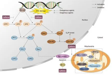
Figure 10 Caffeine influences the DNA repair process. When exogenous and endogenous agents attack DNA, DNA can be damaged, which immediately activates the DNA repair process. In this process, a sensor detects the damage and then causes the phosphorylation of ataxia-telangiectasia mutated and ataxia telangiectasia and Rad3-related. Caffeine can inhibit the activation of both ataxia-telangiectasia mutated and ataxia telangiectasia and Rad3-related. Phosphorylated ataxia telangiectasia and Rad3-related and ataxia-telangiectasia mutated can activate cyclin-dependent kinase 1, which induces G2/M arrest and p53. p53 then modulates its downstream target p21, which can influence the cell cycle by inhibiting Cdk1 and Cdk2. Moreover, Bax is also downstream of p53, and when it enters the cytosol, it can initiate the apoptosis process. Caffeine can promote apoptosis by inhibiting the translocation of Bax from the nucleus to the mitochondria and also by promoting the apoptosis inhibitor Bcl-2. ATM: Ataxia-telangiectasia mutated; ATR: Ataxia telangiectasia and Rad3-related; Chk: Cyclin-dependent kinase.
 World Journal of Gastrointestinal Oncology2020年2期
- World Journal of Gastrointestinal Oncology的其它文章
- Biomarkers for detecting colorectal cancer non-invasively: DNA, RNA or proteins?
- FOLFIRINOX vs gemcitabine/nab-paclitaxel for treatment of metastatic pancreatic cancer: Single-center cohort study
- Prognostic scoring system for synchronous brain metastasis at diagnosis of colorectal cancer: A populationbased study
- Neuropathy experienced by colorectal cancer patients receiving oxaliplatin: A qualitative study to validate the Functional Assessment of Cancer Therapy/Gynecologic Oncology Group-Neurotoxicity scale
- Clinical value evaluation of serum markers for early diagnosis of colorectal cancer
- Yttrium-90 radioembolization for unresectable hepatic metastases of breast cancer: A systematic review
