
Anticancer activity of Mahonia leschenaultii methanolic root extract and berberine on Dalton’s ascitic lymphoma in mice
2019-07-13 01:45:24LathaTMChozhavelRajanathanAmeerKhusroChidambaranathanPaulAgastianSankaranarayananNagarajan
R Latha, TM Chozhavel Rajanathan, Ameer Khusro, N Chidambaranathan, Paul Agastian?,Sankaranarayanan Nagarajan
1Research Department of Plant Biology and Biotechnology, Loyola College, Chennai-600034, India
2K. M. College of Pharmacy, Uthangudi, Madurai-625107, Tamil Nadu, India
3Department of Chemistry, National Institute of Technology, Imphal-795004, Manipur India
Keywords:Mahonia leschenaultii Berberine Dalton's ascitic lymphoma Methanolic extract
ABSTRACT Objective: To evaluate the effects of methanol root extract of Mahonia leschenaultii and berberine of Mahonia leschenaultii on Dalton's ascitic lymphoma in Swiss Albino mice.Methods: The methanol root extracts of Mahonia leschenaultii (200 and 400 mg/kg) were given orally, and berberines (10 and 20 mg/kg) were injected intra-peritoneally for 14 successive days in tumor bearing mice. Hematological parameters (white and red blood cells,haemoglobin level, granulocytes, and agranulocytes), lipid parameters (total cholesterol and triglycerides), serum enzymes (serum glutamate pyruvate transaminases, serum glutamate oxaloacetate transaminases, and alkaline phosphatise) and mean survival and solid tumor growth were determined and compared with untreated mice. 5-fluorouracil (20 mg/kg) was used as a reference standard drug.Results: Mahonia leschenaultii and berberine reduced the hematocrit significantly.Furthermore, Mahonia leschenaultii and berberine improved the survival of mice significantly and restored the affected hematological and lipid parameters similar to the normal levels.Conclusions: These observations show a strong anticancer effect of methanol root extract of Mahonia leschenaultii and berberine in suppressing Dalton's ascitic lymphoma cancer cell growth in a mouse model by controlling haematological, lipid, serum enzymes, and other derived parameters effectively.
1. Introduction
Cancer is still a foremost health problem in the world. Hence, it is an urgency to discover new vigorous chemotherapy medicines which can act as pilot compounds for efficient drug development against cancer. Lymphoma is malignancy derived from hematopoietic cells.Lymphoma might cause ascites by preventing the lymphatic system.When ascites is caused by cancer, it is called as malignant ascites.Malignant ascites is more common among the people affected from breast cancer, colon cancer, and gastrointestinal tract cancers.
Herbal medicines are known to show anticancer effects by inducing antioxidant action, retarding cancer inducing hormones and enzymes, exciting DNA repair mechanism, boosting the construction of defensive enzymes, and enhancing immunity[1].Phytochemicals and their derivatives employ a crucial task in anticancer drug development. Since ancient times, ethno medicinal herbs have played a crucial role in treatment of variety of human cancers[2]. It is reported that alkaloids, phenols, and flavonoids have been extensively reported to demonstrate anti-proliferation effects against various types of cancers[3]. In particular, the most significant anticancer drugs such as podophyllotoxins, camptothecin,vincristine, and taxol are obtained from plants.
The genus Mahonia belongs to the Berberidaceae family. Around 70 species of the Mahonia are distributed worldwide, and have pharmacologically potential bioactive compounds. It is reported that leaves, roots, stems, and barks of Mahonia spp. have potential antibacterial, antifungal, anticancer, and anti-inflammatory effects,irrespective of curing dermatological disorders[2,3]. Mahonia (M.)leschenaultii is used by the ethnic tribes, especially Toda of Nilgiris for their medical practices. Stem bark paste of M. leschenaultii is used not only in postnatal treatment for women, fever, cold, and jaundice but also to arrest other complications of Nilgiri tribe Todas, India. In spite of berberine (a major component), other compounds such as 3',8,8-trimethoxy-3-piperidyl-2,2'-binaphthalene-1,1',4,4'-tetrone,canadine, oxoberberine, oxyberberine, 1-diphenylmethylsilyloxy-4-methoxybenzene, and griseofulvin were isolated from bark extract of this plant[4]. In view of the vast medicinal properties of M. leschenaultii, the present context aimed to explore the anticancer effect of methanol root extract of M. leschenaultii (MEML) and its isolated propitious compound berberine on Dalton's ascitic lymphoma (DAL) induced cancer in mice.
2. Materials and methods
2.1. Chemicals
Chemicals, kits, and reagents used in this study were purchased from Sigma-Aldrich, USA. In this context, COBAS MIRA PLUS-S auto analyzer (Roche Switzerland) was used for investigating biochemical parameters. COBAS MICROS OT 18 from Roche and hematology analyzer sysmex XS800i (Sysmex corporation, USA)was used to observe hematological parameters. The blood sample was analyzed using MAX MAT.
2.2. Procurement of plant materials
The fresh root of M. leschenaultii was collected from Toda mandir area, Udhagamandalam, India. The plant was identified by Dr. G. Jayajothi, Taxonomist, Department of Plant Biology and Biotechnology, Loyola College, Chennai. The voucher specimen(LCH-301) was stored in Loyola College Herbarium, Chennai for future reference.
2.3. Extraction
Roots were shade dried, chopped, and coarsely powdered. Three kilograms of powder were subjected to successive extraction by cold percolation method using hexane, ethyl acetate, methanol, and ethanol at room temperature for 72 h and the extract was filtered.The extraction was repeated three times. Filtrates were concentrated and preserved at 4 ℃ for further experiments purposes.
2.4. Isolation and identification of active compound
The methanol root extract (25 g) was subjected to chromatography using silica gel column in hexane. The column was eluted sequentially with hexane, chloroform, ethyl acetate, and methanol.Each fraction was spotted on a thin layer chromatography (TLC)plates over silica gel 60F254. About 82 fractions were collected and pooled into 18 major fractions based upon their Rfvalues in TLC profile and solvents were removed under reduced pressure. From fraction 15, a yellow colour needle was isolated and showed a single spot on TLC using methanol: acetic acid: water (8:1:1) as developing system.
TLC plates were observed under visible light, ultra violet (UV)light, exposed to iodine vapour, by spraying Dragandroff's solution,and vanillin sulphuric acid for compound visualization as per the methodology described by Alam and Gupta[5]. The UV spectrum,infrared (IR) spectrum,1H, and13C nuclear magnetic resonance(NMR) were used for analysis, and further compared with the reports as described by Adithya et al[6]. Post comparison with an authentic sample, the identity was confirmed.
2.5. Experimental animals
Swiss Albino mice (20-25 g) (C57BL/6) were used in this study.They were procured from animal experimental laboratory and kept at an ambient environment (25±2) ℃with typical laboratory diet(Pranav Agro Industries Ltd., Maharashtra) and water ad libitum.The study was carried out post clearance from Institutional Animal Ethical Committee, followed by the guideline of Committee for the Purpose of Control and Supervision of Experiments on Animals(cpcsea.nic.in), under the Ministry of Environment, Forest and Climate Change, Government of India (Protocol No.R.Latha/Ph.D/LC/IAEC/KMCP/178/2014-2015). Mice were quarantined as per the standard practice for one fortnight before commencing the investigations.
2.6. Toxicity study
To estimate the toxicity of MEML and berberine, the acute toxicity study was carried out using the method of Porwal et al[7]. Ten groups of fasted healthy rats (6 per group) were orally treated with the MEML and berberine at varied doses of 0.5 to 2.5 g/kg and 50 to 250 mg/kg, respectively. The control group was treated with distilled water. The mice were monitored up to 4 h for 15 d in order to check physical signs of toxicity such as palpitation, gasping, writhing, and reduced respiratory rate or death.
2.7. Initiation of DAL cancer
The DAL cancer cells were procured from Amala Cancer Research Centre, Trissur, Kerala, India, and maintained in vivo in Swiss albino mice by intra-peritoneal transplantation. The DAL cells were suctioned from peritoneal cavity of the mice using saline.After counting the cells, it was diluted up to 1×10⁶dilution, and administered intra-peritoneally. The day of cell implantation was designed as day zero. The mice were observed for 7 days for the tumor growth before further investigation.
2.8. Treatment schedule
A total of 42 Swiss Albino mice were divided into 7 groups,containing 6 mice in each group. All the animals in 6 groups were injected with DAL cells and the remaining as control group(vehicle + normal diet and water). Ten millilitres per kilogram of normal saline was used as vehicle according to the methods of Sathiyanarayanan et al[8].
Group 1 (G1): normal control mice treated with vehicle alone;Group 2 (G2): DAL control mice treated with vehicle alone; Group 3 (G3): DAL mice were given MEML 200 mg/kg of body weight orally; Group 4 (G4): DAL mice were given MEML 400 mg/kg of body weight orally; Group 5 (G5): DAL mice were given berberine at 10 mg/kg of body weight intra-peritoneally; Group 6 (G6):DAL mice were given berberine at 20 mg/kg of body weight intraperitoneally; Group 7 (G7): DAL mice were given 5-fluorouracil at 20 mg/kg of body weight intra-peritoneally and served as positive control.
2.9. Biochemical and histopathological studies
Mice were weighed on the day of tumor inoculation and once in 3 d thereafter. Treatment was started on the 14thday of tumor inoculation and continued for 14 days. On day 15, 6 animals of each group were sacrificed by euthanasia. Blood was reserved from each mouse by retro orbital plexus method to study the tumor growth parameters such as haematological parameters, packed cell volume, lipid profile and serum enzyme analysis (biochemical parameters), mean survival time, solid tumor volume, and histopathological studies. Experiments were repeated six times.
Liver was excised and 10% (w/v) homogenate was prepared in 10% (w/v) potassium chloride solution under ice-cold condition.This solution was centrifuged at 1 500 rpm at 4 ℃ for 15 min and obtained supernatants were used for the evaluation of the biochemical parameters.
The dissected liver pieces from all the groups were fixed in paraffin. The embedded liver paraffin blocks were sectioned at 3 μm-5 μm intervals and counter stained with Ehrlich's haematoxylin with eosin for histological examinations. The slides were mounted using DPX mountant and observed at 40 × magnification[9].
2.10. Haematological parameters
White blood cell (WBC) and red blood cell (RBC) counts were estimated according to the methodology of Sarkar et al.[10].Haemoglobin (Hgb) in the blood was estimated based on the method of Drabkin and Austin[11]. Granulocytes and agranulocytes counts were analyzed using hematologyanalyzer Sysmex XS800i (Sysmex corporation, USA).
2.11. Lipid profile and serum enzyme
Total cholesterol in the plasma and tissues was evaluated according to the method as described by Adekiya et al[12]. A serum triglyceride determination kit was used to determine the level of triglycerides.Biochemical parameters including serum glutamate pyruvate transaminases and serum glutamate oxaloacetate transaminases activities were determined as per the method of Reitman and Frankel[13]. Alkaline phosphatase (ALP) activity was estimated as per the methodology of Kind and King[14].
2.12. Derived parameters/tumor growth parameters
Mean survival time was determined according to the methods of Mary et al[15] and Bala et al[16]. DLA-induced solid tumor volume studies were done following the methods of Rajeshkumar[17].
2.13. Statistical analysis
Statistical analysis was performed using one way ANOVA and Newman-Keul's multiple range tests. Results were expressed as mean±SD and P<0.01 was considered as significant value.
3. Results
3.1. Identification of active compound
The active compound berberine was isolated using column chromatography, and confirmed its identity by physical as well as spectroscopic analysis. Post comparison with an authentic sample,the identity was confirmed as shown in Supplementary Figures.UV: λMeOHmaxnm238, 267, 349,430 (Supplementary Figure 1). IR:cm-1: 3433 (Hydroxyl, OH stretching), 2927, 2863 (CH Stretching), 1620, 1522 (aromatic, C=C stretching), 1382 (O-CH3,CH bending), 1261, 1035, 896. Yellow needles EI-MS (m/z): 337(M+, 100, C20H19O4N+) 307, 278, 207, 153, 95, 73 (Supplementary Figure 2).1H NMR δ CD3OD, 500MHz): 3.20 (2H, m, H-5),4.09 & 4.20 (9-OMe), 4.90 [2H, m (merged with solvent)], 6.10(2H, s, O-CH2-O), 7.15 (1H, s, H-4), 7.63 (1H, s, H-1), 8.08 (1H, d,J=7.5Hz, H-12) 8.25 (1H, d, J=7.5Hz,H-11), 8.75 (1H, s, H-13), 9.75(1H, brs, 8-OH) (Supplementary Figure 3).13C NMR (δCD3OD,125MHz): 104.14 (C-1), 148.23 (C-2), 148.5 (C-3), 108.64 (C-4),130.48 (C-4a), 26.25 (C-5), 56.2 (C-6), 145.4 (C-8), 121.75 (C-8a),143 (C-9), 150.32 (C-10), 126.64 (C-11), 123.14 (C-12), 133.72(C-12a), 119.55 (C-13), 137.53 (C-13a), 120.09 (C-13b), 102.28(O-CH2-O), 61.15 and 56.26 (2×OMe) (Supplementary Figure 4).
The active compound from MEML was identified as berberine. It showed the molecular ion at m/z 337 Da which was subsequent to the molecular formula (M+, 100, C20H19O4N+) (Supplementary Figure 5).
3.2. Toxicity study
The observations showed no harmful effects on the common activities or appearance of the mice, and all the mice were alive during the whole experimental stages by the treatment of varied doses of MEML and berberine up to doses of 1.5 g/ kg and 250 mg/kg, respectively. Animals which could survive the lethal effects up to 3 d were able to survive and continued even up to 14 d. Extracts and berberine were not causing great harm or damage at the maximum single oral dose of 1.5 g/kg of body weight and 250 mg/kg of body weight, respectively.
3.3. Hematological parameters
Table 1 elucidate that RBC, Hgb, platelets, lymphocytes, and monocytes were decreased whereas WBC, neutrophils and eosinophils count were significantly (P<0.01) elevated in the DAL control group compared to the normal control group. Treatment with MEML and berberine at doses of 200 mg/kg and 400 mg/kg of body weight and 10 mg/kg and 20 mg/kg of body weight, respectively augmented significantly (P<0.01) the platelets, Hgb, and RBC count,and also significantly (P<0.01) declined the packed cell volume,WBC, neutrophils count and eosinophils count more or less similar to the standard with respect to the DAL control group.
3.4. Biochemical parameters
Results exhibited significant (P<0.01) elevated level in total cholesterol, AST, ALT, and ALP in the DAL control animals,whereas the administration of MEML and berberine at two different doses affected these abnormal conditions towards the normal level significantly (P<0.01). The standard (5-fluorouracil) treatment exhibited similar results. Table 2 shows the effect of MEML and berberine on biochemical parameters.
3.5. Tumor parameters
Anticancer activity of M. leschenaultii was designated by the significant (P<0.01) increase in mean survival time [(16.82±2.57) dto (25.74±0.80) d] of animals treated with MEML and berberine at doses of 200 mg/kg and 400 mg/kg of body weight and 10 mg/kg and 20 mg/kg of body weight, respectively when compared to DAL mice (Figure 1).
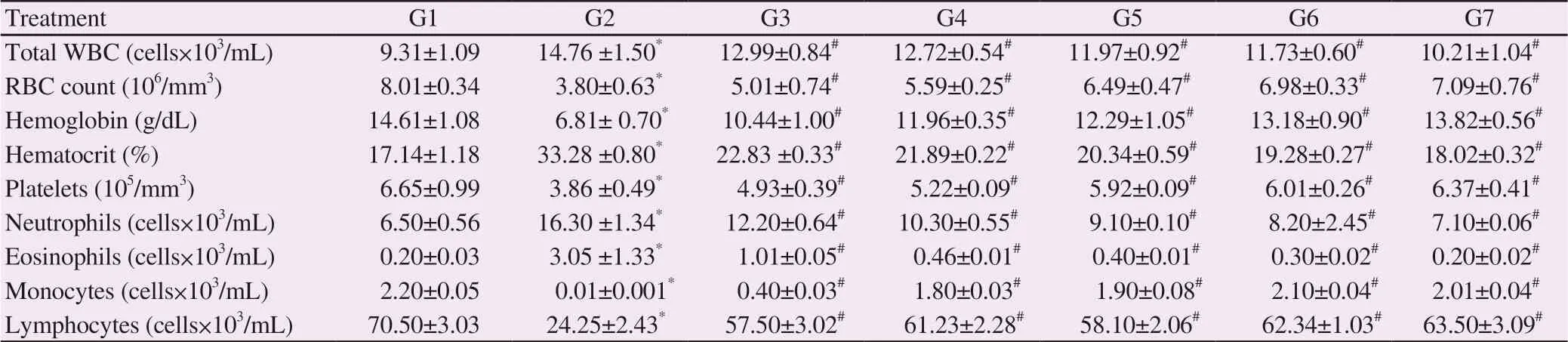
Table 1. Effect of methanol root extracts of Mahonia leschenaultii and berberine on hematological parameters.
Table 2. Effect of methanol root extracts of Mahonia leschenaultii and berberine on lipid proteins and serum enzymes.

Figure 1. Effect of methanol root extracts of Mahonia leschenaultii and berberine on mean survival of tumor-bearing mice. All values are expressed as mean±SD (n=6). *:Values are significantly different (P<0.01) from control(G1); #:Values are significantly different (P<0.01) from cancer control (G2).
3.6. Effect on solid tumor growth
From day 15 to day 30, the reduction in the tumor volume of mice treated with MEML and berberine was reported. Among all the treated groups, only Group 4 and Group 6 revealed pronounced reduction in the solid tumor growth with respect to DAL control(Group 2). Hence, Table 3 comprised the data expressing solid tumor growth inhibitory potentialities of these two groups only with respect to DAL control. On 30thday, tumor volume of DAL control animals was (6.10±0.21) cm3, whereas the MEML and berberine administered groups were found to be (0.040±0.002) and(0.020±0.003) cm3, respectively (Table 3).
Table 3. Effect of methanol root extracts of Mahonia leschenaultii and berberine on solid tumor volume (cm3).
3.7. Histopathological observations
The effect of plant extract and compound on the structural integrity of tissues was observed by histological analysis of the liver tissues using a light microscope (Figure 2A). The study exhibited that there was no alteration found in the organs excised from control animals and the treated animals (Figure 2A). However, the liver of MEML and berberine treatment at the lower and higher doses as well as 5-fluorouracil treatment produced normal histological form.
Figure 2. Haematoxylin-eosin stained sections of the liver from the rat groups fed with saline, DAL, 5-fluorouracil, methanolic root extract, and berberine of Mahonia leschenaultii. A: G1 - Normal control; B: G2 - Toxic control (DAL); C: G7 - Standard control (5-fluorouracil20 mg/kg of body weight) + DAL; D:G3- MEML 200 mg/kg of body weight + DAL; E: G4- MEML 400 mg/kg of body weight + DAL; F: G5- berberine 10 mg/kg of body weight + DAL; G: G6-berberine 20 mg/kg of body weight + DAL. Arrow indicates a small area of lymphomatous cells. Stained section was observed at 40××magnification. DAL:Dalton's ascitic lymphoma.
The liver histological analysis of normal control showed the liver structure with patches of parenchymatous tissues alienated by sinusoids cartial vein and portal triad appearance, while DAL control showed cords of hepatocytes and a small area of lymphmatous cells (Figure 2B). The standard control group showed cords of hepatocytes without tumor (Figure 2C). The treatment groups of MEML and berberine showed liver parenchyma with cords of hepatocytes without tumor in a dose dependent manner (Figures 2D, 2E, 2F, and 2G). However, the liver cells had a comparable characteristic as the controls. The extract and berberine treatment at two doses sustained the biochemical changes observed for AST and ALT in tumor animals. Thus histological study undoubtedly designated the liver tissues that were injured by DAL inoculation,elucidating recovery with MEML, berberine, and 5-fluorouracil treatments.
4. Discussion
In DAL tumor, the spreading of cancer to the abdominal lining(peritoneum) can result in irritation. To soothe this irritation, the fluid is produced by peritoneum within the abdomen. While cancer spreads to the portal vein, blood force elevates inside the liver and results into the damage circulation, thereby causing gathering of fluid inside the abdomen. Fewer blood proteins are produced if the liver is damaged. This causes the disruption of fluid balance in the body, resulting fluids to accumulate in body tissues and the tummy. When the excess fluid cannot be efficiently drained after the blockage of lymphatic system due to tumor growth, it results in fluid accumulation inside the abdomen. Hence, a quick increase in the volume of ascites tumor was observed in DAL tumor developing mice. Tumor growth is directly proportional to the amount of ascites fluid because ascites fluid serves as a direct dietary supply for cancer cells[18].
The toxicity study elucidates that the extracts and berberine were not causing great harm or damage at the maximum single oral dose of 1.5 g/kg and 250 mg/kg of body weight, respectively. Further the treatments exhibited the effects of DAL control, MEML (400 mg), and berberine (20 mg) that suppressed tumor development with respect to the packed cell volume from (33.28±0.80)% to(21.89±0.22)% and (19.28±0.27)%, decreased WBC cell counts from 14.76 cells/mL×× 103to 12.72 cells /mL××103and 11.73 cells/mL××103, increased RBC from 3.80 cells/Mill/cumm to 5.59 cells/Mill/cumm and 6.98 cells/Mill/cumm, respectively.
Myelo suppression and anaemia are the main obstacles found in the chemotherapy treatment of cancer. A significant decline in haemoglobin proportion in tumor mice causes anaemia due to drastic changes in haemolytic, iron deficiency or myelopathic condition[21].Two different doses of methanolic root extract and berberine of M.leschenaultii treatment restored the Hgb level, red blood cells, WBC count, packed cell volume, platelet count, neutrophils, lymphocytes,eosinophils, and monocytes counts similar to the normal and standard control levels. Thus, the study indicates that methanolic root extract and berberine of M. leschenaultii hold defensive effect on the blood streaming structure. Furthermore, the reduction in packed cell volume and improved survival strategy of the mice depicted that the extract and the compound induced a hindrance in vascular permeability to the cells[19]. It is reported that stem extract of Adenanthera pavonina while treated with DAL induced mice,reduced the viable cancer cells, and restored the Hgb and RBC levels to near normal levels when compared to control[22].
Several reports suggest that cancer cells in the experimental animals or in the human body destroy different metabolic processes of the liver. In our observation, the tumor induced animals showed the increased levels of total cholesterol, ALT, ALP, and AST in serum which confirmed a loss in the consistency of character of cell membrane as well as liver injury. A significant (P<0.01) restoration of these changes near the normal control by the treatment of methanolic root extract and berberine of M. leschenaultii indicated hepatoprotective nature.
Besides, it was examined among many groups at the extent to which toxicities would be identified and predicted using animal models for these specific chemical compounds[20]. Any discovered antitumor drug should extend the survival time of animals. It may be concluded that methanolic root extract and berberine of M.leschenaultii at doses of 200 mg/kg and 400 mg/kg of body weight and 10 mg/kg and 20 mg/kg of body weight, respectively suppressed the tumor growth, and increased the life duration of DAL host. This shows that berberine being also a part of the crude extract shows less efficient results than when found to be in the extract. However, this study suggests that in the crude plant extract, berberine may suffer some hindrance in its action by some other compounds. At the same time, we inferred that methanolic root extract and berberine of M.leschenaultii at doses of 200 mg/kg and 400 mg/kg of body weight and 10 mg/kg and 20 mg/kg of body weight, respectively have antitumor effect in opposition to DAL bearing mice.
Treatment with MEML (400 mg) and berberine (20 mg) of M.leschenaultii reduced the tumor volume from (6.10±0.21) cm3(DAL control) to (0.040±0.002) cm3and (0.020±0.003) mL,respectively. In addition, the liver histological observations exhibited normalization of tissue damages in the mice with the treatment of the MEML extract and berberine. Remarkable anomalous conditions in the DAL inoculated hosts elucidated the lethal effect of tumor in these histological studies. Whereas the studied toxic effects were reduced when observed in the tissues treated with MEML extract and berberine, which supported the potent antioxidant and hepatoprotective effects. Anticancer activities such as inhibition of tumor invasion and metastasis of berberine are based on its ability to control cancer cell proliferation. Interestingly, this pro-oxidant effect has been recognized in carcinoma cell lines and not on normal cells[23]. Manoharan et al.[24] reported that berberine has remarkable chemopreventive effects in hamster buccal pouch carcinogenesis. It has been reported that berberine is a promising agent for clinical use in human breast cancer chemotheraphy[25].
According to the report of Eom et al[26], berberine inhibits tumor angiogenesis by arresting the establishment of activated B cells and stimulates the construction of intracellular ROS in cancer cells. It also shows effect on cell cycle arrest and apoptosis. Berberine has potential ability to induce antioxidant defenses. Thus, berberine plays a central role in initiating caspase dependent apoptotic events along with endoplasmic reticulum stress.
Tong et al.[27] concluded that the mixture of doxorubicin and berberine synergistically produced anticancer effect in human lung carcinoma and human cervix carcinoma cells in vitro, perhaps mediated by apoptosis induction. The combination of doxorubicin with berberine is a new approach that has potential in cancer treatments. However, Bao et al.[28] reported that berberine induced a prominent hermetic dose response, in which cancer cell growth is stimulated when berberine is provided at low dose, in turn it is potentially effective as anticancer agent at high dose.
Berberine was suggested to be used in the clinical development of cytotoxic compound since it played a vital role in alterations in cell proliferation, cell cycle progression, DNA synthesis, and apoptosis in melanoma cells (K1735-M2) of mouse as well as in human melanoma malignant cell lines[29]. Mitani et al.[30] stated that berberine combined with camptothecin as anticancer agent showed significant tumor growth inhibition and arrested the lymphatic metastasis.
In our observations, the biochemical assessment of DAL inoculated animals exhibited remarkable altered conditions due to the lethal effect of the tumor. The MEML and berberine administration enhanced the mice's survival time and reduced the tumor volume.The normalization of these intoxication alterations in the serum and liver treated with methanolic root extract and berberine at two different doses of 200 mg/kg and 400 mg/kg of body weight and 10 mg/kg and 20 mg/kg of body weight, respectively suggested a strong antioxidant effect, thereby preventing the formation of tumors and damage to liver. Thus, the plant extract and the compound were found to be potent anticancer agents in treating DAL cancers.
Conflict of interest statement
We declare that we have no conflict of interest.
Acknowledgements
Authors like to extent their gratitude to Dr. K. Balakrishna(Research officer), Dr. P. Saravana Kumar (Entomology Research Institute), Dr. I.V.S. Nimal Christhudas (Loyola College), and Dr.P. Praveen Kumar (Loyola College) for support during this research work.
 Asian Pacific Journal of Tropical Medicine2019年6期
- Asian Pacific Journal of Tropical Medicine的其它文章
- Community-acquired pneumonia with Acinetobacter radioresistens bacteremia in an immunocompetent host: A case report
- Optimized combinations of statins and azoles against Acanthamoeba trophozoites and cysts in vitro
- Symptoms of dengue at the acute and post-infection stage in the Western Province, Sri Lanka: A cross-sectional study
- Epidemiology and immunodiagnostics of Strongyloides stercoralis infections among migrant workers in Malaysia
- Laboratory diagnosis of schistosomiasis mansoni: Current status and future trends