Three-dimensional printing technique assisted cognitive fusion in targeted prostate biopsy
Yn Wng,Xu Go,Qingsong Yng,Hifeng Wng, Ting Shi,Yifn Chng,Chunling Xu,Yingho Sun,*
aDepartment of Urology,Changhai Hospital,Second Military Medical University,Shanghai,China
bDepartment of Radiology,Changhai Hospital,Second Military Medical University,Shanghai,China
Three-dimensional printing technique assisted cognitive fusion in targeted prostate biopsy
Yan Wanga,Xu Gaoa,Qingsong Yangb,Haifeng Wanga, Ting Shia,Yifan Changa,Chuanliang Xua,Yinghao Suna,*
aDepartment of Urology,Changhai Hospital,Second Military Medical University,Shanghai,China
bDepartment of Radiology,Changhai Hospital,Second Military Medical University,Shanghai,China
Prostate cancer;
Objective:To explore the effect of 3-dimensional(3D)printing-assisted cognitive fusion on improvement of the positive rate in prostate biopsy.
Methods:From August to December 2014,16 patients with suspected prostatic lesions detected by multiparametric magnetic resonance imaging(MRI)were included.Targeted prostate biopsy was performed with the use of prostate 3D reconstruction modeling,computersimulated biopsy,3D printing,and cognitive fusion biopsy.All patients had received 3.0 T multiparametric MRI before biopsy.The DICOM MRI files were imported to medical imaging processing software for 3D reconstruction modeling to generate a printable.stl file for 3D printing with use of transparent resin as raw material.We further performed a targeted 2-to 3-core biopsy at suspected lesions spotted on MRI.
Results:For the 16 patients in the present study,3D modeling with cognitive fusion-based targeted biopsy was successfully performed.For a single patient,1-2 lesions(average:1.1 lesions)were discovered,followed by 2-6 cores(average:2.4 cores)added as targeted biopsy.Systematic biopsies accounted for 192 cores in total,with a positive rate of 22.4%;targeted biopsies accounted for 39 cores in total,with a positive rate of 46.2%.Among these cases,10 patients(62.5%)were diagnosed with prostate adenocarcinoma,in which seven were discovered by both systematic and targeted biopsy,one was diagnosed by systematic biopsy only,and two were diagnosed by targeted biopsy only.For systematic biopsy,Gleason score ranged from 6 to 8(average:7),while that for targeted biopsy ranged from 6 to 9(average: 7.67).Among the seven patients that were diagnosed by both systematic and targeted biopsy, three(42.8%)were reported with a higher Gleason score in targeted therapy than in systematic biopsy.
Conclusion:3D printing-assisted cognitive fusion technique markedly promoted positive rate in prostate biopsy,and reduced missed detection in high-risk prostate cancer.
?2015 Editorial Office of Asian Journal of Urology.Production and hosting by Elsevier (Singapore)Pte Ltd.This is an open access article under the CC BY-NC-ND license(http:// creativecommons.org/licenses/by-nc-nd/4.0/).
1.Introduction
Transrectal ultrasound(TRUS)guided prostate biopsy is the first choice in the diagnosis of prostate cancer[1].However,the traditional method has difficulty in avoiding missed diagnosis,for as high as 22%-47%of prostate cancer are missed in the initial biopsy[2].The standard TRUS guided prostate biopsy is mainly for sampling in the peripheral zone where the incidence of cancer is high,but this conventional approach is poor in sampling cancers at the anterior,midline,and apex of the prostate,leading to underdiagnosis of clinically significant diseases[3].Magnetic resonance imaging(MRI)has higher sensitivity in finding clinically significant prostate cancer[4].Functional MRI technique as dynamic contrast-enhancement(DCE)and diffusion weighted imaging(DWI)may provide more accurate space orientation on the basis of qualitative diagnosis. It has now become an important problem regarding the use of image positioning to guide prostate biopsy so as to effectively improve the biopsy positive rate in the early diagnosis of prostate cancer.MRI-TRUS fusion targeted biopsy and MRI-guided targeted biopsy are effective approaches to improve biopsy positive rate and avoid missed diagnosis of prostate cancer[5,6],but both have higher requirement for hardware facilities,and need complicated skills in operation;therefore,it is rather difficult to apply these methods in conventional examination.
Currently,3-dimensional(3D)printing technique is fast developing and has infiltrated into multiple industries including healthcare industry.Now its application in medical field is mainly in implant design,surgery simulation,skill training and others.There has not been much report from urinary surgery in this regard.This study explores 3D printing technique assisting cognitive fusion for design of prostate biopsy regimen,and evaluates its feasibility and efficacy in improvement of positive rate of prostate biopsy.
2.Patients and methods
2.1.Patients
This prospective study was conducted in the Changhai Hospital Affiliated to Second Military Medical University (Shanghai,China).The code of ethics was reviewed by the Institutional Review Board of Changhai Hospital and approval was obtained.Informed consents were obtained from patients eligible for this study,and any potential harms and benefits regarding to the study were elaborated. Study enrollment began in 2014.
2.2.Multi-parameter MRI examination
All patients had received 3.0 T multiparametric MRI (Siemens Magnetom Skyra,Germany)before biopsy.The scan sequence included T1 weighted,T2 weighted,DCE and DWI.Two radiologists identified and located lesions suspicious for cancer according to the MRI sequences(Fig.1A). Both radiologists were blinded to pre-imaging serum prostate specific antigen(PSA)values and digital rectal examination(DRE)status.
2.3.3D reconstruction and 3D printing
Digital Imaging and Communications in Medicine(DICOM) format file of MRI was introduced into medical image processing software accordingly.Prostate and tumor images were introduced for 3D model reconstruction and smoothly processed to generate printable.stl format files(Fig.1B,C). By means of SLA,RS-450,3D printer,the.stl files were printed in accordance with a thickness of 0.1 mm.The printing material was transparent resin(Fig.1D).
2.4.Systematic prostate biopsy
All patients received 12-core systematic prostate biopsy under TRUS guidance.The 12-core biopsy was performed on the basis of the traditional 6-core biopsy by adding three cores on both sides of the lateral peripheral zones.
2.5.Targeted biopsy under 3D printing assisted cognitive fusion
The suspected lesions found by multi-parametric MRI further underwent a targeted 2-3-core biopsy.Before biopsy,the location of the suspected focus in the prostate was determined according to the 3D reconstruction data, followed by computer simulation 12-needle systematic biopsy performed(Fig.2A)to estimate whether the suspected focus could be detected in the course of biopsy.If the suspected focus was in the anterior,midline,and apex, because of the limitations of biopsy angle or biopsy depth, systematic biopsy might not be able to take samples.Then computer simulation of targeted biopsy procedures (Fig.2B)should be done to select biopsy sections,needling angles and needling depth at triggering.
3.Results
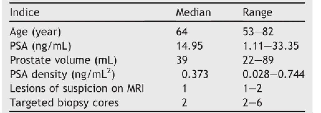
From August to December,2014,16 patients(median age 64 years)successfully underwent 3D modeling with cognitive fusion-based targeted biopsy.Patient and biopsycharacteristics are described in Table 1.Median PSA at biopsy was 14.95 ng/mL(range 1.11-33.35 ng/mL),and 14 of 16(87.5%)patients had a negative DRE.
For a single patient,one to two lesions(average:1.1 lesions)were discovered,followed by 2-6 cores(average: 2.4 cores)added as targeted biopsy.Eight patients had suspected lesions in the peripheral zone,and the remaining eight had lesions in atypical zones such as the prostate apex and central gland.Systematic biopsies accounted for 192 cores in total,with a positive rate of 22.4%;targeted biopsies accounted for 39 cores in total,with a positive rate of 46.2%.No patient required hospitalization for fever or sepsis after biopsy.
Of the 16 patients,10 patients(62.5%)were diagnosed with prostate adenocarcinoma(Table 2),in which seven were discovered by both systematic and targeted biopsy, one was diagnosed by systematic biopsy only,and two were diagnosed by targeted biopsy only.For systematic biopsy, Gleason score ranged from 6 to 8(average:7),while that for targeted biopsy ranged from 6 to 9(average:7.67).
Among the seven patients that were diagnosed by both systematic and targeted biopsy,three(42.9%)were reported with a higher Gleason score in targeted therapy than in systematic biopsy.
4.Discussion
How to effectively avoid false negative results in biopsy is an important problem in the early diagnosis of prostate cancer.The development of MRI techniques provides more and more accurate localization diagnosis on the basis of its qualitative diagnosis.With rational use of imaging localization information,performing targeted biopsy on suspected cancer foci is an effective method to improve the positive rate of diagnosis.At present,fusion technique based on MRI-identified foci includes MRI/TRUS fusion,MRI/MRI fusion and cognitive fusion.Sonn et al.[5]utilized Artemis device to perform biopsy on 105 cases,in which PSA continued to rise after initial negative biopsy.By performing subsequent targeted biopsy with MRI/TRUS fusion on these cases,the positive rate was 34%.Of the positive targeted biopsy cases,91%were of clinical significance (Gleason score≥7),and that of systematic biopsy was 54%. Hoeks et al.[6]performed MR-guided biopsy on patients with elevated PSA and one or more previous negative TRUS biopsy sessions.In a total of 117 patients,cancer detection rate was 41%,and the majority of detected cancers were clinically significant(87%).The two methods are both able to improve the positive rate and avoid missed diagnosis of cancer in prostate biopsy,but requirements for image fusion device are high,which was inconvenient to operate, hence is not conducive to extended promotion on large scale.

Table 1Patient demographics.

Table 2Prostate cancer grades found by systematic and targeted biopsies.
In cognitive fusion,the operator selects suspected regions by reading the MRI image and then performing biopsyunder TRUS guidance.Its efficacy is still in controversy. Puech et al.[7]believed that the result of targeted biopsy in terms of cognitive fusion is not obviously different from that of systematic biopsy in terms of MRI/TRUS fusion; Delongchamps et al.[8]thought that targeted biopsy by cognitive fusion did not have obvious advantage over systematic biopsy(p=0.66).Cognitive fusion depends greatly on the operator’s experience,which is easy and simple to handle but without accurate methods.Its efficacy and repeatability is relatively poor.Therefore,to improve cognitive fusion method and raise its efficacy in prostate biopsy is of high importance for the improvement of biopsy positive rate and avoidance of missed diagnosis of high-risk prostate cancer.
3D printing technique performs accurate modeling in terms of multi-parameter MRI localization diagnostic information that makes use of computer software simulated biopsy and objectively evaluates systematic biopsy capability in the diagnosis of the suspected areas.In conventional systematic biopsy,the needle is triggered the moment it touches the prostatic capsule.If the tumor is located at the tip of the prostate or close to the urethra or at other atypical areas,conventional systematic biopsy may have difficulty in sampling these suspected areas;therefore,an individualized biopsy plan can be worked out in terms of computer simulated needling angles and depths. For the patients in the present study,3D print assisting cognitive fusion targeted biopsy avoided the missed diagnosis of two cases(20%),effectively improving the biopsy positive rate.Besides,of the seven patients found with cancer by both system and target biopsies,three(42.9%) were with higher points of Gleason score by targeted biopsy than by systematic biopsy;high-risk prostate cancer was effectively found.Two patients in this group were found with cancer only by targeted biopsy;both had a history of one prior negative prostate biopsy,multi-parametric MRI found suspected lesions in transition zone,thus it is clear that this technique is of marked significance in the diagnosis of cancer in non-peripheral zones.
3D printing technique can accurately reproduce 3D image,and application of transparent resin material can intuitively show the location,size and morphology of the tumor.Before biopsy,the operator can observe the 3D model of the tumor from multiple angles,thus evaluating the possibility of sampling by systematic biopsy.If the suspected focus is located in the peripheral zone,sampling can be done by systematic biopsy,for which the corresponding needle position for transrectal 12-needle systematic biopsy would be determined.In cases of nonperipheral zone and larger-sized cancers,it is impossible for the biopsy needle to sample in the suspected area that triggers the moment it touches the envelope of the prostate.The needing depth can thus be adjusted so as to break through the envelope and get close to the suspected area prior to triggering.Each suspected area can be further determined with 2-3 biopsies to avoid tumor omissions.In this study,the single needle positive rate for targeted biopsy was 46.2%,which was markedly higher than the 22.4% one for systematic biopsy.
The application of 3D printing technique effectively improves the efficacy of cognitive fusion,and avoids the drawback of depending too much on the operator’s experience.It is of certain accuracy and repeatability,but there is still much room to be desired in this approach. Firstly,the prostate is a soft tissue organ,so MRI data image processing is more difficult than that for bone,teeth and other tissues.In the data modeling phase,adequate acknowledgment of pelvic anatomy is very much required. With the assistance of the image practitioner,only by being clear of the suspected area in MRI image,can one perform relatively accurate modeling of the prostate and the suspected focus.Secondly,prostate cancer is likely to be characterized by multiple foci,so multi-parametric MRI cannot find small-sized foci.Therefore,based on various fusions of multi-parametric MRI,targeted biopsy cannot take place of systematic biopsy completely.Development of targeted biopsy relies on advances in more sensitive and specific imaging technologies.
Currently,3D printing technique is fast developing and integrating in multiple industries including healthcare industry.Its major application in the medical field is now involved in implant design,surgery simulation,training and others[9,10].There have been just a few 3D print applications in the urology field.Zhang et al.[11]reported research on 3D printing technique applied in the planning of renal tumor surgery and believed that it was of great significance for doctor’s surgery planning and doctor-patient communication.With its development and the advent of new materials,this technology will surely be applied more extensively in the urology surgery field.
5.Conclusion
The development of new technologies and their crossfusion with multi-discipline is an important driving force for medical development.In this study,we applied 3D printing technique assisting cognitive fusion in the early diagnosis of prostate cancer,which markedly improved the positive rate of biopsy and avoided missed diagnosis of high-risk prostate cancer.This technical operation proves to be easy and simple.The increased number of needling in targeted biopsy does not increase the incidence of complication.Its application and popularization will surely benefit more patients on a larger scale in the future.
Conflicts of interest
The authors declare no conflict of interest.
[1]Hodge KK,McNeal JE,Terris MK,Stamey TA.Random systematic versus directed ultrasound guided transrectal core biopsies of the prostate.J Urol 1989;142:71-5.
[2]Taira AV,Merrick GS,Galbreath RW,Andreini H, Taubenslag W,Curtis R,et al.Performance of transperineal template-guided mapping biopsy in detecting prostate cancer in the initial and repeat biopsy setting.Prostate Cancer Prostatic Dis 2010;13:71-7.
[3]Moore CM,Robertson NL,Arsanious N,Middleton T,Villers A, Klotz L,et al.Image-guided prostate biopsy using magnetic resonance imaging-derived targets:a systematic review.Eur Urol 2013;63:125-40.
[4]Puech P,Potiron E,Lemaitre L,Leroy X,Haber GP,Crouzet S, et al.Dynamic contrast-enhanced-magnetic resonance imaging evaluation of intraprostatic prostate cancer:correlation with radical prostatectomy specimens.Urology 2009;74: 1094-9.
[5]Sonn GA,Chang E,Natarajan S,Margolis DJ,Macairan M, Lieu P,et al.Value of targeted prostate biopsy using magnetic resonance-ultrasound fusion in men with prior negative biopsy and elevated prostate-specific antigen.Eur Urol 2014;65: 809-15.
[6]Hoeks CM,Schouten MG,Bomers JG,Hoogendoorn SP,Hulsbergen-van de Kaa CA,Hambrock T,et al.Three-Tesla magnetic resonance-guided prostate biopsy in men with increased prostate-specific antigen and repeated,negative,random, systematic,transrectal ultrasound biopsies:detection of clinically significant prostate cancers.Eur Urol 2012;62: 902-9.
[7]Puech P,Rouviere O,Renard-Penna R,Villers A,Devos P, Colombel M,et al.Prostate cancer diagnosis:multiparametric MR-targeted biopsy with cognitive and transrectal US-MR fusion guidance versus systematic biopsy-prospective multicenter study.Radiology 2013;268:461-9.
[8]Delongchamps NB,Peyromaure M,Schull A,Beuvon F, Bouazza N,Flam T,et al.Prebiopsy magnetic resonance imaging and prostate cancer detection:comparison of random and targeted biopsies.J Urol 2013;189:493-9.
[9]Cousley RR,Turner MJ.Digital model planning and computerized fabrication of orthognathic surgery wafers.J Orthod 2014;41:38-45.
[10]Rohner D,Guijarro-Martinez R,Bucher P,Hammer B.Importance of patient-specific intraoperative guides in complex maxillofacial reconstruction.J Craniomaxillofac Surg 2013;41: 382-90.
[11]Zhang Y,Ge HW,Li NC,Yu CF,Guo HF,Jin SH,et al.Evaluation of three-dimensional printing for laparoscopic partial nephrectomy of renal tumors:a preliminary report.World J Urol 2015 Apr 5[Epub ahead of print].
Received 28 August 2015;received in revised form 3 September 2015;accepted 8 September 2015
Available online 18 September 2015
*Corresponding author.
E-mail address:sunyh@medmail.com.cn(Y.Sun).
Peer review under responsibility of Shanghai Medical Association and SMMU.
http://dx.doi.org/10.1016/j.ajur.2015.09.002
2214-3882/?2015 Editorial Office of Asian Journal of Urology.Production and hosting by Elsevier(Singapore)Pte Ltd.This is an open access article under the CC BY-NC-ND license(http://creativecommons.org/licenses/by-nc-nd/4.0/).
Prostate biopsy;
3D printing
 Asian Journal of Urology2015年4期
- Asian Journal of Urology的其它文章
- GUIDE FOR AUTHORS
- Ureteral stent technology:Drug-eluting stents and stent coatings
- Stellate scar sign of renal cell carcinoma
- Laparoscopic ureterolysis with simultaneous ureteroscopy and percutaneous nephroscopy for treating complex ureteral obstruction after failed endoscopic intervention:A technical report
- Implication of ultrasound bladder parameters on treatment response in patients with benign prostatic hyperplasia under medical management
- Prostate chronic inflammation type IV and prostate cancer risk in patients undergoing first biopsy set:Results of a large cohort study
