Precision radiotherapy for brain tumors A 10-year bibliometric analysis☆
Ying Yan, Zhanwen Guo, Haibo Zhang, Ning Wang, Ying Xu
Department of Radiotherapy, Shenyang Northern Hospital, Shenyang 110016, Liaoning Province, China
Precision radiotherapy for brain tumorsA 10-year bibliometric analysis☆
Ying Yan, Zhanwen Guo, Haibo Zhang, Ning Wang, Ying Xu
Department of Radiotherapy, Shenyang Northern Hospital, Shenyang 110016, Liaoning Province, China
OBJECTIVE:Precision radiotherapy plays an important role in the management of brain tumors. This study aimed to identify global research trends in precision radiotherapy for brain tumors using a bibliometric analysis of the Web of Science.
DATA RETRIEVAL:We performed a bibliometric analysis of data retrievals for precision radiotherapy for brain tumors containing the key words cerebral tumor, brain tumor, intensity-modulated radiotherapy, stereotactic body radiation therapy, stereotactic ablative radiotherapy, imaging-guided radiotherapy, dose-guided radiotherapy, stereotactic brachytherapy, and stereotactic radiotherapy using the Web of Science.
SELECTION CRITERIA:Inclusion criteria: (a) peer-reviewed articles on precision radiotherapy for brain tumors which were published and indexed in the Web of Science; (b) type of articles: original research articles and reviews; (c) year of publication: 2002-2011. Exclusion criteria: (a) articles that required manual searching or telephone access; (b) Corrected papers or book chapters.
MAIN OUTCOME MEASURES:(1) Annual publication output; (2) distribution according to country; (3) distribution according to institution; (4) top cited publications; (5) distribution according to journals; and (6) comparison of study results on precision radiotherapy for brain tumors.
RESULTS:The stereotactic radiotherapy, intensity-modulated radiotherapy, and imaging-guided radiotherapy are three major methods of precision radiotherapy for brain tumors. There were 260 research articles addressing precision radiotherapy for brain tumors found within the Web of Science. The USA published the most papers on precision radiotherapy for brain tumors, followed by Germany and France. European Synchrotron Radiation Facility, German Cancer Research Center and Heidelberg University were the most prolific research institutes for publications on precision radiotherapy for brain tumors. Among the top 13 research institutes publishing in this field, seven are in the USA, three are in Germany, two are in France, and there is one institute in India. Research interests including urology and nephrology, clinical neurology, as well as rehabilitation are involved in precision radiotherapy for brain tumors studies.
CONCLUSION:Precision radiotherapy for brain tumors remains a highly active area of research and development.
Cerebral tumor; brain tumor; intensity-modulated radiotherapy; stereotactic body radiation therapy; stereotactic ablative radiotherapy; imaging-guided radiotherapy; dose-guided radiotherapy; stereotactic brachytherapy; stereotactic radiotherapy
Research Highlights
(1) We performed a bibliometric analysis of data retrievals for precision radiotherapy for brain tumors from 2002 to 2011 using the Web of Science.
(2) We analyzed the articles by annual publication output, distribution according to country, distribution according to institution, top cited publications, distribution according to journals, and made a comparison of study results on precision radiotherapy for brain tumors.
(3) We found that precision radiotherapy for brain tumors is still an area of active research in the past 10 years and Chinese radiologists should be encouraged to write more high-quality papers to participate in and enlarge academic exchange worldwide.
INTRODUCTION
Radiotherapy plays an increasingly dominant role in the comprehensive multidisciplinary management of cancer. About half of all cancer patients will receive radiotherapy either as a part of the initial treatment with curative intent or as palliative treatment. Methods for improving the therapeutic ratio by increasing the radiation dose to the relevant target and /or decreasing the volume of irradiated normal tissue are therefore desirable. Precision radiotherapy refers to the precise delivery of radiation to targeted areas, such as brain tumors, by combined utilization of radiotherapy, computing and physics[1]. It differs from normal radiation regimes in the following four aspects: (1) the delivery of very high radiation doses to targeted tumors; (2) very little normal tissue is exposed to the radiation; (3) the dose homogeneity and conformity index are used to evaluate plan quality; (4) precision localization of the targeted tumor area and radiation delivery. These four aspects should result in an improved therapeutic ratio, namely, that more patients will be cured with fewer side effects. The best examples of high-precision radiotherapy are intensity-modulated radiotherapy (IMRT), stereotactic radiotherapy and imaging-guided radiotherapy (IGRT).
Stereotactic radiotherapy is a way of targeting radiotherapy very precisely at the tumor. The stereotactic radiotherapy treatment is usually divided into between 6 and 25 daily doses called fractions[2]. The objective of IGRT is to take into account the inter- and/or intrafraction anatomic variations (organ motion and deformations) in order to improve treatment accuracy. The IGRT enables direct or indirect tumor visualization during radiation delivery and is realized by different types of devices which can vary in principle as well as in their implementation: from linear accelerators with onboard kV or MV-cone beam computed tomography (CBCT), helical tomotherapy, Cyberknife? and Novalis?with stereoscopic kV X-ray imaging systems[3-4]. These techniques have led to a more rational choice of planning target volume (PTV). Three dimensional conformal radiation therapy (3D-CRT) is a sophisticated irradiation technique which allows a high dose delivered to the tumor while keeping the dose to the adjacent normal tissues below tolerance. This improves cure rates and decreases chances of treatment related complications. In recent years, 3D-CRT has become quite popular and widely available due to recent advancements in computer technology[5].
The diagnosis and management of brain tumors has been confounded by the challenge of determining the extent of the tumor and elucidation of the function of the surrounding brain tissue. F-18 fluorodeoxyglucose (FDG) positron emission tomography (PET) has been evaluated in the planning of radiation with radiosurgery and IMRT with simultaneous integrated boosts[6]. Popperlet al[7]evaluated the value of O-(2-[18F]fluoroethyl)-L-tyrosine PET (FET-PET) for the diagnosis of recurrent glioma. In a group of 63 patients with clinically suspected recurrence after initial therapy, FET-PET was positive in all cases. FET-PET reliably distinguished between post-therapeutic benign lesions and tumor recurrence after initial treatment of low- and high-grade gliomas. Chaoet al[8]have shown that the sensitivity and specificity of FDG-PET was 75% and 81%, respectively.
DATA SOURCES AND METHODOLOGY
Data retrieval
In this study, we used bibliometric methods to quantitatively and qualitatively investigate research trends in studies of precision radiotherapy for brain tumors. Web of Science, a research database of publications and citations that are selected and evaluated by the Institute for Scientific Information in Philadelphia, PA, USA, using the key words cerebral tumor, brain tumor, intensitymodulated radiotherapy, stereotactic body radiation therapy, stereotactic ablative radiotherapy, imaging-guided radiotherapy, dose-guided radiotherapy, stereotactic brachytherapy, and stereotactic radiotherapy. We have limited the period of publication from 2002 to 2011, found 260 results, and downloaded the data on June 02, 2012.
Inclusion criteria
(a) Peer-reviewed articles on precision radiotherapy for brain tumors which were published and indexed in the Web of Science; (b) type of articles: original research articles and reviews; (c) year of publication: 2002-2011.
Exclusion criteria
(a) Articles that required manual searching or telephone access; (b) we excluded a number of corrected papers or book chapters from the total number of articles.
The searching results were analyzed by (1) annual publication output; (2) distribution according to country; (3) distribution according to institution; (4) top cited publications; (5) distribution according to journals; and (6) comparison of study results on precision radiotherapy for brain tumors.
RESULTS
Search results of publications addressing precision radiotherapy for brain tumors from 2002 to 2011 (Table 1)
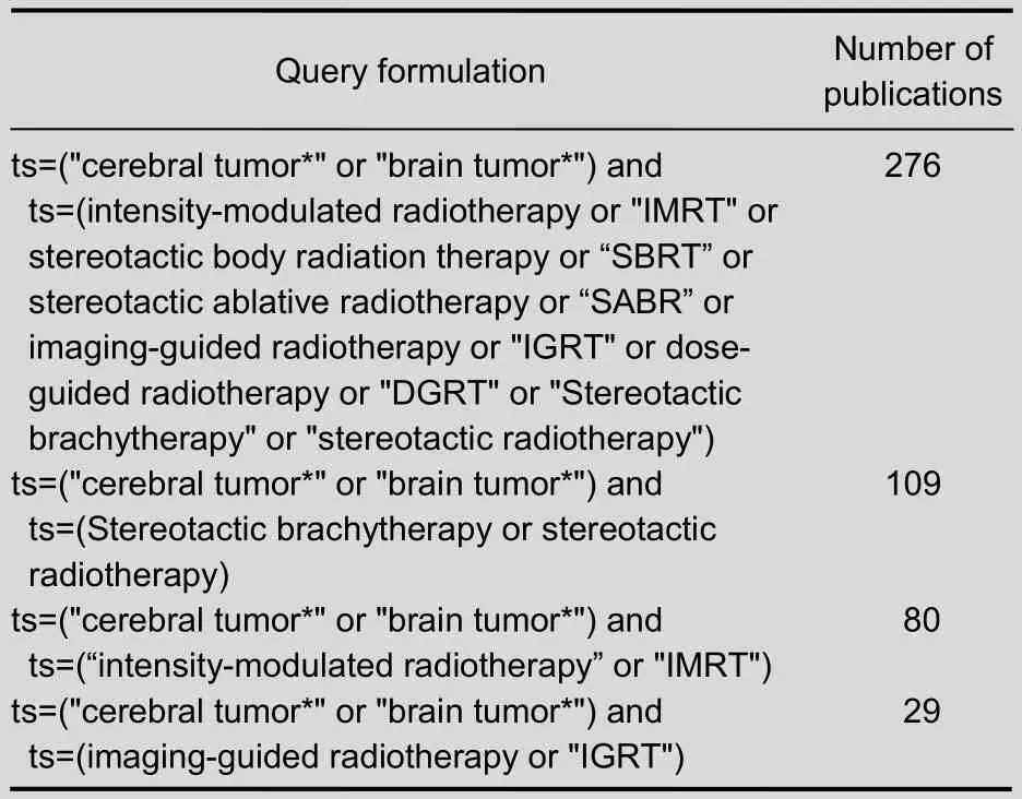
Table 1 Number of publications addressing precision radiotherapy for brain tumors included in the Web of Science from 2002 to 2011
Annual publication output of precision radiotherapy for brain tumors from 2002 to 2011 (Figure 1)
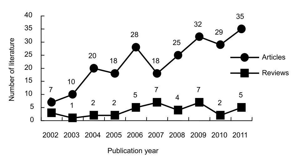
Figure 1 Annual number of publications on precision radiotherapy for brain tumors in the Web of Science from 2002 to 2011.
There were 260 publications on precision radiotherapy for brain tumors in the Web of Science from 2002 to 2011, including 222 articles and 38 reviews. The number of publications on precision radiotherapy for brain tumors has gradually increased over the past 10 years. However, there was a slight decrease in the number of papers published in 2005 and 2007.
Publication distribution of countries and institutes based on precision radiotherapy for brain tumors from 2002 to 2011 (Tables 2, 3)
The contribution analysis of different countries for publications was based on journal articles in which the address and affiliation of at least one author were provided. The total number of articles analyzed by country and institute publications was 260.
From Table 2, it can be seen that the USA published the most papers on precision radiotherapy for brain tumors. Germany published 51 papers that accounted for 19.62% of the total, which was much higher than the number of publications by other countries. France ranked third with 22 papers that accounted for 8.46%.

Table 2 Top nine countries in terms of number of studies on precision radiotherapy for brain tumors included in the Web of Science from 2002 to 2011

Table 3 Top 13 institutions publishing studies on precision radiotherapy for brain tumors in the Web of Science from 2002 to 2011
European Synchrotron Radiation Facility, German Cancer Research Center and Heidelberg University were the most prolific research institutes for publications on precision radiotherapy for brain tumors. Among the top 13 research institutes publishing in this field, seven are in the USA, three are in Germany, two are in France, and there is one institute in India.
The most-cited papers from researchers at European Synchrotron Radiation Facility from 2002 to 2011 were:Prolonged survival of Fischer rats bearing F98 glioma after iodine-enhanced synchrotron stereotactic radiotherapy, by Adamet al[9], published inInternational Journal of Radiation Oncology Biology Physics, with 42 citations.
Enhanced survival and cure of F98 glioma-bearing rats following intracerebral delivery of carboplatin in combination with photon irradiation, by Rousseauet al[10], published inClinical CancerResearch, with 31 citations.
Gadolinium dose enhancement studies in microbeam radiation therapy, by Prezadoet al[11], published inMedical Physics, with 10 citations.
Intracerebral delivery of 5-iodo-2'-deoxyuridine in combination with synchrotron stereotactic radiation for the therapy of the F98 glioma, by Rousseauet al[12], published inJournal of Synchrotron Radiation, with 10 citations.
The most-cited papers from researchers at Heidelberg University from 2002 to 2011 were:Differentiation of radiation necrosis from tumor progression using proton magnetic resonance spectroscopy, by Schlemmeret al[13], published inNeuroradiology, with 59 citations.
Efficacy of fractionated stereotactic reirradiation in recurrent gliomas: long-term results in 172 patients treated in a single institution, by Combset al[14], published inJournal of Clinical Oncology, with 51 citations.
PET and SPECT for detection of tumor progression in irradiated low-grade astrocytoma: a receiver-operating-characteristic analysis, by Henzeet al[15], published inJournal of Nuclear Medicine, with 26 citations.
Follow-up gliomas after radiotherapy: 1H MR spectroscopic imaging for increasing diagnostic accuracy, by Lichyet al[16], published inNeuroradiology, with 16 citations.
The most-cited papers from researchers at Harvard University from 2002 to 2011 were:Exciting new advances in neuro-oncology: the avenue to a cure for malignant glioma, by Van Meiret al[17], published inCA-A Cancer Journal for Clinicians, with 107 citations.
Advantage of protons compared to conventional X-ray or IMRT in the treatment of a pediatric patient with medulloblastoma, by St Clairet al[18], published inInternational Journal of Radiation Oncology Biology Physics, with 82 citations.
Stereotactic radiotherapy for localized low-grade gliomas in children: final results of a prospective trial, by Marcuset al[19], published inInternational Journal of Radiation Oncology Biology Physics, with 36 citations.
Most cited articles on precision radiotherapy for brain tumors from 2002 to 2011
According to bibliometric “l(fā)aw”, the main index for evaluating the quality of an article is the amount of citations it garners. Scientometrics has shown that references are considered as “classical references” once an article is cited four or more times[20]. In our analysis, the top 13 citations are listed in Table 4; therefore, they are classical references in the precision radiotherapy for brain tumors field.

Table 4 Articles with more than six citations average per year on precision radiotherapy for brain tumors in the Web of Science from 2002 to 2011
Among the 13 articles with more than six citations per year, the article “Exciting new advances in neuro-oncology the avenue to a cure for malignant glioma[17]”, published in 2010, was on average cited 35.67 times per year, which was more over than any other article.
Journals that published on precision radiotherapy for brain tumors from 2002 to 2011 (Table 5)
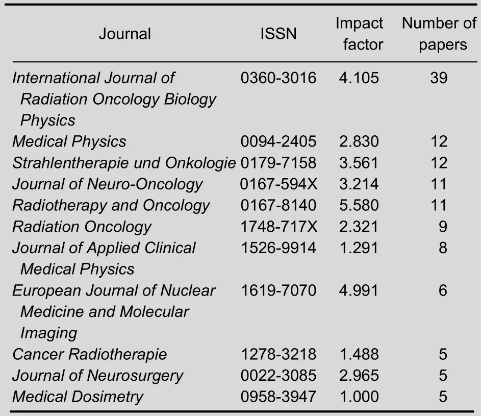
Table 5 Top 11 journals that published studies of precision radiotherapy for brain tumors from 2002 to 2011
In Table 5, it is evident that most papers on precision radiotherapy for brain tumors appeared in journals with a particular focus on oncology research.International Journal of Radiation Oncology Biology Physicspublished 39 papers that accounted for 15.01% of the total number of publications, which was followed byMedical Physicswhich published 12 papers and accounted for 4.62%.
It is disappointing that there are only five papers published by Chinese authors[31-35]though the precision radiotherapy has been widely applied in the treatment of brain tumors. Accordingly, Chinese radiologists should be encouraged to write more high-quality papers to participate in and enlarge academic exchange worldwide.
Analysis of intensity-modulated radiotherapy, stereotactic radiotherapy and imaging-guided radiotherapy for brain tumors (Tables 6-8)
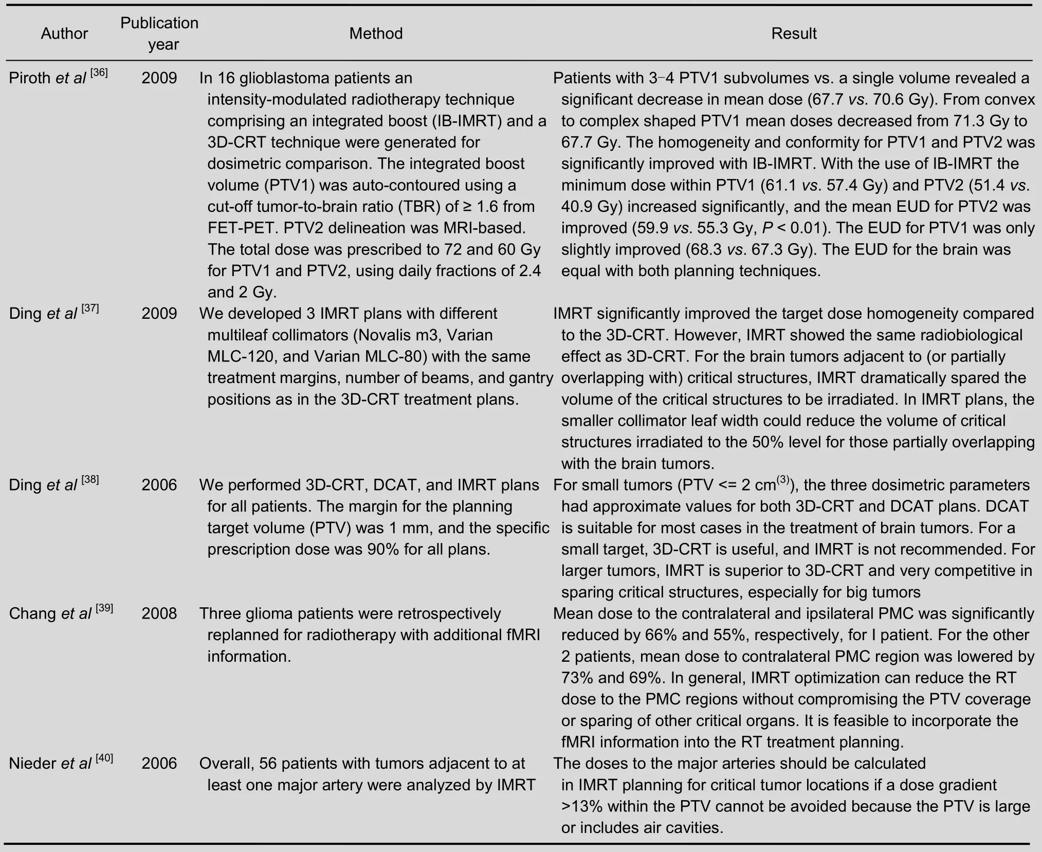
Table 6 Studies on intensity-modulated radiotherapy for brain tumors included in the Web of Science from 2002 to 2011
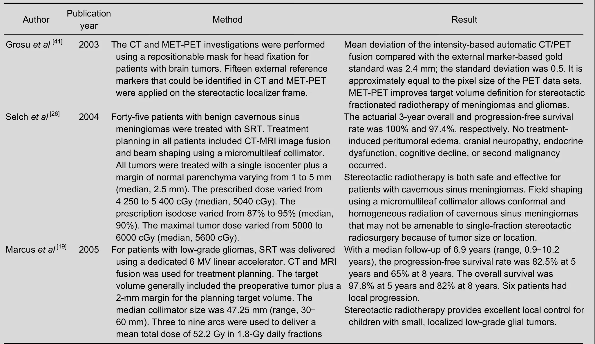
Table 7 Studies on stereotactic radiotherapy for brain tumors included in the Web of Science from 2002 to 2011
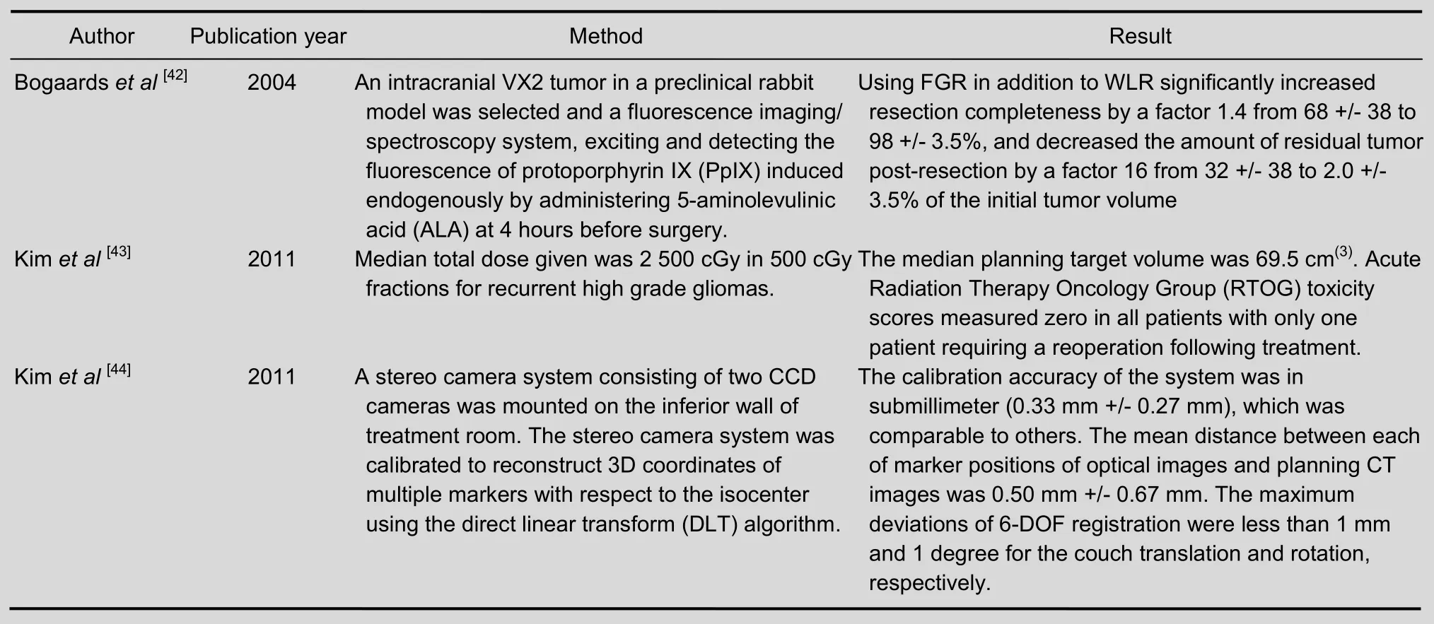
Table 8 Studies on imaging-guided radiotherapy for brain tumors included in the Web of Science from 2002 to 2011
DISCUSSION
Based on our bibliometric results from the Web of Science, we found the following research trends in studies on precision radiotherapy for brain tumors over the past 10 years. There were 260 research articles addressing precision radiotherapy for brain tumors included in the Web of Science.
The USA published the most papers on precision radiotherapy for brain tumors, followed by Germany and France. European Synchrotron Radiation Facility, German Cancer Research Center and Heidelberg University were the most prolific research institutes for publications on precision radiotherapy for brain tumors. Among the top 13 research institutes publishing in this field, seven are in USA, three are in Germany, two are in France, and there is one institute in India. Research interests including urology and nephrology, clinical neurology, as well as rehabilitation are involved in precision radiotherapy for brain tumors studies. Most researchers are focused on stereotactic radiotherapy and intensity-modulated radiotherapy in brain tumors, and fewer on image-guided radiotherapy. Though precision radiotherapy has resulted in major advances in brain tumor treatment in China, there are only five articles by Chinese authors that can be found in the Web of Science. This suggests that Chinese investigators should improve their writing and communication skills as well as increase the number of publications and preferred conference abstracts in order to contribute to and enlarge worldwide academic exchange in the field of precision radiotherapy for brain tumors.
Author contributions: Ying Yan conceived and designed the study. Zhanwen Guo and Haibo Zhang, retrieved the references, extracted the data, and provided technical support. Ning Wang wrote the manuscript. Ying Xu revised the manuscript.
Conflicts of interest: None declared.
[1] Nylén U, Kock E, Lax I, et al. Standardized precision radiotherapy in choroidal metastases. Acta Oncol. 1994;33(1):65-68.
[2] McIver JI, Pollock BE. Radiation-induced tumor after stereotactic radiosurgery and whole brain radiotherapy:case report and literature review. J Neurooncol. 2004; 66(3):301-305.
[3] Oelfke U, Tücking T, Nill S, et al. Linac-integrated kV-cone beam CT: technical features and first applications. Med Dosim. 2006;31(1):62-70.
[4] Miller A, Allen P, Fowler D. In-vivo stereoscopic imaging system with 5 degrees-of-freedom for minimal access surgery. Stud Health Technol Inform. 2004;98:234-240.
[5] de Crevoisier R, Louvel G, Cazoulat G, et al. Image-guided radiotherapy: rational, modalities and results. Bull Cancer. 2009;96(1):123-132.
[6] Solberg TD, Agazaryan N, Goss BW, et al. A feasibility study of 18F-fluorodeoxyglucose positron emission tomography targeting and simultaneous integrated boost for intensity-modulated radiosurgery and radiotherapy. J Neurosurg. 2004;101 Suppl 3:381-389.
[7] P?pperl G, G?tz C, Rachinger W, et al. Value of O-(2-[18F]fluoroethyl)- L-tyrosine PET for the diagnosis of recurrent glioma. Eur J Nucl Med Mol Imaging. 2004; 31(11):1464-1470.
[8] Chao ST, Suh JH, Raja S, et al. The sensitivity and specificity of FDG PET in distinguishing recurrent brain tumor from radionecrosis in patients treated with stereotactic radiosurgery. Int J Cancer. 2001;96(3):191-197.
[9] Adam JF, Joubert A, Biston MC, et al. Prolonged survival of Fischer rats bearing F98 glioma after iodine-enhanced synchrotron stereotactic radiotherapy. Int J Radiat Oncol Biol Phys. 2006;64(2):603-611.
[10] Rousseau J, Boudou C, Barth RF, et al. Enhanced survival and cure of F98 glioma-bearing rats following intracerebral delivery of carboplatin in combination with photon irradiation. Clin Cancer Res. 2007;13(17):5195-5201.
[11] Prezado Y, Fois G, Le Duc G, et al. Gadolinium dose enhancement studies in microbeam radiation therapy. Med Phys. 2009;36(8):3568-3574.
[12] Rousseau J, Adam JF, Deman P, et al. Intracerebral delivery of 5-iodo-2'-deoxyuridine in combination with synchrotron stereotactic radiation for the therapy of the F98 glioma. J Synchrotron Radiat. 2009;16(Pt 4):573-581.
[13] Schlemmer HP, Bachert P, Henze M, et al. Differentiation of radiation necrosis from tumor progression using proton magnetic resonance spectroscopy. Neuroradiology. 2002; 44(3):216-222.
[14] Combs SE, Thilmann C, Edler L, et al. Efficacy of fractionated stereotactic reirradiation in recurrent gliomas:long-term results in 172 patients treated in a single institution. J Clin Oncol. 2005;23(34):8863-8869.
[15] Henze M, Mohammed A, Schlemmer HP, et al. PET and SPECT for detection of tumor progression in irradiated low-grade astrocytoma: a receiver-operatingcharacteristic analysis. J Nucl Med. 2004;45(4):579-586.
[16] Lichy MP, Plathow C, Schulz-Ertner D, et al. Follow-up gliomas after radiotherapy: 1H MR spectroscopic imaging for increasing diagnostic accuracy. Neuroradiology. 2005;47(11):826-834.
[17] Van Meir EG, Hadjipanayis CG, Norden AD, et al. Exciting new advances in neuro-oncology: the avenue to a cure for malignant glioma. CA Cancer J Clin. 2010;60(3):166-193.
[18] St Clair WH, Adams JA, Bues M, et al. Advantage of protons compared to conventional X-ray or IMRT in the treatment of a pediatric patient with medulloblastoma. Int J Radiat Oncol Biol Phys. 2004;58(3):727-734.
[19] Marcus KJ, Goumnerova L, Billett AL, et al. Stereotactic radiotherapy for localized low-grade gliomas in children:final results of a prospective trial. Int J Radiat Oncol Biol Phys. 2005;61(2):374-379.
[20] Zhou JY, Sun T. A Quantitative analysis of digital library research article based on the Web of Science. Qingbao Kexue. 2005;23(10):1521-1525.
[21] Hu LS, Baxter LC, Smith KA, et al. Relative cerebral blood volume values to differentiate high-grade glioma recurrence from posttreatment radiation effect: direct correlation between image-guided tissue histopathology and localized dynamic susceptibility-weighted contrastenhanced perfusion MR imaging measurements. AJNR Am J Neuroradiol. 2009;30(3):552-558.
[22] Chan JL, Lee SW, Fraass BA, et al. Survival and failure patterns of high-grade gliomas after three-dimensional conformal radiotherapy. J Clin Oncol. 2002;20(6):1635-1642.
[23] MacDonald SM, Safai S, Trofimov A, et al. Proton radiotherapy for childhood ependymoma: initial clinical outcomes and dose comparisons. Int J Radiat Oncol Biol Phys. 2008;71(4):979-986.
[24] Vees H, Senthamizhchelvan S, Miralbell R, et al. Assessment of various strategies for 18F-FET PET-guided delineation of target volumes in high-grade glioma patients. Eur J Nucl Med Mol Imaging. 2009;36(2):182-193.
[25] Rock JP, Hearshen D, Scarpace L, et al. Correlations between magnetic resonance spectroscopy and image-guided histopathology, with special attention to radiation necrosis. Neurosurgery. 2002;51(4):912-919.
[26] Selch MT, Ahn E, Laskari A, et al. Stereotactic radiotherapy for treatment of cavernous sinus meningiomas. Int J Radiat Oncol Biol Phys. 2004;59(1):101-111.
[27] Minniti G, Saran F, Traish D, et al. Fractionated stereotactic conformal radiotherapy following conservative surgery in the control of craniopharyngiomas. Radiother Oncol. 2007;82(1):90-95.
[28] Pirzkall A, Li X, Oh J, Chang S, et al. 3D MRSI for resected high-grade gliomas before RT: tumor extent according to metabolic activity in relation to MRI. Int J Radiat Oncol Biol Phys. 2004;59(1):126-137.
[29] Shaffer R, Nichol AM, Vollans E, et al. A comparison of volumetric modulated arc therapy and conventional intensity-modulated radiotherapy for frontal and temporal high-grade gliomas. Int J Radiat Oncol Biol Phys. 2010; 76(4):1177-1184.
[30] Wagner D, Christiansen H, Wolff H, et al. Radiotherapy of malignant gliomas: comparison of volumetric single arc technique (RapidArc), dynamic intensity-modulated technique and 3D conformal technique. Radiother Oncol. 2009;93(3):593-596.
[31] Zhang X, Zhang W, Fu LA, et al. Hemorrhagic pituitary macroadenoma: characteristics, endoscopic endonasal transsphenoidal surgery, and outcomes. Ann Surg Oncol. 2011;18(1):246-252.
[32] Hu W, Ye J, Wang J, et al. Use of kilovoltage X-ray volume imaging in patient dose calculation for head-and-neck and partial brain radiation therapy. Radiat Oncol. 2010;5:29.
[33] Gu Z, Qin B. Nonrigid registration of brain tumor resection mr images based on joint saliency map and keypoint clustering. Sensors (Basel). 2009;9(12):10270-10290.
[34] Wan H, Chihiro O, Yuan S. MASEP gamma knife radiosurgery for secretory pituitary adenomas: experience in 347 consecutive cases. J Exp Clin Cancer Res. 2009; 28:36.
[35] Liu BJ, Law MY, Documet J, et al. Image-assisted knowledge discovery and decision support in radiation therapy planning. Comput Med Imaging Graph. 2007; 31(4-5):311-321.
[36] Piroth MD, Pinkawa M, Holy R, et al. Integrated-boost IMRT or 3-D-CRT using FET-PET based auto-contoured target volume delineation for glioblastoma multiforme-a dosimetric comparison. Radiat Oncol. 2009;4:57.
[37] Ding M, Newman F, Chen C, et al. Dosimetric comparison between 3DCRT and IMRT using different multileaf collimators in the treatment of brain tumors. Med Dosim. 2009;34(1):1-8.
[38] Ding M, Newman F, Kavanagh BD, et al. Comparative dosimetric study of three-dimensional conformal, dynamic conformal arc, and intensity-modulated radiotherapy for brain tumor treatment using novalis system. Int J Radiat Oncol Biol Phys. 2006; 66(4):S82-86.
[39] Chang J, Kowalski A, Hou B, et al. Feasibility study of intensity-modulated radiotherapy (IMRT) treatment planning using brain functional MRI. Med Dosim. 2008; 33(1):42-47.
[40] Nieder C, Grosu AL, Stark S, et al. Dose to the intracranial arteries in stereotactic and intensity-modulated radiotherapy for skull base tumors. Int J Radiat Oncol Biol Phys. 2006;64(4):1055-1059.
[41] Grosu AL, Lachner R, Wiedenmann N, et al. Validation of a method for automatic image fusion (BrainLAB System) of CT data and 11C-methionine-PET data for stereotactic radiotherapy using a LINAC: first clinical experience. Int J Radiat Oncol Biol Phys. 2003;56(5):1450-1463.
[42] Bogaards A, Varma A, Collens SP, et al. Increased brain tumor resection using fluorescence image guidance in a preclinical model. Lasers Surg Med. 2004;35(3):181-90.
[43] Kim B, Soisson E, Duma C, et al. Treatment of recurrent high grade gliomas with hypofractionated stereotactic image-guided helical tomotherapy. Clin Neurol Neurosurg. 2011;113(6):509-512.
[44] Kim H, Park YK, Kim IH, et al. Development of an optical-based image guidance system: technique detecting external markers behind a full facemask. Med Phys. 2011;38(6):3006-3012.
Cite this article as:Neural Regen Res. 2012;7(22):1752-1759.
Ying Yan☆, M.D., Associate chief physician, Master’s supervisor, Department of Radiotherapy, Shenyang Northern Hospital, Shenyang 110016, Liaoning Province, China
Ying Yan, Department of Radiotherapy, Shenyang Northern Hospital, Shenyang 110016, Liaoning Province, China
yanyingdoctor@sina.com
2012-04-14
2012-08-02
(N20120806007/ZLJ)
Yan Y, Guo ZW, Zhang HB, Wang N, Xu Y. Precision radiotherapy for brain tumors: a 10-year bibliometric analysis. Neural Regen Res. 2012;7(22):1752-1759.
www.crter.cn
www.nrronline.org
10.3969/j.issn.1673-5374. 2012.22.011
(Edited by Ruan XZ/Zhao LJ/Song LP)
- 中國神經(jīng)再生研究(英文版)的其它文章
- Early hyperbaric oxygen therapy inhibits aquaporin 4 and adrenocorticotropic hormone expression in the pituitary gland of rabbits with blast-induced craniocerebral injury**★
- Transgene expression and differentiation of baculovirus-transduced adipose-derived stem cells from dystrophin-utrophin double knock-out mouse********☆
- Thrombospondins and synaptogenesis***★
- Stem cell transplantation for treating Duchenne muscular dystrophy A Web of Science-based literature analysis*
- Changes in expression and secretion patterns of fibroblast growth factor 8 and Sonic Hedgehog signaling pathway molecules during murine neural stem/progenitor cell differentiation in vitro***☆
- ldentification of α7 nicotinic acetylcholine receptor on hippocampal astrocytes cultured in vitro and its role on inflammatory mediator secretion***★

