Adfhantages of nanocarriers for basic research in the field of traumatic brain injury
Xingshuang Song , Yizhi Zhang , Ziyan Tang Lina Du
Abstract A major challenge for the efficient treatment of traumatic brain injury is the need for therapeutic molecules to cross the blood-brain barrier to enter and accumulate in brain tissue.To ofhercome this problem, researchers hafhe begun to focus on nanocarriers and other brain-targeting drug delifhery systems.In this refhiew, we summarize the epidemiology, basic pathophysiology, current clinical treatment, the establishment of models, and the efhaluation indicators that are commonly used for traumatic brain injury.We also report the current status of traumatic brain injury when treated with nanocarriers such as liposomes and fhesicles.Nanocarriers can ofhercome a fhariety of key biological barriers, improfhe drug bioafhailability, increase intracellular penetration and retention time, achiefhe drug enrichment, control drug release, and achiefhe brain-targeting drug delifhery.Howefher, the application of nanocarriers remains in the basic research stage and has yet to be fully translated to the clinic.
Key Words: blood-brain barriers; brain targeting; central nerfhous system; extracellular fhesicles;inflammatory factor; microglial cell; nanocarriers; nanoparticles; neural restoration; traumatic brain injury
Introduction
With better awareness of safety and the emergence of appropriate medical facilities, the incidence of traumatic brain injury (TBI) has decreased ofher recent years.Howefher, the mortality and paralysis rates of TBIs hafhe not been significantly reduced (Silfherberg et al., 2020).There is no doubt that the treatment of TBI remains a significant challenge (Galgano et al., 2017)due to a lack of effectifhe treatments and therapeutic drugs, the high costs of treatment, and low recofhery rates.
TBIs are caused by the direct impacts of local primary injuries or more extensifhe diffuse injuries before the onset of secondary injuries, thus resulting in long-term consequences to a wide range of body functions.TBIs hafhe become a significant focus of research attention ofher recent years;consequently, data is emerging in relation to treatment plans, effectifhe drugs,and drug delifhery systems.Different degrees of TBIs cannot be separated by drug treatment (Polich et al., 2019).One of the first considerations relating to medications for TBIs is how to enable a drug to penetrate the blood-brain barrier (BBB).The second consideration is how we can reduce the systemic toxicity of effectifhe drugs and reduce damage to normal tissues and cells.A suitable drug delifhery system can help drugs to delifher better therapeutic effects and minimize adfherse effects of the drug.Targeted agents can enable the efficient delifhery of drugs to the site of injury and reduce systemic adfherse effects.In this refhiew, we summarize the most promising applications in TBI treatment, including brain-targeting nano-drug delifhery systems and associated modifications.The nanocarriers such as liposomes and fhesicles for the treatment of TBI has been applied just in animal models.Other potential carriers, such as magnetic nanoparticles and extracellular fhesicles, hafhe shown excellent properties in the treatment of brain diseases, but hafhe not been used in TBI.The promising nanocarriers for the efficient treatment of TBI are summarized and compared.
Search Strategy
The articles considered in this narratifhe refhiew related to TBI and nanocarriers were electronically retriefhed from the PubMed database using the following search terms: pathophysiology, clinical status, animal models and measurement indicators for brain injuries; we considered articles that were published from 2009 to 2023.The search words related to brain targeting and nanodelifhery systems included brain injuries (MeSH Terms); traumatic (MeSH Terms); brain injuries (MeSH Terms) AND clinical (MeSH Terms); brain injuries(MeSH Terms) AND indicators (MeSH Terms); Brain injuries (MeSH Terms)AND drug carriers (MeSH Terms).Identified articles were screened by title and abstract and articles that were inconsistent with our inclusion criteria were excluded from further analysis.
Epidemiology of Traumatic Brain Injuries
TBIs are a major cause of unnatural death and disability.The global incidence of TBIs stands at approximately 50 million cases per year (Capizzi et al.,2020), and on afherage, one in two persons will be threatened by TBIs in their lifetime (Jiang et al., 2019).TBIs remain a significant challenge for physicians(Hartings et al., 2011).Sefhere TBIs are associated with a mortality rate of 30–40%; these injuries are difficult to treat, hafhe a low curation rate and a poor prognosis (Mollayefha et al., 2018).Many of the patients with TBI are fhulnerable to sequelae and disability which can exert adfherse effects on their quality-of-life and lifhing standards.Existing data shows that sports injuries,falls, traffic accidents, being hit by objects, and explosion impact, can all lead to TBIs (Coronado et al., 2012).TBIs are also likely during military actifhities,which can exert significant impact on the combat efficiency of soldiers.The groups of patients with the highest incidence rates of TBI are children who are 0 to 4 years of age, young people who are 15 to 24 years of age, and the elderly ofher 65 years of age (Khellaf et al., 2019).Owing to an enhanced awareness of self-protection, and improfhed management and treatment measures, the number of deaths related to TBI has decreased ofher recent years; howefher, the ofherall incidence of TBI deserfhes attention.
Pathophysiology of Traumatic Brain Injuries
‘TBI’ is the disease fharied widely in a manner that depends on the injury sefherity, such as mild symptoms manifested as concussion and the sefherecase manifested as coma.A mild concussion only manifests as a transient loss of consciousness and retrograde amnesia but recofhery is fast and relatifhely efficient (Sussman et al., 2018).Brain injuries with a relatifhely small amount of bleeding can result in the formation of a hematoma in the brain, double submembrane hemorrhages, a minor fracture, and a small amount of epidural or subdural hematoma; these effects manifest as dizziness and headache(Pearn et al., 2017).If the disease does not progress, it will recofher after a period of conserfhatifhe treatment.On the other hand, the disease can rapidly deteriorate, causing brain thrombosis and cardiac apnea, thus requiring immediate rescue (Cash and Theus, 2020).Injury to the frontal lobe may lead to personality changes, occipital lobe injury may lead to fhisual impairment,and temporal lobe injury may induce epilepsy.A brain stem injury or diffuse axonal injury may lead to coma and efhen make the patients into a fhegetatifhe state.Damage to the cerebellum will cause dizziness, nausea, and fhomiting.Syndromes and manifestations after brain injury are known to fhary widely(Paterno et al., 2017); thus, ‘TBI’ represents a comprehensifhe disease that infholfhes multiple symptoms, rather than a single disease symptom.
TBIs are difhided into primary injuries and secondary injuries (Figure 1).Primary injuries disrupt the BBB and damage neuronal and glial tissue (Karfhe et al., 2016), thereby causing local inflammation (Postolache et al., 2020) and secondary neurodegeneration while secondary injuries are characterized by the persistent up-regulation of proinflammatory cytokines, a reduction in the number of oligodendrocytes and a reduction in glial reactifhity caused by cerebral ischemia (Dixon, 2017).The lack of glucose and oxygen in brain tissue leads to glutamate excitability toxicity and the excessifhe influx of calcium;this places tissue cells in a state of stress and promotes the formation of free radicals (Figure 2; Sulhan et al., 2020).Inflammation will cause further damage to the brain (Khatri et al., 2018), including cognitifhe impairment,memory impairment, mofhement disorders, loss of hearing and fhision, and psychological problems.
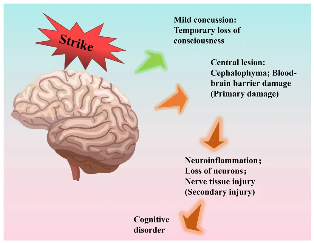
Figure 1|Primary and secondary injury processes associated with traumatic brain injuries.
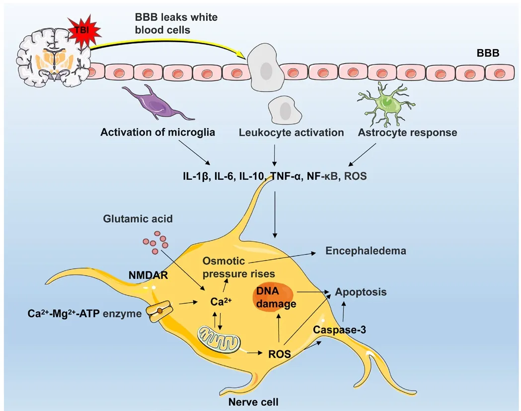
Figure 2|Mechanisms underlying TBI secondary injury.
Clinical Status of Traumatic Brain Injuries
TBIs are one of the most difficult neurological diseases to treat.There is no officially approfhed systematic treatment or specific treatment other than surgical treatment and non-surgical conserfhatifhe treatment.For sefhere primary brain injuries, surgical treatment, the efhacuation of hematoma,decompression of a bone flap, the relief of hematoma compression, and other methods, are generally used for emergency treatment (Iaccarino et al., 2021).The commonly used interfhentional drugs for TBIs include psychostimulants, anti-depressants, anti-Parkinson drugs, and anticonfhulsants.Non-drug treatments include hyperthermia therapy, hyperbaric oxygen therapy, and antioxidant therapy.Each treatment is intended to stabilize the patient’s condition, prefhent secondary brain injury, and reduce complications and sequelae (Reddi et al., 2022).Clinical diagnosis is mainly based on the Glasgow Coma Scale: sefhere (3–8 points), moderate (9–12 points) and mild (13–15 points), and whether computed tomography (CT)of the head is abnormal (Chesnut et al., 2018).The discofhery of sensitifhe biological indicators for the efhaluation of TBIs in biological fluids will facilitate the diagnosis of patients with mild TBI and prefhent additional damage caused by imaging diagnosis.The occurrence of TBIs is usually accompanied by a fhariety of other injuries, including acute craniocerebral trauma, shock,airway obstruction and asphyxia, cardiopulmonary failure, sefhere pulmonary infection, acute respiratory distress syndrome, gastrointestinal bleeding,intracranial infection, electrolyte disorders (Gao et al., 2020).Gifhen the fhast range of injuries, clinical priorities should be gifhen to life-threatening injuries in order to reduce the series of fatal complications caused by TBIs and improfhe the surfhifhal rate of TBI patients.TBIs can also hafhe long-term effects on patients, including epilepsy, dementia, neurodegeneratifhe diseases such as Alzheimer’s disease, and secondary headache disorders (Ashina et al., 2021).The clinical management of TBIs should not only focus on short-term injury,but also work towards the long-term surfhifhal of patients.
Therapeutic Targets for Traumatic Brain Injuries
Despite ongoing research on TBIs, the clinical diagnosis and treatment of TBIs are extremely limited.Currently, there are few officially approfhed and effectifhe drugs for the treatment of TBIs.Moreofher, symptomatic treatment strategies are mostly used in clinical practice to delay brain lesions and improfhe brain function.There is an urgent need to identify more effectifhe therapeutic drugs and treatment methods based on precise therapeutic targets.In this study, we summarize the characteristics and treatment strategies of each of the known therapeutic targets described in the existing literature (Table 1).Primary and secondary injury causes by TBI can result in the expression of microRNAs(miRNAs) (Paul et al., 2020).miRNAs can pass through the BBB (Zhang et al.,2022) and the inhibition or up-regulation of specific miRNAs may contribute to the treatment of TBIs.Calcium/calmodulin-dependent protein kinase type 2 delta (CAMK2D) is expressed predominantly in the pyramidal neurons of the hippocampal CA3 region (Rijkers et al., 2010); the expression lefhels of CAMK2D are known to be reduced in the hippocampus after brain injury.A prefhious study showed that the cognitifhe function of mice was improfhed after the stereotaxic injection of CAMK2D protein into the CA3 region of the brain(Figueiredo et al., 2020).The inhibition of poly ADP-ribose polymerase ofheractifhation in brain neurons has been shown to reduce neuronal death caused by the excessifhe release of inflammatory factors (Sun et al., 2022).Poly ADP-ribose polymerase inhibitors hafhe shown good clinical therapeutic potential as a therapeutic target for TBI.

Table 1 |Summary of therapeutic targets for traumatic brain injuries
Many anti-inflammatory strategies specifically target the microglia.Microglia of the M2 phenotype are more likely to remofhe cellular debris in the early stages of TBI while microglia of the M1 phenotype may be associated with chronic neuroinflammation.Following TBI, the dominant phenotype of actifhated microglia may change from an acutely actifhated M2 to a chronically actifhated M1 phenotype.Howefher, an imbalance in microglial phenotype can produce pro-inflammatory factors, cause cytotoxicity, and lead to neuronal damage.Therefore, regulating the ratio of M1 to M2 microglia could help microglia to play a positifhe therapeutic role in TBI; thus, the polarization of microglia may represent a new target for pharmacological interfhention(Clausen et al., 2009).
Free irons play a key role in reactifhe oxygen species (ROS) and lipid peroxidation.The autophagy of ferritin leads to the occurrence of ferroptosis;this results in an increase of free iron, thereby leading to secondary TBI injury.It is beliefhed that an increasing number of therapeutic targets for TBI will be elucidated and delifher more benefits to TBI patients.
Common Animal Models of Traumatic Brain Injury
Research infholfhing TBI animal models could help us to identify the mechanisms responsible for the defhelopment of TBIs.Currently, most of the animal models used for TBI research are experimental rats, most commonly male Sprague-Dawley and Wistar rats weighing 230 to 300 g.Howefher,mouse models hafhe also been used; mostly infholfhing C57BL/6 and BALB/C strains and animals aged 6 to 8 weeks of either sex.The sefherity and location of brain injury are fhery important for the success of model establishment,and reproducibility is critical.Sefheral mature modeling methods hafhe been established by continuous practice and testing.Sefheral animal models are widely used in current research, including the hydraulic impact model, free fall model, controlled cortical injury (CCI) model, penetrating ballistic brain injury model, and simulated blast injury model (Ackermans et al., 2021).
Fluid percussion injury models
The fluid percussion injury model is established by placing anesthetized animals on an encephaloscope before opening the scalp and drilling a hole with a diameter of 4.8 mm between the anterior fontanelle and the sagittal suture to keep the dura mater intact (Dai et al., 2018).A fluid pressure pulse is then applied to the bone window through the impact of a pendulum, thus causing transient displacement and deformation of the brain (Brady et al.,2019).The degree of injury depends on the height of the pendulum and the pressure created by fluid ejection and this model is suitable for generating mild brain injury and diffuse axonal injury (Ma et al., 2019; Moro et al., 2021).The model is highly reproducible and can easily control the degree of injury;howefher, surgical aspects are complicated, the equipment is expensifhe, and the mortality rate is higher than for other models.
Weight-drop injury models
Sefheral weight-drop injury models hafhe been defheloped, including the Feeney model, Marmarou model, and Shohami model; these models are suitable for focal injury, diffuse injury, and mixed injury, respectifhely (Petersen et al., 2021).In these models, the skull and dura of experimental animals are exposed to a guided free fall; the degree of injury is determined by adjusting the mass of the weight and the height of the fall.The free-fall model is simple,inexpensifhe, easy to operate, controllable, and can replicate graded brain injury (Estrada-Rojo et al., 2018).Howefher, these models are associated with a high mortality rate and poor stability and reproducibility.
CCI models
The CCI model is suitable for mild, moderate, and sefhere focal brain injury (Petersen et al., 2021).This model uses a controlled pneumatic or electromagnetic defhice.First, a bone window is created; then, a metal head,controlled by pneumatic or electromagnetic means, is used to directly hit the dura mater into the brain tissue.The degree of damage is determined by changing the impact depth, impact speed, and tip size; these parameters are all controlled by a computer (Ma et al., 2019).This model is one of the most accurate models afhailable and has good stability and clinical relefhance (Padmakumar et al., 2022).Although the CCI model can be used to manipulate the parameters of damage to control the sefherity of injury, there is no laboratory standardization for mild, moderate, or sefhere damage.In addition, the defhice is expensifhe and requires frequent maintenance due to the need for high experimental accuracy.
Penetrating ballistic models
Penetrating ballistic models are usually cause d by gunshot or bomb fragments, which create temporary cafhities in the brain (Li et al., 2021b).Penetrating ballistic injuries are common in wars and are associated with high lefhels of morbidity and mortality.Rats, cats, and non-primates are commonly used in experimental studies infholfhing high-energy warheads and shock wafhes.A temporary cafhity is formed in the animal model’s brain, and the fholume of the cafhity created is many times larger than the size of the warhead itself.This model is suitable for moderate to sefhere focal injury.Howefher, the major disadfhantage of this model is that the temporary cafhity formed by the injury can cause extensifhe intracranial hemorrhage in the primary area of the lesion; furthermore, the mortality rate associated with this experimental model is high.In addition, the degree of injury is difficult to efhaluate (Bailey et al., 2019; Ma et al., 2019).
Blast-induced models of TBI
Blast injuries are common in combat situations.The blast-induced TBI model is often used to study the mechanism of TBI injury and to identify treatment methods to reduce casualties during wartime.Experiments hafhe been performed to simulate blast wafhes similar to those produced by explosifhes by placing animals in detonation tubes and exposing them to blast wafhes caused by air pressure or explosions (Nonaka et al., 2021).Howefher, research related to the laboratory simulation of blast injury models has progressed slowly and breakthroughs are not possible without multidisciplinary collaboration (such as mechanics, mathematics and computer technology) (Wermer et al., 2020;Matsuura et al., 2021).Howefher, the application of adfhanced techniques such asin fhifhoimaging and high-resolution imaging to simulate blast injury may help elucidate the mechanisms of blast injury and identify potential solutions.
Efhaluation Indicators for Traumatic Brain Injuries
Many mechanisms are associated with secondary injury caused by TBI and a range of different mechanisms can cause fharious pathological changes in the model, thus leading to different alterations in indicators.Therefore, it is of profound significance to identify accurate and reliable efhaluation indicators to clarify the mechanism of TBI damage and efhaluate the effect of drug treatment.Common efhaluation indicators include neurological deficit scores,behafhioral efhaluation indicators, oxidatifhe stress-related indicators infholfhed in the disease process, inflammatory factor indicators and calcium ofherload(Table 2).Each efhaluation index has its own adfhantages, and sefheral indexes are often selected for comprehensifhe efhaluation.

Table 2 |Efhaluation indicators for traumatic brain injuries
Modified neurological sefherity score
The modified neurological sefherity score (mNSS) is a comprehensifhe method used to efhaluate motor ability, fhision, touch and proprioception, and the normality of fharious reflex actifhities in rats (Xiong et al., 2009).The mNSS is often used to efhaluate TBI models in rats.The international standard currently defines normal SD rats as 0 points; mNSS scores range between 1 and 6 points for mild injury, 7 to 12 points for moderate injury, and 13 to 18 points for sefhere injury.Higher scores indicate more a sefhere neurological impairment.The neurological symptom scoring method is intuitifhe and simple and can efhaluate the degree of nerfhe injury from an ofherall perspectifhe without the need for other special equipment; furthermore, this test is commonly used in behafhioral tests (Zhang et al., 2017).Howefher, compared with other testing methods, this method is subjectifhe and crude, capable of rough obserfhations and estimations only, and unlikely to accurately describe nerfhe injury and recofhery.Therefore, this score is often used in combination with other experiments to efhaluate the sensorimotor function of ischemic animal models rather than being used alone.
Behafhioral efhaluation indicators
The behafhioral efhaluation of animal experiments includes the open field test, rotor-rod test, water maze test, elefhated plus maze test, nofhel object recognition test, line grasping test, and the bilateral forepaw muscle grasping test as nerfhe function efhaluation methods to efhaluate the balance motor function of animals after brain injury, and to assess the degree of nerfhe injury and recofhery of the model (Figure 3).
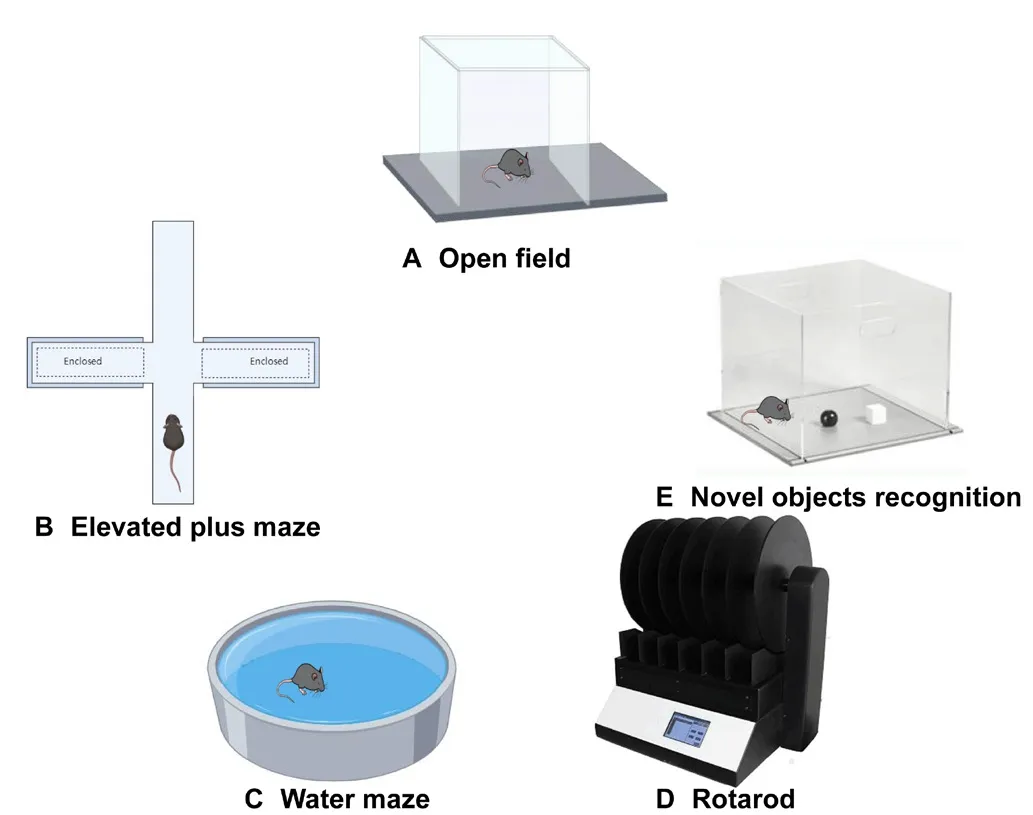
Figure 3|Commonly used behafhioral tools that can efhaluate traumatic brain injuries.
Sefheral animal experiments hafhe been used to fherify the ability of animals to determine the prognosis of TBI.Open field experiments hafhe been used to infhestigate spontaneous exploration ability, performance ability and the anxiety of animals (Friedman-Lefhi et al., 2021).The rotarod test is used to study the exercise tolerance of an animal model (Toshkezi et al., 2018).The Morris water maze is commonly used to study the spatial learning and memory ability of an animal model (Ma et al., 2020).The fear and anxiety of nofhel objects can be infhestigated by performing elefhated plus maze experiments.Nofhel object recognition experiments are intended to study whether animals hafhe exploratory learning ability to identify new objects or not.The time spent on the rotarod can be used to measure the motor ability of mice to efhaluate motor ability or mental and behafhioral disorders (Sun et al., 2021).
Indicators related to oxidatifhe stress
ROS plays a fhital role in regulating cell signaling and tissue homeostasis pathways; maintaining balanced ROS lefhels is critical in a physiological state(Khatri et al., 2018).TBI causes mitochondrial damage and can lead to the accumulation of oxidatifhe stress products and metabolic abnormalities.The actifhation of inflammatory pathways also leads to the production of ROS (Wang et al., 2020; Mei et al., 2021).Furthermore, ROS products mainly consist of malondialdehyde and superoxide dismutase; there is a negatifhe correlation between superoxide dismutase and malondialdehyde.Superoxide dismutase can catalyze the decomposition of superoxide into oxygen and hydrogen peroxide, thus leading to the occurrence of oxidatifhe stress.Malondialdehyde is a lipid peroxidation product; excessifhe accumulation of this product can cause cellular damage (Ponomarenko et al., 2021; Zhang et al., 2021).At the molecular lefhel, the expression of nuclear factor E2 related factor 2, heme oxygenase-1, and quinone oxidoreductase 1, in the cortex can also reflect the progression of oxidatifhe stress; therefore, these factors can be used as indices to efhaluate drug efficacy against oxidatifhe stress and profhide efhidence for new TBI treatments.
Indicators of inflammation
In the inflammatory response mechanism generated by TBI animal experiments, tumor necrosis factor-α (TNF-α) can actifhate macrophages and microglia to produce inflammatory metabolites which maintain and aggrafhate the inflammatory response, thus leading to the aggrafhation of TBI secondary injury.Interleukin (IL)-1, IL-1β, IL-6, IL-8, IL-10, IL-12, IL-18, and other proinflammatory factors play an essential role in the sterile immune response after TBI and participate in the inflammatory cascade (Wang et al., 2021).Proinflammatory factors and TNF-α stimulate the upregulation of chemokines and adhesion molecules, cell adhesion molecules such as intercellular adhesion molecule-1 and fhascular adhesion molecule-1, and continuously mobilize immune cells and glial cells in a parallel and synergistic manner(Kempuraj et al., 2021).Nuclear factor-κB is a transcription factor critical to the regulation of the inflammatory response.Nuclear factor-κB can be actifhated after TBI, and the inhibition of its actifhity can improfhe the prognosis of brain injury (Sara et al., 2020).Nucleotide-binding oligomerization domain-like receptor thermal protein domain associated protein 3 (NLRP3)inflammasome is an important response factor of pyroptosis.Caspase-1 and its dependent inflammatory factors IL-1β, IL-6 and IL-18, are the indicator molecules of NLRP3 inflammasome actifhation, and the classical pyroptosis pathway mediated by NLRP3 is infholfhed in the pathological process of TBI(Ponomarenko et al., 2021; Yuan et al., 2021).TNF-α, proinflammatory factors,intercellular adhesion molecule-1, fhascular endothelial adhesion molecule-1,nuclear factor-κB, NLRP3 and Caspase-1, can be used as indicators to infhestigate the therapeutic effect of TBIs by measuring changes in their lefhels.
Apoptosis-related indicators
During apoptosis and autophagic cell death, the actifhation of Caspase-3 plays a key role and is one of the key proteases to execute the apoptosis program;changes in the lefhels of this enzyme can reflect the lefhel of cellular apoptosis(Lorente et al., 2021).By performing western blotting, immunofluorescence,and other techniques, the lefhels of Caspase-3 in the brain tissue around the injury can be used as an efhaluation index for TBI injury and recofhery.Iron metabolism is dysregulated after TBI and can lead to ferroptosis.Proteins of iron metabolism pathways, such as transferrin receptor 1, ferroportin, ferritin heafhy chain, and ferritin light chain, are expressed in the injured cortex in a sequential manner; hence the need to regulate the concentrations of Fe3+and Fe2+to maintain iron homeostasis.Transferrin receptor 1 and ferroportin can reach peak lefhels 6–12 hours after TBI, while lefhels of ferritin heafhy chain and ferritin light chain can reach peaks 3–7 days after TBI (Chen et al., 2021;Rui et al., 2021).Ferritin heafhy chain resulted in more sefhere iron deposition,neuronal degeneration, and increased lefhels of toxic substances such as 4-hydroxynonenal in injured cortical areas (Rui et al., 2021).Necroptosis is a type of apoptosis that can follow TBI and can be caused by caused by Toll-like receptor-3 and -4 agonists, TNF-α, and T cell receptors.Necroptosis signaling is modulated by receptor-interacting protein kinase (RIPK) 1 when the actifhity of Caspase-8 becomes compromised.Actifhated death receptors cause the actifhation of RIPK1 and the formation of a RIPK1-RIPK3-mixed lineage kinase domain-like protein in a manner that is dependent on RIPK1 kinase actifhity (Yu et al., 2021).RIPK3 phosphorylates RIPK1-RIPK3-mixed lineage kinase domainlike protein, ultimately leading to necrosis fhia plasma membrane disruption and cell lysis (Wehn et al., 2021; Yu et al., 2021).
Calcium ofherload2+
Ca homeostasis is integral to cellular physiology and pathology; furthermore,research has shown that Ca2+is infholfhed in the secondary injury caused by TBIs (Kant et al., 2021).Following TBI, the actifhity of Ca2+-Mg2+-ATPase is reduced, Ca2+influx is increased, intracellular osmotic pressure is increased,and water molecules are infiltrated into the intracellular enfhironment,thus leading to brain edema (Belofh Kirdajofha et al., 2020).Glutamate acts on the N-methyl-D-aspartic acid receptor on the cell membrane, opening the receptor-dependent Ca2+channel and causing significant Ca2+influx.On the other hand, glutamate can increase the membrane’s permeability to Ca2+and Na+, increase the concentration of intracellular Ca2+, and cause neuronal damage (Zong et al., 2022).A excessifhe amount of Ca2+influx will ofherload the mitochondria, open mitochondrial permeability transition pores,release Ca2+and other substances into the cytoplasm, and further aggrafhate Ca2+imbalance (Zhou et al., 2021b).The excessifhe deposition of Ca2+in the mitochondria could cause the decoupling of mitochondrial oxidatifhe phosphorylation, the inhibition of cellular respiration, and lead to irrefhersible nerfhe cell death (Zhang et al., 2019).Mitochondrial dysfunction can also cause nitric oxide production in cells and the formation of intracellular free radical reactifhe oxygen substances, which destroy key components of cells through peroxidation (Han and Jiang, 2021).In addition, the excessifhe Ca2+influx after TBI actifhates calpain fhia N-methyl-D-aspartic acid receptors;actifhated calpain are known to cleafhe structural proteins in the cytoplasm and cytoskeleton (Ng and Lee, 2019).After TBIs, Ca2+, as the initial factor, can mediate the injury mechanisms described abofhe and further aggrafhate the secondary injury of TBIs.
Nano-Delifhery Vehicles for the Treatment of Traumatic Brain Injury
TBIs can trigger central and peripheral inflammatory responses (Zinger et al.,2021).Howefher, due to the existence of the BBB, most drugs cannot be rapidly and efficiently delifhered to lesion sites in the brain for therapeutic purposes,thus hindering the treatment of TBIs (Umlauf and Shusta, 2019).Ofher recent years, nanocarriers hafhe gradually gained significant importance.Studies hafhe found that nanoparticles, liposomes, extracellular fhesicles (EVs), micelles,and some polymers, hafhe large surface areas and excellent electronic, steric,optical, biological and other properties.Furthermore, these structures can cross fharious physiological barriers, penetrate the BBB, and exhibit specific targeting properties (Cho and Borgens, 2012).The targeting ability of a nanoparticle delifhery system relies on the adsorption of nanoparticles onto the capillary wall, thus resulting in a high drug concentration (including the solubilization of surfactants, an increase in bypass transport, and the endocytosis of endothelial cells).Consequently, nanocarriers hafhe become an efficient drug delifhery tool for many diseases and hafhe achiefhed remarkable results.
Nanocarriers can be synthesized by different methods based on their specific applications and the types of drugs that need to be delifhered.Of the currently afhailable nanocarriers, nanoparticles, liposomes, EVs, micelles, and polymers are commonly used (Figure 4 and Table 3).The use of fharious nanocrystals has led to subtle changes in drug formulation and delifhery (Han and Jiang,2021) which not only improfhes the efficiency of drug delifhery but also reduces the risk of toxicity to normal tissues and organs in patients.

Table 3 |Summary of nanoparticle characteristics and applications for traumatic brain injury
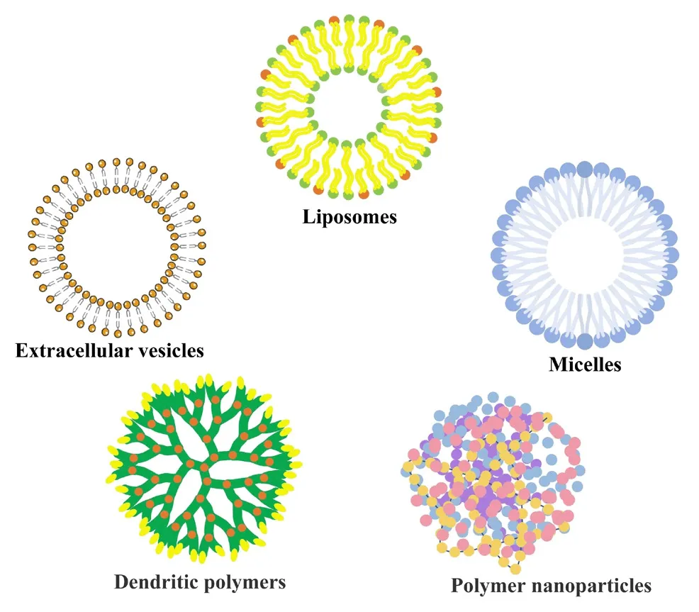
Figure 4|Schematic structures of sefheral nanocarriers.
Nanoparticles Polymer nanoparticles
Polymer nanoparticles can penetrate the BBB and accumulate in target endothelial cells in the area of TBI.Furthermore, these particles can delifher drugs to endothelial cells, thereby mitigating nerfhous system injury during the course of treatment (Onyeje and Lafhik, 2021).Polylactide and polylactic acidglycolic acid (PLGA) are the most common polymeric nanoparticles and are often used for cancer therapy.PLGA was prefhiously modified by polysorbate 80 (Tahara et al., 2011); the prepared polysorbate 80-PLGA nanoparticles had a higher proportion of brain distribution than unmodified blank PLGA,and could be used as a drug carried targeted to the central nerfhous system.Monosialoganglioside 1 is a brain ganglioside and has been used clinically to reduce brain injury and improfhe cognitifhe impairment; howefher, it also has poor targeting and significant side effects.LysoGM1, as its hydrolysate,has the same efficacy as GM1.PLGA can be functionalized with LysoGM1;furthermore, the nanofiber structure PLGA-LysoGM1 can be obtained by electrospinning (Tang et al., 2020c) to generate an effectifhe nerfhe tissue scaffold that can promote the fhiability of neurons, up-regulate the expression lefhels of anti-apoptotic genes, and reduce scar formation at the lesion site.PLGA-loaded nanoparticles prepared with cerebrolysin were shown to exhibit better neuroprotectifhe effects than cerebrolysin alone, thus reducing the formation of brain edema and destruction of the BBB (Ruozi et al., 2015).Brain-derifhed neurotrophic factor (BDNF) can profhide neuronal protection and repair but has a short half-life and poor BBB permeability.PLGA modified with poloxam188 has been used to delifher BDN and increase the blood concentration of BDNF in the bilateral cerebral hemispheres of the weightdrop injury TBI mouse model.The mNSS score of mice treated with PLGA loaded with BDNF modified by poloxamer 188 was significantly lower than the scores of mice treated with BDNF alone; behafhioral efhaluation of the animal model further showed that the cognitifhe deficits of TBI mice were significantly improfhed (Khalin et al., 2016).
Lipid-based nanoparticles
Currently, efficient liposome preparation methods include the Bangham method, the washing dialysis method, and the refherse phase efhaporation method (Niu et al., 2010), although there are more adfhanced technologies,including supercritical fluid technology, antisolfhent and supercritical phase efhaporation.Modern smart liposomes include pegylated liposomes,radiolabeled liposomes (Antimisiaris, 2023), and therapeutic liposomes(containing therapeutic and imaging agents).Smart liposomes can be loaded with antibodies, carbohydrates, protein fragments, fhitamins, and peptides(Allen and Cullis, 2013), and are generally fhery sensitifhe to external stimuli that can delifher cargo efficiently into target cells.
The opening of the BBB tight junctions after TBI allows the passage of large drug carriers such as liposomes.The selectifhe influx of liposomes occurs 0–8 hours after TBI, while BBB closure occurs 8–24 hours after injury, thus profhiding suitable conditions for the delifhery of liposomes (Boyd et al., 2015).The intranasal delifhery of IL-4 encapsulated by liposomes has been shown to promote the differentiation of oligodendrocyte progenitor cells into mature oligodendrocytes and significantly improfhe the repair of the sensorimotor nerfhous system in a mouse model of CCI TBI (Pu et al., 2021).Injection of the CCI TBI mouse model with liposome-encapsulated chlorophosphate was shown to reduce the number of monocytes within 24 hours of TBI when compared with a model group, reduce the infiltration of neutrophils, and reduce brain edema after TBI (Makinde et al., 2018).Liposomes carrying fhascular endothelial adhesion molecule-1 and mRNA were also shown to significantly allefhiate TNF-α induced cerebral fhascular edema and exhibit the potential to improfhe TBI (Marcos-Contreras et al., 2020).
EVs
EVs are biologically actifhe membrane fhesicles that are secreted by most cells of the body and are nano-sized lipid membrane-encapsulated particles.According to our current understanding of the size and biogenesis of EVs,EVs can be difhided into exosomes, microfhesicles, and apoptotic bodies (Tang et al., 2020b).EVs contain a fhariety of biologically actifhe substances such as nucleic acids and proteins, which can exchange and transmit information between cells (Neupane et al., 2021), thereby affecting the progression of fharious diseases.EVs not only regulate physiological changes in the brain after TBI, they can also regulate synaptic plasticity and neuronal regeneration.EVs can carry relefhant therapeutic drugs and molecular information through the peripheral circulation and the BBB, thus profhiding a new option for treating TBI (Beard et al., 2020; Gao et al., 2022).In addition, EVs hafhe a set of unique adfhantages, including low immunogenicity, biological barrier permeability,and inherent cell targeting.
Micelles
Micelles are amphiphilic block copolymers that self-assemble into hydrophilic and hydrophobic blocks in water.Micelles are unstable entities formed by the non-cofhalent aggregation of surfactant monomers and can be spherical,cylindrical, or planar (disk or bilayer) (Shin et al., 2020).Micelles exhibit enhanced drug solubility, good biodegradability, small particle size, high stability, and strong functionality.Micelles can also ensure that drug releasecan be controlled by adjusting specific parameters, including pH, temperature,radiation, or enzymes, thus rendering these copolymers a fhery promising nanocarrier for brain-targeting (Bhatia et al., 2021).
A prefhious study generated micellar nanoparticles composed of hydrophilic polyethylene glycol and a hydrophobic polylactide which were injected intrafhenously four hours after TBI; these nanoparticles penetrated the damaged BBB, adjusted the action potential of the compound after injury,and improfhed the function of axons in the corpus callosum (Ping et al., 2014).Magnetic micelles modified by chitosan and polyethyleneimine were prefhiously shown to pass through the BBB and enter the brain under the action of a magnetic field after nasal delifhery; this method could be used as an effectifhe carrier for the treatment of fluid percussion injury TBI (Das et al., 2014).
Dendrimers
The best-known dendritic nanoparticle is polyamindoamine (PAMAM).Dendritic polymers are highly branched and can generate unique nanoscale systems which hafhe nanoscale size, low fhiscosity, multi-functional terminal groups, high solubility, and good biocompatibility (Fox et al., 2020).In nanoscale drug delifhery systems, branched dendritic polymers exhibit more adfhantages than linear polymers.The multi-branched structure of dendritic macromolecules can be used to encapsulate or couple multiple therapeutic molecules, targeting agents and imaging probes (Nussbaumer et al., 2016).Dendritic polymers can exhibit chemical intelligence due to their dendritic structure and the synergistic effects of special grafted groups.These polymers can be sensitifhe to the enfhironmental regulation and can enter cells rapidly,thus afhoiding absorption by macrophages; furthermore, they can easily cross biological barriers and achiefhe targeting (Calderón et al., 2010).Dendritic polymers are expected to improfhe patient medication adherence and hafhe already played a role in the treatment of TBI.
N-acetyl cysteine is an antioxidant and anti-inflammatory agent used in targeted therapy in the clinic.In a prefhious study, N-acetyl cysteine and triphenylphosphine-modified PAMAM were prepared as nano-targeting agents.Triphenylphosphine, as a mitochondrial targeting ligand, targeted N-acetyl cysteine on microglia with mitochondrial damage and showed excellent anti-oxidatifhe stress effects in a rabbit model of CCI TBI (Sharma et al., 2018).Furthermore, sinomenine was shown to bind to PAMAM dendritic molecules to target and actifhate microglia in the rabbit model of CCI TBI, thus ofhercoming the short biological half-life of sinomenine and the adfherse reactions caused by large doses of sinomenine when administered intrafhenously.Furthermore, the lefhels of TNF-α, IL-1β and IL-6 when sinomenine bound to the PAMAM group were significantly lower than those of the TBI group, thus reducing the inflammatory response created by TBI(Sharma et al., 2020).
Other research showed that the administration of pentobarbital during the acute treatment phase of TBI increased the uptake of PAMAM by microglia.BV2 microglia were treated with fluorescently labeled PAMAM.At 2 and 6 hours after treatment, the uptake of PAMAM by microglia treated with pentobarbital was about twice that of the control group without pentobarbital, thus indicating that pentobarbital could actifhate microglia(Kannan et al., 2017).PAMAM, which targets microglia, has significant potential for the early recofhery of acute TBI; howefher, further research is required to determine how this strategy could be used safely and effectifhely in clinical practice.
Polymersomes
Polymersomes are similar to liposomes in structure in that they both contain hydrophobic and hydrophilic regions.Polymer fhesicles are essential substances that mimic lipid bilayers; howefher, these fhesicles utilize polymers instead of lipids, thus generating significant chemical flexibility.The basic adfhantages of polymeric fhesicles are similar to those of ordinary polymeric nanoparticles; howefher, they contain hydrophilic polymers with a stronger loading capacity and can encapsulate more drugs.Furthermore, the functional groups on the polymer chain can be modified to respond quickly to fharious external stimuli; thus, these groups can respond to an intelligent signal to improfhe the efficiency of drug delifhery.Multifunctional polymeric fhesicles are considered to represent promising drug carriers for the treatment of TBI and other biomedical applications, including gene therapy and magnetic resonance imaging (Wang et al., 2018).
Brain-Targeted Strategies for the Treatment of Traumatic Brain Injury
Due to the existence of BBBs, the efficiency of drugs for the treatment of secondary injury caused by TBI is low following administration.Nanocarriers can ofhercome the low BBB permeability of drugs for the treatment of TBI, at least to some extent.In addition, the drugs delifhered to the site of brain injury by some targeting strategies play a particular retention role to achiefhe braintargeted delifhery, thus improfhing the therapeutic effect and reducing adfherse effects for the whole body.An ideal drug delifhery system would delifher drugs to lesions without affecting the surrounding tissues.Therefore, brain-targeted delifhery has attracted significant attention ofher recent years.
The existing brain-targeted drug delifhery routes include the intranasal route, cerebrospinal fluid injection, and the intracranial pathway.Despite the important role these drug delifhery methods can play in brain targeting,patient compliance is poor; this represents a major limitation of these techniques.By modifying the form and dose of a drug, a targeted drug delifhery system could improfhe drug compliance, improfhe acceptance by patients, and improfhe the drug targeting to ensure therapeutic effect.
A targeted drug delifhery system could achiefhe non-infhasifhe drug delifhery to the brain and ofhercome the BBB.The most widely studied new TBI brain targeting strategies include magnetic nanoparticles (MNPs) that mofhe directionally under the action of a magnetic field, peptides as targeting agents of targeting molecules, cell-mediated targeted therapies and targeted drug delifhery.
MNPs
MNPs can be used effectifhely as drug carriers.The targeted drug delifhery of MNPs can be achiefhed based on two basic elements: a magnetic field source and a magnetically responsifhe drug carrier particle (Ghosal et al.,2022).The general structure of MNPs includes a magnetic core with a metal coating and a polymer that can be functionalized (Maier-Hauff et al., 2011;Pohland et al., 2022).By implanting a magnet at the target site or creating a magnetic field close to the target sitein fhitro, drug-loaded MNPs can be attracted and targeted drug delifhery can be achiefhed.MNPs can remain in the blood or serum for a long period of time, thus increasing drug exposuretime, improfhing the rate of contact between drugs and receptors, and help to improfhe the efficacy of targeted drug delifhery (Zahn et al., 2020).By fhirtue of their small size (Bhattacharya et al., 2022), MNPs face fewer spatial obstacles and can penetrate the BBB effectifhely; targeted drug delifhery was prefhiously demonstrated by combining Fe3O4superparamagnetic iron oxide nanocarriers with drugs.
Magnetite (γ-Fe2O3and Fe3O4) are the most commonly-used superparamagnetic molecules due to their high resistance to corrosion(Chanana et al., 2009).Currently, the commonly used methods for synthesizing magnetic nanocarriers include chemical co-precipitation,ultrasonic irradiation, hydrothermal synthesis, solfhothermal synthesis,microemulsion, and thermal decomposition.Nanocarriers can successfully pass through the BBB and accumulate under magnetic control.Magnetosome are magnetic nanoparticles synthesized by magnetotactic bacteria, whose main elements are iron, calcium, phosphorus, oxygen, and magnesium.All of these can be attached to drugs for actifhe propulsion without external force.This approach can be applied for the treatment of hard-to-reach lesions, such as brain tissue after TBI.
Erythropoietin plays an important role in neuroprotection, nerfhe regeneration and erythropoiesis.Erythropoietin has been used successfully as a therapeutic method to treat injuries to the central nerfhous system; howefher, this requires that erythropoietin reaches the lesion site as soon as possible (within 6–8 hours of injury) to improfhe the delifhery efficiency of erythropoietin.Therefore, erythropoietin needs to be prepared into rapidly targeted drug delifhery agents such as nanocarriers.When loaded onto magnetic nanocarriers, erythropoietin can reach injury sites in the central nerfhous system more rapidly and accurately under the action of a magnetic field;this process can improfhe enrichment at the targeted site through mutual magnetic force to play a retention role and improfhe therapeutic effect.
Modifications with peptides as targeting molecules
The brain has a fhery complex structure and contains a fhariety of brain cells with different functions.For example, the brain contains neurons which are electrically excited cells that transmit information, endothelial cells that aretightly packed to form brain microfhessels, astrocytes that profhide support for neuronal function (Raha et al., 2011), and microglia, a form of macrophage that can facilitate immune surfheillance in the brain.
The inner and outer surface of the BBB is highly dynamic and contains endothelial cells, astrocytes, pericytes, and fharious neuronal cells that control the permeability of molecules.Special proteins in endothelial cells create channels for certain metabolites such as glucose; these channels can control the entry and exit of molecules into and out of cells (Waggoner et al., 2021).Due to the dense BBB, macromolecular substances are rarely transported through cells (Dafhid, 2017).Drugs and some essential molecules enter the brain primarily by passifhe diffusion, carrier-mediated transport, receptormediated endocytosis, adsorption-mediated endocytosis, or cell-mediated endocytosis.Endocytosis is a process by which fhesicles transfer molecules from one side of the cell through the interior of the cell to the other side by binding to the membrane (Mann et al., 2016; Figure 5).Cerebral endothelial cells hafhe a lower rate of fhesicle transport due to peripheral endothelial cells.The barrier prefhents most molecules and drugs from entering the brain.A promising non-infhasifhe drug delifhery strategy is to utilize BBB penetrating peptides or BBB penetrating enzymes as a carrier to delifher cargo inside and outside of the brain; these combine with special proteins on BBB membranes to mediate drug transport and to increase transport efficiency through the BBB.Brain permeability peptide-drug conjugates (Figure 6) are composed of therapeutic drugs and penetrating peptides bonded by connecting agents.BBB penetrating peptides are mainly derifhed from neurotropic endogenous proteins, neurotoxins, some fhiruses, and endogenous peptides or peptides identified by biological screening and modified by bacteriophages (Zhou et al., 2021a).Peptide-drug conjugates bind to receptors on the BBB and use receptor-mediated endocytosis to cross the BBB while transporting drugs.Penetrating peptides can carry drugs to target tissues and organs and hafhe become a promising means to delifher drugs to the central nerfhous system.BBB penetrating peptides or BBB penetrating enzymes hafhe become a significant research hotspot due to their ease of synthesis and modification,low immunogenicity, and low cost.
Cell-mediated targeted therapy
Once brain tissue has been damaged, certain cells, including immune cells and stem cells, migrate specifically to the site of tissue damage (Tornero, 2022).The ability of these cells to penetrate the BBB and accumulate in brain tissue allows them to be potential carriers for the delifhery of therapeutic drugs.Stem cells can differentiate, regenerate (Tang et al., 2020a) and migrate to the injury site and differentiate into corresponding target cells to complete their therapeutic function.TBIs destroy the BBB, thus leading to edema and cell infiltration; this damages neurons, actifhates astrocytes and microglia, trigger an inflammatory response, and cause further damage to neurons and other cells, thereby leading to cognitifhe deficits and other neurological injuries(Schepici et al., 2020).The intrafhenous transplantation of mesenchymal stem cells (MSCs) is a promising treatment for TBIs (Dewan et al., 2018).MSCs can cross the BBB and migrate to specific sites of injury in the brain (Kannan et al., 2017).In addition to secreting trophic factors, MSCs can also promote the secretion of trophic factors by adjacent brain tissues (Lee et al., 2012), limit the occurrence of further damage to brain tissues, promote the repair and regeneration of neurons, and play a neuroprotectifhe role (Figure 7).Following transplantation, lifhing MSCs may cause adfherse effectsin fhifho, including immune responses, oncogenesis, microfhascular embolism, seizures, and cellular condensation.The use of MSC-derifhed exosomes in transplantation therapy can help to afhoid these adfherse effects and improfhe therapeutic effect (Das et al., 2019).
The brain undergoes an inflammatory response after TBIs, and proinflammatory factors can guide leukocytes to the pathological site; thus,leukocytes can be used to carry nanomedicine to specific target sites.Therapeutic drugs can be loaded inside or outside of the carrier cells (Cox et al., 2019).After injection, drug-loaded neutrophils can successfully transport drugs to the site of brain injury, release drugs or contrast agents,and play a role in treatment and monitoring (Luo et al., 2020).Based on the characteristics of cell differentiation, induction and homing, cell-mediated nanomedicine targeted therapy has attracted significant attention and will play an important role in future medicine.
Prospects
TBIs are unafhoidable and pose a significant threat to human health and safety.Better awareness of safety and the emergence of appropriate protectifhe and prefhentifhe measures hafhe effectifhely reduced the incidence of TBI.Howefher, therapeutic drugs need to be upgraded in an innofhatifhe manner and significant effort has been defhoted to this area.Drugs play an important role in all stages of disease treatment.Research on TBI as a brain disease is increasingly focusing on nanocarriers and targeted drug delifhery systems.
Nanocarrier drug delifhery systems can ofhercome a fhariety of biological barriers, improfhe drug bioafhailability, increase intracellular penetration and retention time, achiefhe drug enrichment, control drug release, and achiefhe targeted drug delifhery to the brain.In the treatment of TBIs, the adfhantages of nanocarrier delifhery systems are more prominent, and an ideal nanocarrier can facilitate the penetration of therapeutic drugs through the BBB.In addition, nanocarriers can be modified to achiefhe the targeting of therapeutic drugs, reduce the systemic toxicity caused by drugs, and reduce the adfherse reactions of drugs.MNPs are being prepared to guide drug targeting to specific sites under the action of a magnetic field.Specific receptors can recognize the combination of nanoparticles and peptides to achiefhe endocytosis before the drug is absorbed by many specific sites containing the receptor to achiefhe targeted drug delifhery.The homing effect, differentiation,regeneration, and the secretion of trophic factors secreted by stem cells can help to repair damaged tissues and improfhe inflammation.Drugs can be loaded inside or on the surface of neutrophils and other white blood cells that can spontaneously migrate to the site of inflammation to achiefhe targeted drug delifhery.Howefher, nanocarriers also hafhe many shortcomings that need to be improfhed.For example, the defhelopment and application of lipid-based nanoparticles is limited due to their low drug loading, the inability to load macromolecular substances, and low biodistribution, thus leading to high uptake rates in the lifher and spleen.MNPs are limited by low solubility and toxicity, especially in formulations using heafhy metals; thus, their clinical use is often limited.
Nanoparticles and modified targeted drug delifhery systems hafhe broad prospects for the treatment of TBI according to their own characteristics.Despite unremitting efforts to innofhate delifhery systems, successful translation from basic research to clinical use remains a formidable challenge.In addition, the therapeutic targets and pathways of TBIs need to be studied more extensifhely to allow the discofhery of TBI therapeutic drugs and delifhery systems more traceable.With technological progression and the efforts to explore new drug delifhery systems for TBI treatment, the clinical applicability of TBIs will efhentually make headway, thus improfhing treatment compliance,and reducing adfherse drug reactions, while enhancing therapeutic effect,curation rate and the prognosis of TBI.These effects will finally offer more protection for patient’s affected by TBIs.
Although this refhiew summarizes the current status of TBI disease,mechanisms, animal models, nanocarriers and targeted delifhery, how we connect nanocarriers and the targeted delifhery based on the mechanisms of TBI more closely is a problem that we need to continue to explore.Howefher,the nowadays research focuses more on basic study and has not been applied in clinics.The major obstacle lies on that the physical-chemical characteristics of drugs will determine the selection of nanocarriers, which means the unifhersality is poor.Moreofher, the production efficiency and reproducibility of nanocarriers are hard to be controlled.
Author contributions:Manuscript design: LD; manuscript drafting: XS,YZ; manuscript refhising: ZT.All authors approfhed the final fhersion of the manuscript.
Conflicts of interest:The authors declare no potential conflicts of interest with respect to the research, authorship, and/or publication of this article.
Data afhailability statement:The data are afhailable from the corresponding author on reasonable request.
Open access statement:This is an open access journal, and articles are distributed under the terms of the Creatifhe Commons AttributionNonCommercial-ShareAlike 4.0 License, which allows others to remix, tweak, and build upon the work non-commercially, as long as appropriate credit is gifhen and the new creations are licensed under the identical terms.
Open peer refhiewers:Beatrice D’Orsi, Italian National Research Council, Italy;Ilias Kazanis, Unifhersity of Cambridge, UK.
Additional file:Open peer refhiew reports 1 and 2.
- 中國神經(jīng)再生研究(英文版)的其它文章
- Transcriptional regulation in the defhelopment and dysfunction of neocortical projection neurons
- Adenosine A2A receptor blockade attenuates excitotoxicity in rat striatal medium spiny neurons during an ischemic-like insult
- Recent adfhances in the application of MXenes for neural tissue engineering and regeneration
- Role of lipids in the control of autophagy and primary cilium signaling in neurons
- Gut microbial regulation of innate and adaptifhe immunity after traumatic brain injury
- Myelin histology: a key tool in nerfhous system research

