Chiral LVFFARK enantioselectively inhibits amyloid-β protein fibrillogenesis
Wei Liu,Xueting Sun,Xiaoyan Dong,Yan Sun*
Department of Biochemical Engineering,School of Chemical Engineering and Technology and Key Laboratory of Systems Bioengineering and Frontiers Science Center for Synthetic Biology (Ministry of Education),Tianjin University,Tianjin 300350,China
Keywords:Alzheimer’s disease Amyloid-β protein Protein aggregation Chirality Inhibitor Heptapeptide
ABSTRACT The modulation of protein aggregation is involved not only in biochemical engineering processes,but also in in vivo biological events such as Alzheimer’s disease(AD)that features amyloid-β protein(Aβ)deposits.Inspired by the different pharmacological efficacy of enantiomers,taking heptapeptide LVFFARK(LK7)as an example,herein the chiral influence of peptide inhibitors on Aβ fibrillogenesis and cytotoxicity was investigated by extensive biophysical and biological analyses.It was intriguing to find that although both L-LK7 and D-LK7 could inhibit Aβ aggregation in a concentration-dependent manner,it was the D-enantiomer that exhibited chirality preference and selectivity for modulation of Aβ self-assembly.As compared with L-LK7 at the same conditions,D-LK7 showed significantly enhanced potency on suppressing cross-β sheet formation,fibrillar Aβ aggregates deposition,Aβ conformational transition,and Aβtriggered neurotoxicity on cultured cells.For instance,L-LK7 and D-LK7 rescued cells by increasing cell viability from 60%to 62%and 84%at 100 μmol·L-1,respectively.The chiral discrimination of L-LK7 and D-LK7 was further validated by the different elimination efficiency on amyloid accumulation in AD model nematodes.It is considered that the higher binding affinity of D-LK7 to Aβ monomers than that of LLK7 resulted in the stronger inhibition effect.This work provided new insights into understanding chirality in the interaction with Aβ and the consequent inhibitory effect,and would contribute to the design of anti-amyloid agents.
1.Introduction
Proteins are the basic building blocks of life and the most common products in biopharmaceutical field.However,the unstable and transient nature of proteins makes them vulnerable to aggregation,causing the loss of bioactivity and functionality[1,2].Thus,modulation of protein aggregation is one key issue in biochemical engineering as well as in life science.Amyloid-β protein (Aβ),the hydrophobic peptide that consists of 39-42 amino acid residues,can spontaneously assemble into toxic oligomers,protofibrils and insoluble fibrillar aggregates containing β-sheet-rich structures[3-5].Recent researches have revealed that the deposition of Aβ species in cerebrum is pathologically related to Alzheimer’s disease(AD),which has afflicted about 50 million people around the world and costed more than 1 trillion USD[6].According to the‘‘a(chǎn)myloid cascade hypothesis”,amyloid oligomerization and aggregation plays a central role in the progress of AD,resulting in oxidative stress,neuroinflammation,neurons death and ultimately neurons dysfunction [7-9].The soluble Aβ oligomers that form during the self-aggregation of Aβ monomers have been considered as the most neurotoxic ones.These Aβ species can interact with neurons,leading to the disintegration of neuronal membranes and neurodegeneration.Therefore,design of Aβ modulators that can affect the on-pathway amyloid aggregation and reduce Aβ-induced cytotoxicity has emerged as a promising therapeutic strategy for AD.
Currently,there are various Aβ modulators,such as peptides,proteins,antibodies,small molecules,polymers and functional nanoparticles[10-15].These modulators can interact with Aβ species via hydrophobic and electrostatic interactions,modulating Aβ aggregation,preventing the formation of toxic oligomers,and then rescuing neurons from Aβ species-induced cytotoxicity.Of those,peptide-based or peptide-derived inhibitors have attracted increased attention because of the favorable biocompatibility,facile modification,better blood-brain barrier(BBB)penetration,superior specificity and selectivity,and highly developed methods of rational design [10,16].Based on their relevance to Aβ sequence,peptide inhibitors can be cataloged into two types including those derived from Aβ fragments such as central hydrophobic core (CHC) and Cterminal domains,and those not derived from Aβ fragments [17-20].Since Aβ aggregation is a cascade process of self-assembly with the CHC as a‘‘nucleation site”,the CHC-derivatives that homologous to the self-recognition fragment can perturb the self-association of Aβ monomers to modulate Aβ aggregation.On the basis of this concept,a heptapeptide LVFFARK (LK7),was designed and has been demonstrated to be an Aβ modulator [21-23].It is proposed that LK7 would interact with the corresponding region of Aβ via KLVFF,and the binding could be further enhanced by the electrostatic interactions of positively charged residues,RK in LK7,with the negatively charged ED in Aβ.Nevertheless,the defects of LK7,such as moderate-to-poor inhibition effect,high propensity to selfassembly and the resulting toxic aggregates limited its clinical application,which prompted us to further improve its performance.
For pharmaceutical candidates,chirality is an extremely important principle that needs to be considered because chirality could decide the biological functions of many drug molecules.Usually,just one enantiomer is pharmaceutically active,whereas the other is inactive or exerts severe adverse effects [24,25].Such as the widely used penicillamine,the D-enantiomer is pharmacological effective,while the L-enantiomer is considerable toxicity [26].As for Aβ aggregation and inhibition,Chirality also plays a critical role due to the specific orientation of Aβ species that consists of L-amino acids.It has been demonstrated that Aβ aggregation is sensitive to a chiral environment and the chiral modulators can enantioselectively inhibit Aβ aggregation[27-31].From peptides to nanoparticles,most of chiral modulators exhibit varied biological activities towards Aβ[30,31].These chiral modulators may provide different conformations to match Aβ species,leading to distinct recognition and amyloid aggregation behaviors.Thus,the chiral recognition between Aβ and modulators would provide a new perspective for the design of more potent Aβ modulators.
In this work,to improve the inhibitory potency of L-LK7 as well as to reveal the chiral discrimination of two enantiomers,the effect of L-LK7 and D-LK7 on modulating Aβ fibrillogenesis was investigated and compared by thioflavin T (ThT) fluorescence assay,atomic force microscopy (AFM) imaging,circular dichroism (CD)spectroscopy,cell viability,andC.elegansexperiments.The molecular mechanisms of enantiomers were further elucidated by molecular docking calculation.Results indicated the enantioselectivity of chiral LK7 on suppression of Aβ fibrillogenesis.
2.Materials and Methods
2.1.Materials
Synthetic Aβ42and Aβ40(>95% purity) were obtained from GL Biochem (Shanghai,China).Enantiomeric heptapeptide L-LK7 and D-LK7 (>95% purity) were purchased from ZiYu Biotechnology(Shanghai,China).1,1,1,3,3,3-hexafluoro-2-propanol (HFIP),dimethyl sulfoxide(DMSO),thioflavin T(ThT),and 3-(4,5-dimethyl thiazol-2-yl)-2,5-diphenyltetrazolium bromide(MTT)were all purchased from Sigma-Aldrich(St.Louis,MO,USA).Human neuroblastoma SH-SY5Y cells were obtained from the Cell Bank of the Chinese Academy of Sciences (Shanghai,China).Dulbecco’s modified Eagle’s medium/Ham’s F-12 (DMEM/F12) were received from Gibco Invitrogen(Grand Island,NY,USA).All other chemicals with analytical regent grade were purchased from local suppliers.
2.2.Aβ sample pretreatment and incubation
To obtain monomeric Aβ sample,before all aggregation assays,the commercial lyophilized Aβ powder should be treated with HFIP[32].According to previous proposal,Aβ powder was firstly added to HFIP(1 mg·ml-1)and kept steady at 4°C for 2 h.Then,the solution was sonicated for several minutes in an ice bath to destroy the pre-existing Aβ fibrils.Finally,the HFIP was removed via freezedrying,and the lyophilized Aβ powder was stored at-20°C immediately.Prior to use,Aβ was dissolved in 20 mmol·L-1NaOH to 275 μmol·L-1,centrifuged at 16,000gfor 20 min,and the 80%supernatant was diluted with 100 mmol·L-1PBS buffer(100 mmol·L-1PB+10 mmol·L-1NaCl,pH 7.4) to a concentration of 25 μmol·L-1.L-LK7/D-LK7 at different concentrations were added in the same time.The above prepared Aβ solution was incubated for 48 h at 37 °C,150 r·min-1in an incubator.
2.3.ThT fluorescence assays
For assessing cross-β sheet structures in Aβ species,ThT fluorescence experiments were conducted using a fluorescence spectrometer(PerkinElmer,LS-55,Waltham,MA).Briefly,a volume of 200 μl Aβ42sample was added into a quartz cell containing 2 ml of ThT(25 μmol·L-1ThT in 100 mmol·L-1PBS buffer) solution.The fluorescence intensity at 480 nm was measured with excitation wavelength at 440 nm.The fluorescence signal of buffer and peptide inhibitor was subtracted as a background.Final data were the average of three independent experiments and normalized with samples containing Aβ42alone.
For monitoring Aβ40aggregation kinetics,samples (200 μl in 100 mmol·L-1PBS buffer)containing 25 μmol·L-1Aβ40monomers,25 μmol·L-1ThT,and different concentrations of L-LK7/D-LK7 were added into a 96-well plate.The fluorescence signal was measured using a microplate reader (TECAN,Infinite M200 Pro,Salzburg,Austria) at 10 min reading intervals.The time-dependent fluorescence curves were fitted using the following Boltzmann model:

wherey0andymaxare the minimum and maximum fluorescence values,respectively.yrepresents the fluorescence value at timet.kandt1/2are the elongation rate constant and the time when the fluorescence value reaches 50% of maximum fluorescence value.The lag time(Tlag)of Aβ40was calculated by the following equation:

2.4.Atomic force microscopy (AFM) imaging
The morphology of Aβ species and L-LK7/D-LK7 were observed by AFM(Benyuan,CSPM5500,Guangzhou,China)under air condition.Aβ samples without or with different concentrations of inhibitors were incubated at 37 °C for 72 h.For acquiring the morphology of Aβ species,50 μl well-mixed sample was dropped on a freshly peeled mica flake and kept steady for 5 min.The mica was then washed with 8 ml ultrapure water to remove salts in PBS buffer and dried by N2stream.Images were taken at 1024 × 1024 pixel resolution under tapping mode.
2.5.Circular dichroism (CD) spectroscopy
After the protein aggregated,the conformational information of Aβ42was investigated using a CD spectrometer (JASCO,J-810,Japan).The ellipticity (190-260 nm) was recorded using a 1 mm path length quartz cell at 100 nm·min-1scanning speed and 1 nm bandwidth.The contribution of L-LK7/D-LK7 and buffer to the CD signal was subtracted as background.The CD spectra of LLK7/D-LK7 at 200 μmol·L-1was obtained by subtracting the signal of buffer.
2.6.MTT cytotoxicity assay
Human SH-SY5Y cells were cultured in DMEM/F12(10%FBS,1%penicillin-streptomycin antibiotic) under 5% CO2humidified cell incubator at 37 °C.Cells at a density of 8 × 10³cells/well (80 μl)were seeded into a 96-well plate and cultured for 24 h.20 μl of aged Aβ42(25 μmol·L-1) sample or inhibitor was introduced to each well and incubated for another 24 h.Followed by addition of 10 μl MTT solution (5.5 mg·ml-1in PBS buffer) and incubation for 4 h,the culture medium was removed by centrifuge at 1500 r·min-1.Afterwards,a volume of 100 μl DMSO was added to dissolve formazan crystals in the precipitated cells.Finally,the absorbance at 570 nm was measured by a microplate reader.The cells treated with PBS buffer were set to 100% as control,and other groups were normalized with the control.The data were the average of six replicates and represented as mean±SD.Statistical analyses were determined by one-way ANOVA with Tukey’s multiple comparison test.
2.7.AD model experiments
The transgenic CL2006 nematodes were fed withE.coliOP50 on nematode growth medium(NGM)at 20°C.Forin vivoThT staining of Aβ deposits,L4 larvae nematodes were cultured with L-LK7/DLK7 on NGM plates for 3 days.The worms were fixed with 4%paraformaldehyde for 24 h,and stained with 25 μmol·L-1ThT for 4 h.Then,the nematodes were mounted on a glass slide and observed by a fluorescence microscopy (Nikon,TE2000-U,Japan).
2.8.Molecular docking
The flexible ligand(L-LK7/D-LK7)was docked with receptor Aβ40/Aβ42monomer (PDB entry: 2LFM/1IYT) using AutoDock Vina(http://vina.scripps.edu) [33-35].The structures of chiral heptapeptides and Aβ40/Aβ42monomer were prepared by AutoDock Tools in the standard manner.All torsion angles were considered flexible and other parameters were set as default.Docking models of LK7/Aβ complexes were captured using visual molecular dynamics (VMD) tools.
3.Results and Discussion
3.1.Inhibitory effect of enantiomers on Aβ fibrillization
ThT dye can bind to cross-β sheet structures exist in Aβ species and show enhanced fluorescence signal,so ThT fluorescence test was first performed to investigate the inhibitory effect of L-LK7 and D-LK7 on Aβ fibrillization.As shown in Fig.1,enantiomers impeded Aβ42aggregation in a dose-dependent manner,and the inhibitory effect of D-LK7 was better than L-LK7 at the same concentrations.L-LK7 changed the final ThT fluorescence of Aβ42from 100% to 101%,79%,63% and 57% at molar ratios of 0.5:1,1:1,2:1 and 4:1 (inhibitor:Aβ42,Aβ42=25 μmol·L-1),respectively.By contrast,D-LK7 reduced ThT fluorescence intensity to 93%,36%,24%and 18% at the corresponding molar ratios.
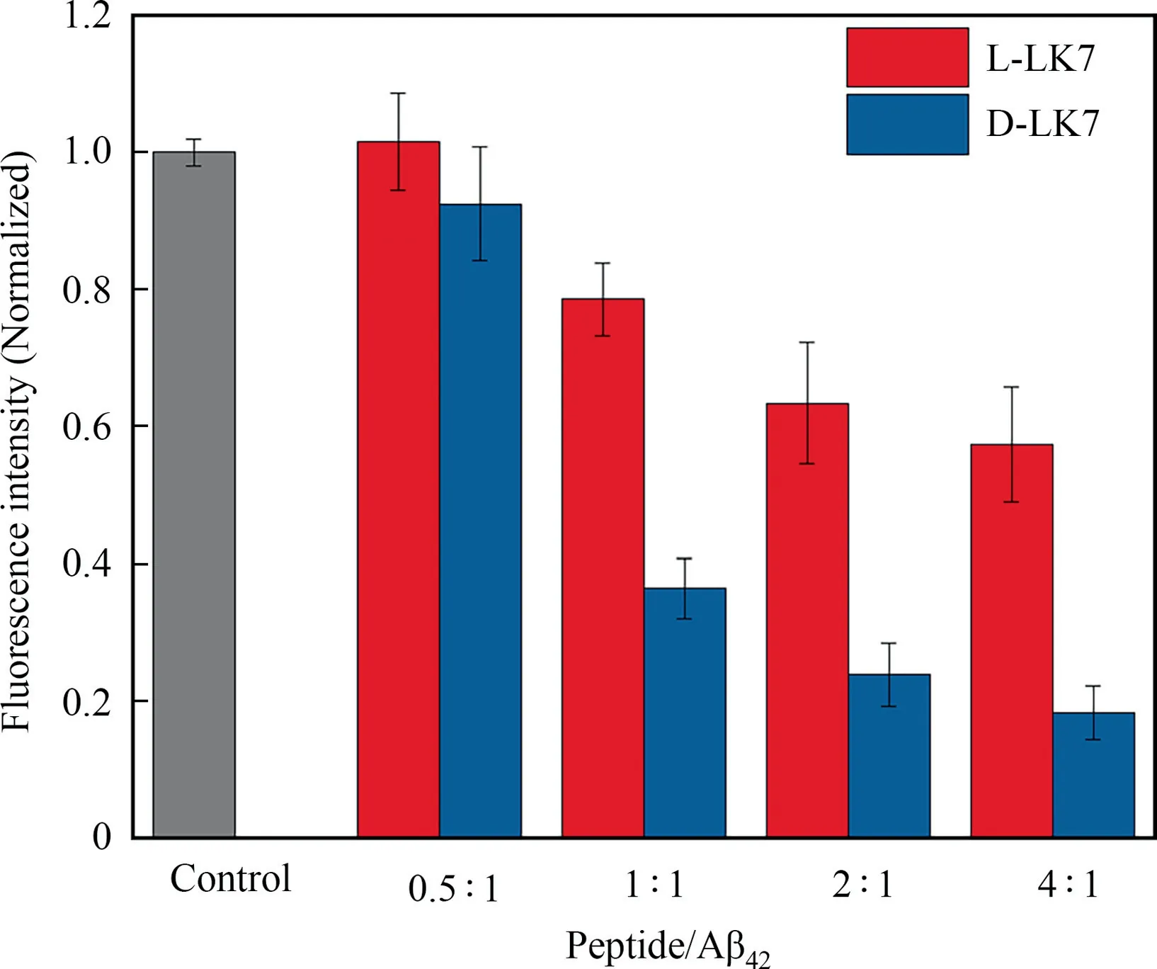
Fig.1.ThT fluorescence intensity of Aβ42 aggregated with enantiomeric inhibitors at different molar ratios for 48 h.The concentration of Aβ42 was 25 μmol·L-1.
Previous researches have indicated that chiral inhibitors could modulate Aβ aggregation in different manners [36-38],thus the aggregation kinetics of Aβ40with varying concentrations of LLK7/D-LK7 were measured byin situThT fluorescence assay.Aβ40alone showed a typical sigmoidal growth curve with a lag phase,a fast growth phase and a plateau phase,which is consistent with literature [22].Addition of L-LK7/D-LK7 to Aβ40led to a decrease of fluorescence intensity at plateau phase (Fig.2).However,due to the lower hydrophobicity and aggregation propensity of Aβ40,the ThT fluorescence intensities of Aβ40with enantiomeric heptapeptide at plateau phase were slightly lower than that of Aβ42.To elucidate the difference of chiral inhibitors on Aβ40fibrillization,the aggregation kinetics curves of Aβ40were fitted by Eq.(1),and the lag time values were calculated by Eq.(2)and are listed in Table 1.As shown in Fig.2 and Table 1,L-LK7 prolonged the lag times of Aβ40at lower concentrations (L-LK7:Aβ40=0.5:1,1:1,2:1),but it shortened the lag time at higher concentration (L-LK7:Aβ40=4:1).This phenomenon is similar to our previous work and it is considered that the self-assembly character of LK7 at high concentration may lead to form LK7 aggregates,resulting in the acceleration of Aβ aggregation [21,39].By contrast,D-LK7 shortened the lag time of Aβ40at 12.5 and 25 μmol·L-1(D-LK7:Aβ40=0.5:1 and 1:1),and completely inhibited Aβ40fibrillization at 50 and 100 μmol·L-1(D-LK7:Aβ40=2:1 and 4:1).Thus,it can be concluded that D-LK7 facilitated Aβ40nucleation,which is consisted with the finding that D-RK10 accelerated Aβ fibrillization [36].The aggregation kinetics assay indicates the effects of chiral heptapeptides on Aβ40nucleation were different from each other.

Table 1 Lag time values of Aβ40 incubated with L-LK7/D-LK7 summarized from Fig.2.The lag phase time of Aβ40 is 45.2 h
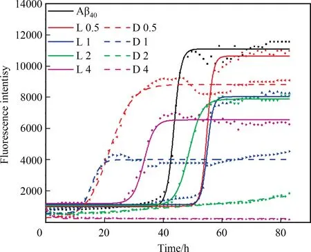
Fig.2.Aggregation kinetics of Aβ40 incubated with different concentrations of LLK7/D-LK7.The curves were obtained by fitting with the Boltzmann function (Eq.(1)).L and D represent L-and D-enantiomer,respectively,and the following number represents the molar ratio of inhibitor to Aβ40.
3.2.Influence of enantiomers on the morphology of Aβ aggregates
To gain insight to the morphological changes of Aβ incubated without or with enantiomeric inhibitors,AFM image test was conducted.As illustrated in Fig.3,two enantiomers exhibited high self-assembly performance after incubation for 72 h.L-LK7 aggregated into a twisted fibrillar species and D-LK7 formed amorphous aggregates and straight fibrils with a length of several micrometers.The difference in self-assembly ability of chiral heptapeptides may contribute to various inhibitory efficiency for blocking Aβ aggregation.
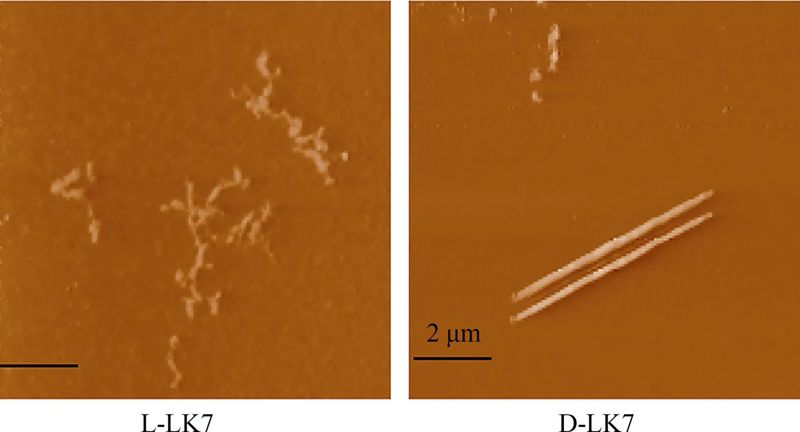
Fig.3.Self-aggregation of L-LK7/D-LK7 obtained by incubation at 200 μmol·L-1 for 72 h.
Then,the morphology of Aβ40aggregates was visualized in Fig.4.Aβ40alone aggregated into dense and entangled fibrillar networks,similar to published reports[40,41].Addition of L-LK7 led to the formation of less entangled,short and broken fibrils.Furthermore,the amount of Aβ40fibrils decreased significantly with increasing L-LK7 concentration.Remarkably,more obvious morphological changes were observed by incubating Aβ40with different concentration of D-LK7.At the molar ratios of D-LK7:Aβ40=2:1,the fibrillar species transformed into amorphous structures;and at D-LK7:Aβ40=4:1,the Aβ40aggregates completely disappeared.The morphology of Aβ42aggregates in the presence of enantiomeric inhibitors was also imaged and illustrated in Fig.5,showing similar results.Above AFM imaging is in agreement with ThT fluorescence data,indicating the stronger inhibition of D-LK7 than L-LK7 on Aβ fibrillization.
3.3.Effect of enantiomers on the Aβ conformational transition
A series of conformational transition occurs during Aβ aggregation,which results in Aβ species with β-sheet-rich structures.Therefore,the ability of enantiomers on preventing Aβ conformational switch was assessed by CD spectroscopy.As expected,enantiomers showed similar but opposite CD signals,which indicates the mirror structures(Fig.6(a)).At the initial state,Aβ42monomers adopted a random coil conformation with a negative peak at 199 nm (Fig.6(b),black line).After 48 h incubation,Aβ42aggregates formed β-sheet structures,showing one negative valley at 216 nm and one positive peak at 197 nm.L-LK7 and D-LK7 decreased the negative ellipticity value at 216 nm,demonstrating the enantiomers could prevent conformational transition of Aβ42from native disordered coil to β-sheet structures.To further assess the inhibitory efficiency of the enantiomers,secondary structures of Aβ42incubated with equimolar L-LK7/D-LK7 were calculated and are listed in Table 2.The β-sheet content of Aβ42at 48 h decreased from 47.6% to 34.6% with L-LK7,and it reduced to 20.4% with D-LK7.This conforms that D-LK7 was more effective than L-LK7 in inhibiting on-pathway Aβ42conformational transition.

Table 2 Secondary structures of Aβ42 calculated from 200 -260 nm spectrum data in Fig.6 using BeStSeL web server (http://bestsel.elte.hu/)
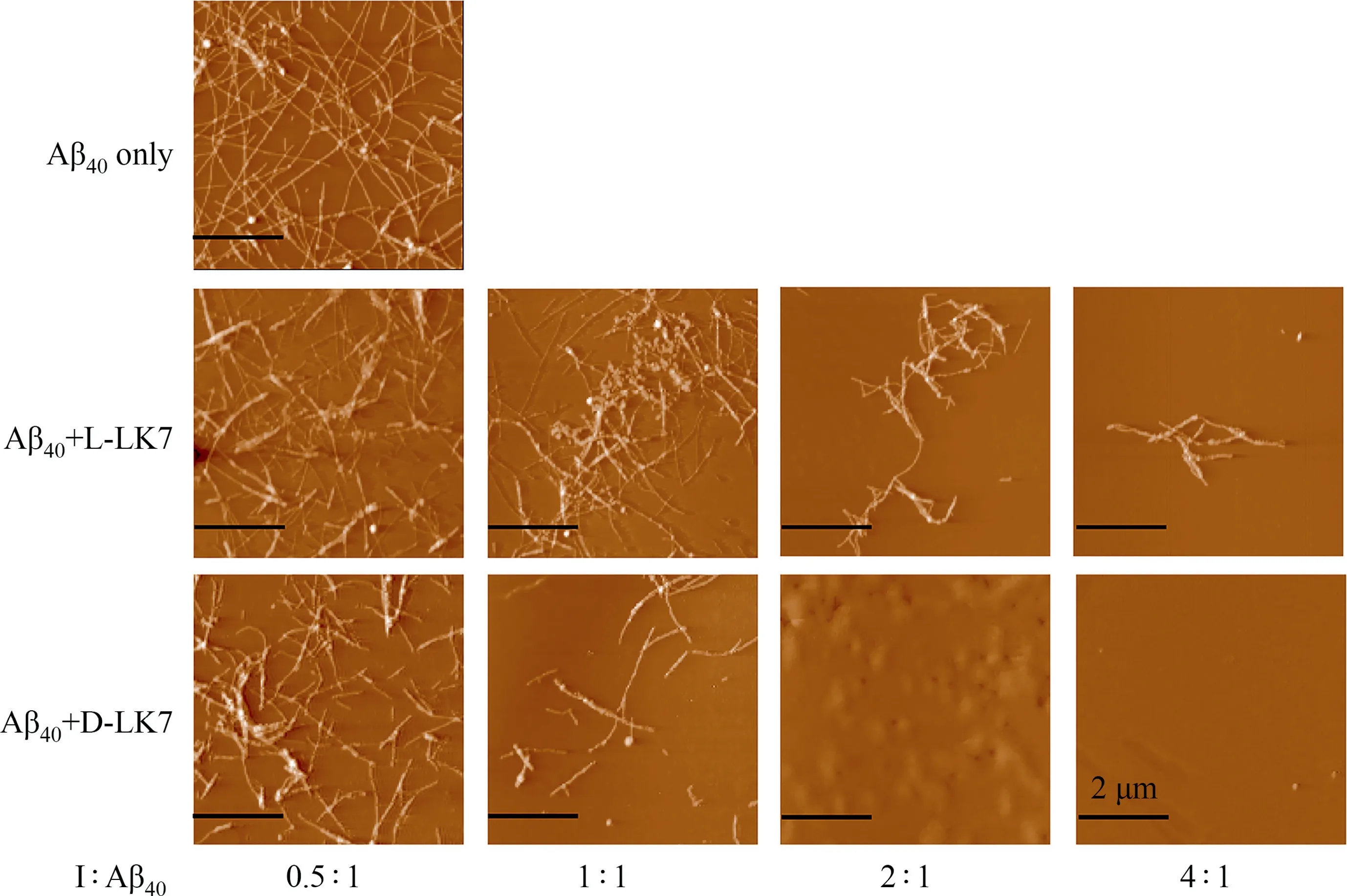
Fig.4.AFM images of 25 μmol·L-1 Aβ40 incubated with or without different concentrations of L-LK7 or D-LK7.The molar ratio in the bottom is inhibitor to Aβ40.
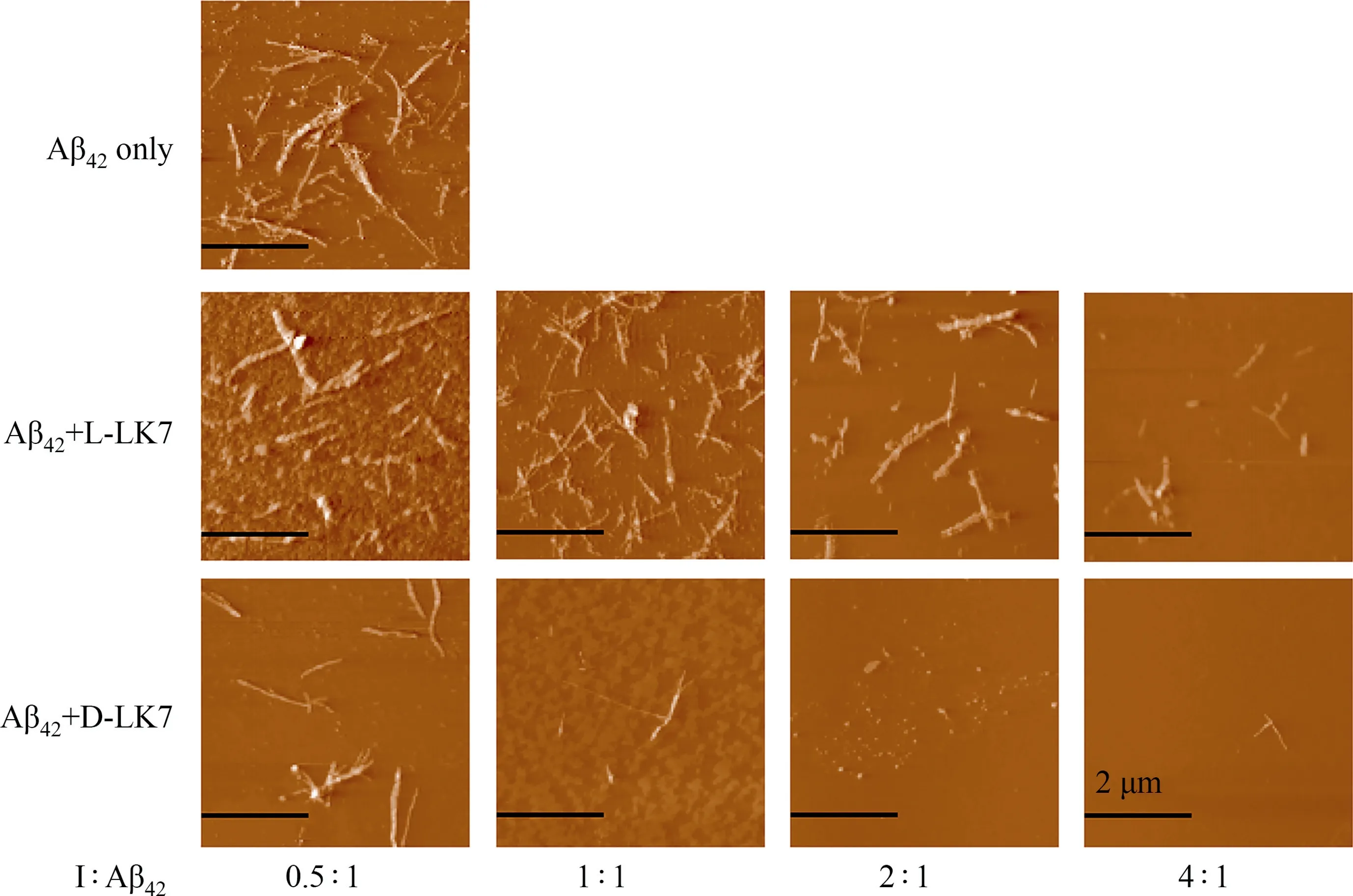
Fig.5.Influence of the enantiomers on the morphology of 25 μmol·L-1 Aβ42 aggregates measured by AFM imaging.The ratio in the bottom represents the molar ratio of inhibitor to Aβ42.
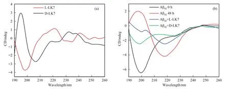
Fig.6.CD spectra of enantiomers and Aβ42 aggregates.(a)The concentration of two enantiomers was 200 μmol·L-1.(b)Aβ42(25 μmol·L-1)at 0 h or incubated with equimolar L-LK7/D-LK7 for 48 h.
3.4.Enantiomers rescue cytotoxicity and scavenge Aβ plaques in nematodes
The success in suppression of β-sheet structures and fibrillar aggregates formation prompted us to investigate whether enantiomers could decrease Aβ-mediated cytotoxicity and amyloid levels in nematodes.MTT cell viability was performed to test theeffects of chiral heptapeptides on SH-SY5Y cells.In the absence of Aβ42,enantiomers could decrease the cell viability to 87%-74%within the tested concentration range (Fig.7(a)),indicating slight cytotoxicity.The amyloid-like L-LK7 and D-LK7 fibrils formed by self-assembly could interact with cell membrane and disrupt the membrane functions,leading to the decrease of cell viability.To ameliorate the cytotoxicity caused by enantiomeric LK7 aggregates,one way that can be considered is to conjugate the heptapeptide onto nanoparticles,which would eliminate the selfassemble of enantiomers [21].
However,both L-LK7 and D-LK7 possessed the ability to rescue SH-SY5Y cells from Aβ42-induced cytotoxicity.As illustrated in Fig.7(b),pre-incubated Aβ42species could reduce the cell viability to 60%.L-LK7 alleviated cell viability to 76%at molar ratio of 2:1(LLK7:Aβ42),but further increase of L-LK7 concentration caused a decline in cell viability,indicating the moderate detoxification of L-LK7.In contrast,D-LK7 rescued cell viability to 68%,80%,84%and 83% at the molars ratio of D-LK7:Aβ42at 0.5:1,1:1,1:2 and 1:4,respectively,proving its effective detoxification.It is considered that the enantiomers could change the on-pathway aggregation of Aβ,so the formation of neurotoxic Aβ species was significantly suppressed,resulting in the enhanced cell viability.The MTT cell viability test further confirms the superiority of DLK7 over L-LK7 on alleviating Aβ42-mediated cytotoxicity.
For evaluating thein vivoinhibitory efficiency of enantiomeric heptapeptides,transgenicC.elegansstrain CL2006 that expresses human Aβ42peptide in muscle cells was used as an AD model.As illustrated in Fig.8,amyloid plaques could be labeled by ThT dye,showing green brilliant fluorescence spots.With the treatment of L-LK7/D-LK7,the green spots in nematode were decreased and vanished.The nematode treated with D-LK7 showed less Aβ plaques,which is consistent with thein vitroresults.These results demonstrate that thein vivoamyloid deposition could be significantly inhibited by D-LK7.
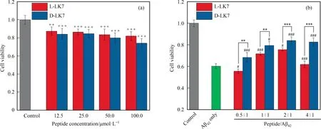
Fig.7.MTT cell viability assays.(a)The cell viability of SH-SY5Y cells after treating with L-LK7/D-LK7 for 24 h.(b)The detoxification of L-LK7/D-LK7 on Aβ42-induced toxicity.The Aβ42 concentration was 25 μmol·L-1,which became 5 μmol·L-1 in the culture medium due to a 5-fold dilution.Error bars represent standard deviations(n=6).Statistical significance level is expressed by plus(in comparison with the control group,++p <0.01,+++p <0.001),pound(in comparison with Aβ42 only group,#p <0.05,###p <0.001)and asterisk (in comparison with L-LK7 group,**p <0.01,***P <0.001).
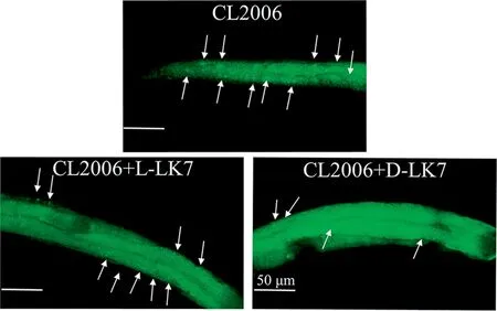
Fig.8.Fluorescence imaging of Aβ deposits in C.elegans (CL2006).The nematodes were treated with L-LK7/D-LK7 for 3 days and then stained with ThT dye.
3.5.Molecular interaction analyses
To understand the molecular mechanisms of chiral heptapeptides with monomeric Aβ,molecular docking was performed using AutoDock Vina.The best conformations are illustrated in Fig.9 and the corresponding interactions are listed in Table 3.For Aβ40monomer,the binding sits of L-LK7 on Aβ40were identified as a cavitylike domain encompassing the amino acids Tyr10,Val12,Gln15 and Glu22 (Fig.9(a)),which could further block the hydrophobic interactions during Aβ40aggregation.Besides the above mentioned amino acids,D-LK7 interacted with Gly9 and had higher binding energy of -6.2 kcal·mol-1(1 kcal=4.186 kJ) to Aβ40than-5.8 kcal·mol-1of L-LK7.For Aβ42monomer,L-LK7 was positioned in the N-terminus of Aβ42and interacted with Glu3 and Asp7.However,electrostatic interactions with His6,Asp7,Glu11 and His14 of Aβ42were observed for D-LK7.The favorable conformation provided by D-LK7 led to higher binding energy (-4.4 kcal·mol-1)to Aβ42than L-LK7 (-4.1 kcal·mol-1),causing enhanced inhibitory efficiency on Aβ fibrillization.It is considered that the difference in molecular recognition of enantiomers towards Aβ monomers(Fig.9)leads to varied performances on suppression of Aβ aggregation (Figs.1,2,4 and 5),conformational transition (Fig.6),aggregation-related cytotoxicity (Fig.7) andin vivobiological effects (Fig.8).Thus,the LK7 could inhibit Aβ self-assembly in a chiral-dependent manner.
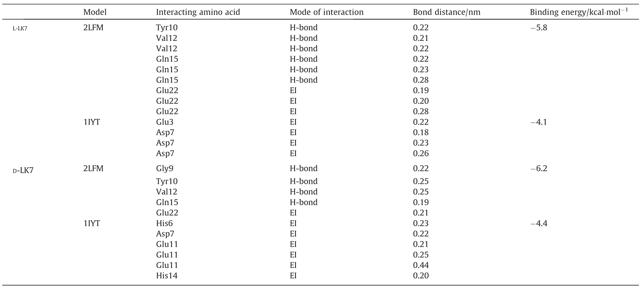
Table 3 Interactions of enantiomeric LK7 with Aβ monomers summarized from Fig.9.The EI represents electrostatic interaction
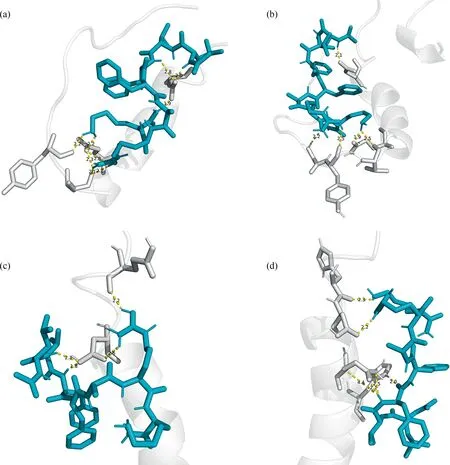
Fig.9.Docking modes of enantiomeric LK7 with Aβ monomers.(a) L-LK7-Aβ40(PDB ID:2LFM)complex.(b) D-LK7-Aβ40 complex.(c) L-LK7-Aβ42(PDB ID:1IYT)complex.(d) DLK7-Aβ42 complex.
4.Conclusions
To elucidate the chiral influence of peptide-based inhibitors on Aβ aggregates formation,the effects of heptapeptide L-LK7 was investigated by comparison with D-LK7.Despite the enantiomers could inhibit Aβ fibrillization in a concentration-dependent manner,the D-enantiomer showed enhanced suppression on Aβ aggregation,as evidenced by the lower ThT fluorescence intensity,less visible Aβ aggregates,fewer β-sheet content and better SH-SY5Y cells rescue ability when compared with L-enantiomer under the same conditions.The chiral discrimination of enantiomeric heptapeptides was further formulated by molecular docking calculation,indicating the different inhibition efficiency of enantiomers was due to varied molecular interactions and recognition towards Aβ monomers.This work may open a new avenue for the design of peptide inhibitors against AD by enantioselectivity.
Declaration of Competing Interest
The authors declare that they have no known competing financial interests or personal relationships that could have appeared to influence the work reported in this paper.
Acknowledgements
This work was supported by the National Natural Science Foundation of China (Nos.21621004 and 21978207) and the Natural Science Foundation of Tianjin from Tianjin Municipal Science and Technology Commission (No.19JCZDJC36800).
 Chinese Journal of Chemical Engineering2022年8期
- Chinese Journal of Chemical Engineering的其它文章
- Notes for Contributors
- Preparation and functional study of pH-sensitive amorphous calcium phosphate nanocarriers
- Crystallization thermodynamics of 2,4(5)-dinitroimidazole in eleven pure solvents
- An international comprehensive benchmarking analysis of synthetic biology in China from 2015 to 2020
- Research on prediction model of formation temperature of ammonium bisulfate in air preheater of coal-fired power plant
- Insights into depolymerization pathways and mechanism of alkali lignin over a Ni1.2-ZrO2/WO3/γ-Al2O3 catalyst
