Multimodal imaging in immunogammopathy maculopathy secondary to Waldenstrom’s macroglobulinemia: a case report
Hui-Rong Xu, Jie Zhu, Xiao-Yan Xie, Jun Zhu, Fang Chen
1Department of Ophthalmology, Northern Jiangsu People’s Hospital, Yangzhou 225001, Jiangsu Province, China
2Dalian Medical University, Dalian 116044, Liaoning Province,China
Dear Editor,
I am Dr. Hui-Rong Xu, from Northern Jiangsu People’s Hospital, Yangzhou, China. I write to present a case of multimodal imaging in immunogammopathy maculopathy secondary to Waldenstrom’s macroglobulinemia (WM).As is known to us all, ocular manifestations can be the initial symptoms of some serious general diseases, clinicians’ prompt detection can even save patients’ lives. WM is a rare kind of non-Hodgkin’s lymphoma whose first symptom could be the blurred vision, the concurrent sign hyperviscosity associated with this disease was life-threatening, while reports about WM that first diagnosed in ophthalmology were limited. Hence we reported a rare case of a woman who got WM first diagnosed in our department.
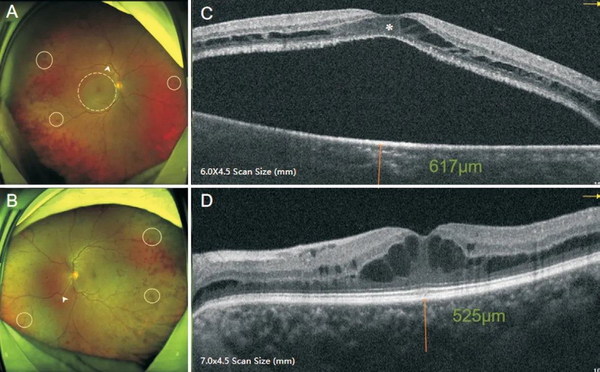
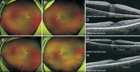
This study followed the tenets of the Declaration of Helsinki and was approved by Northern Jiangsu People’s Hospital.Written informed consent was obtained from the participant.A 53-year-old woman was referred for a 2-month history of blurred vision in her right eye last year. Her medical history included a 3-year hypertension and a cerebral infarction that happened 3 years ago, other than that, she denied any physical discomfort. On examination, the best-corrected visual acuity was 20/125 in the right eye and 20/20 in the left eye. Fundus showed venous dilation and tortuosity with diffuse patchy-like hemorrhages in bilateral peripheral retina (Figure 1A-1B). Optical coherence tomography (OCT) in the right eye demonstrated intraretinal fluid with prominent serous macular detachment, and a marked disorder of the outer retina,schisis-like intraretinal fluid in the left eye, and the subfoveal choroidal thickness was 617 and 525 μm, respectively (Figure 1C-1D). Fundus autofluorescence (FAF) demonstrated hypoautofluorescence in the posterior pole especially in the right eye (Figure 2A-2B). Ultra-widefield fluorescein angiography (UWFA) of both eyes demonstrated diffuse leaking microaneurysms in the mid-periphery, mild leakage of peripheral vessels, and sporadic far peripheral capillary nonperfusion. There was no obvious macular leakage in both eyes,while a leakage of microaneurysms was observed near to the temporal margin of the macula in the left eye (Figure 2C-2D).A workup for these features of multimodal imaging included blood routine and the protein electrophoresis test, which revealed a high concentration of serum globulin (78.0 g/L, normal range, 20-40 g/L), and the IgM (85.5 g/L, normal range,0.4-2.3 g/L) with immunoglobulin kappa light chain (11.2 g/L,normal range, 1.7-3.7 g/L). The patient underwent urgent plasmapheresis after consultation with the hematology team.By the next day, her IgM dropped to 53.0 g/L. Combined with the following results of bone marrow biopsy and DNA sequencing, which showed CD5-CD10-B cell lymphocytosis and gene MYD88-L265P mutation, a diagnosis of WM with hyperviscosity syndrome was confirmed. During the chemotherapy (bortezomib and dexamethasone), her IgM level maintains at 30-40 g/L. Three months following chemotherapy,fundus hemorrhages and tortuous veins were nearly reversed with obvious absorption of intraretinal fluid in both eyes(Figure 3A-3D). At a 7-month follow-up, there was no peripheral hemorrhages in both eyes with the subretinal fluid gradually decreased in the right eye (Figure 3E-3H). While the visual acuity did not improve, the loss of ellipsoid zone was present and the choroidal thickness was barely changed.

Figure 1 Initial presentation of SLO and OCT SLO of both eyes (A, B) shows tortuous and dilated retinal veins (arrowheads) with peripheral patchy-like hemorrhages (solid circles), and there is a circular area of relative translucent with macular fluid involving the fovea (dotted circle)in the right eye (A). OCT of the right eye (C) demonstrates impressive serous macular detachment with intraretinal fluid, and a marked loss of the outer retinal layers in the macula (asterisks). The subfoveal choroidal thickness was 617 μm. OCT of the left eye (D) reveals schisis-like intraretinal fluid with the thickened choroid 525 μm. SLO: Scanning laser ophthalmoscopy; OCT: Optical coherence tomography.

Figure 2 Initial Presentation of FAF and UWFA FAF (A-B) reveals increased hypoautofluorescence in the posterior pole corresponding to the macular detachments especially in the right eye. UWFA in both eyes (C-D) demonstrates diffuse leaking microaneurysms in the midperiphery,mild leakage of peripheral vessels (dotted circle), and sporadic far peripheral capillary non-perfusion zone in the far peripheral retina(arrowheads). There was no leakage of the macula, while in the left eye (D), there was a mild leakage of microaneurysms at the temporal margin of the macula. FAF: Fundus autofluorescence; UWFA: Ultra-widefield fluorescein angiography.

Figure 3 Following up after plasmapheresis and systemic chemotherapy Follow-up SLO from the patients at 3mo after initiating medical treatments shows that the patchy-like hemorrhages and tortuous veins in both eyes (A-B) were nearly reversed, OCT reveals obvious absorption of intraretinal fluid in both eyes (C-D), OCT in the right eye (C) shows the decreased subretinal fluid with the ellipsoid zone partially absent (asterisks), the choroidal thickness of both eyes was 614 and 518 μm, respectively. At 7mo after treatment, SLO shows that there were no peripheral hemorrhages in both eyes (E-F) with the subretinal fluid gradually decreased in the right eye (G), and mild absorption of intraretinal fluid in the left eye (H). And the choroidal thickness of both eyes were 572 and 478 μm, respectively (G-H). SLO: Scanning laser ophthalmoscopy; OCT: Optical coherence tomography.
DISCUSSION
WM is an indolent lymphoproliferative disease characterized by IgM overproduction, over one-third of patients with WM presented as hyperviscosity-related retinopathy with varying degrees[1-2]. There were few reports about WM that first diagnosed in ophthalmology, for whose manifestations were not that easy to be recognized especially when patients do not show their systemic symptoms, one-sidedly and curtly to some signs may cause clinicians to delay in diagnosis, leading worse health issues that could have potentially been preventable.
Fundus manifestations such as optic nerve edema, punctate retinal hemorrhages, tortuous blood vessels and macular detachments are related to hyperviscosity[2-3]. Previous studies have shown that the earliest retinal changes could be observed when the mean IgM level was 54.42 g/L, while the patient was asymptomatic until the level of which reaches 85.15 g/L[4]. Nowadays, limited references are available for the pathophysiology and the diagnosis of visual dysfunction caused by immunogammopathy, while multimodal imaging could promote doctors to identify such patients early. Scanning laser ophthalmoscopy (SLO) can detect fundus abnormalities in small pupils, especially peripheral retinopathy in the early stage, our patient present as unilateral vision loss, and we also found the peripheral retinopathy of the other eye that was unnoticed by the patient. OCT could demonstrate serous macular detachment and schisis-like intraretinal fluid[5-7],UWFA present as mild leakage from peripheral vessels with no obvious non-perfusion zone, and the macular detachment is “silent”[6,8]. The majority of our findings are consistent with those manifestations above, except a little non-perfusion zone in the far-periphery in UWFA. It has been speculated that IgM primarily deposits in the intraretinal space and proceeding into the area of subretinal by gradually destroying the integrity of outer retina, which might be confirmed by the results of autopsy and immunoelectrophoresis for IgM had been detected in the cystoid space, outer plexiform layer,around photoreceptors and within subretinal fluid[5,9-10]. The accumulation of subretinal fluid might due to the osmotic effects of IgM rather than the vascular leakage such as diabetic retinopathy or retinal vein occlusion[6], which can explain the serous detachment in the OCT while the UWFA showed no fluorescence leakage. And we speculate that the left eye of our patient with normal vision was in the asymptomatic stage of the disease, it may deteriorate just as the right eye if there was no timely treatment. Besides, we measured the thickness of choroid that has never been described previously in this condition[6,11], and the deposition of IgM may be responsible for it. Our FAF shows hypoautofluorescence in the area of detachment which is different from previous reports[6],we suspected that it might be relevant to the duration of detachment which lead to the different components in the subretinal fluid. These fundus findings may not that specific for WM, but they could prompt further laboratory testing when patients with atypical multimodal imaging whether unilateral or bilateral.
Nowadays, early initiation of systemic therapy especially plasmapheresis and chemotherapy remain the cornerstone of therapy[5], while the strategies to improve vision are still obscure. In addition to systemic therapy, the treatment of anti-VEGF and dexamethasone implants have been reported previously, which demonstrated that they may be beneficial to the reduction of intraretinal fluid but little effect in the subretinal fluid and the visual acuity[6-7]. While there were several reports showed moderate improvement in vision after plasmapheresis[3]. And Leskov et al reported that a WM patient who was treated with ibrutinib, a specific inhibitor of Bruton’s tyrosine kinase that was recently approved by the FDA,revealed near resolution of intraretinal fluid and subretinal fluid with improvement in visual acuity[2]. Our patient also underwent the plasmapheresis immediately after diagnosis,the retinal hemorrhages and the intraretinal fluid resolved with the improvement of hyperviscosity, but visual acuity did not improve. This may due to the damage of the ellipsoid zone and retinal pigment epithelium in the detachment region resulting from long-term toxicity of exposure to IgM[12-13], which deserves further investigation.
This case demonstrated that multimodal imaging examinations could potentially assist earlier diagnosis in patients with WM,hyperviscosity syndrome should be kept on the differential diagnosis for atypical serous macular detachment and signs of hyperviscosity-related retinopathy, prompt treatment can reverse associated retinopathy and prevent other complications.
ACKNOWLEDGEMENTS
Authors’ contributions:Xu HR was responsible for the acquisition of the clinical information and writing the manuscript. Chen F was responsible for explanations of all the UWFA and OCT results and reviewing the manuscript.Zhu J, Xie XY and Zhu J were responsible for reviewing the manuscript. All authors read and approved the final manuscript.
Conflicts of Interest: Xu HR,None;Zhu J,None;Xie XY,None;Zhu J,None;Chen F,None.
 International Journal of Ophthalmology2022年5期
- International Journal of Ophthalmology的其它文章
- Periorbital necrotizing fasciitis accompanied by sinusitis and intracranial epidural abscess in an immunocompetent patient
- Multimodal imaging in Purtscher-like retinopathy associated with sarcoidosis: a case report
- Can a sneeze after phacoemulsification cause endophthalmitis? A case report
- Persistent macular oedema following Best vitelliform macular dystrophy undergoing anti-VEGF treatment
- Genetic, environmental and other risk factors for progression of retinitis pigmentosa
- lnflammation and dry eye disease—where are we?
