Chest radiological finding of COVID-19 in patients with and without diabetes mellitus: Differences in imaging finding
2022-02-12 01:48:40SunayGangadharanStormParkerFahadWaliAhmed
World Journal of Radiology 2022年1期
lNTRODUCTlON
The world is currently undergoing a significant healthcare crisis due to the severe acute respiratory syndrome coronavirus 2 (SARS-CoV-2) pandemic.In March 2020, World Health Organisation declared a pandemic caused by SARs-CoV-2.SARS-CoV-2 was named novel coronavirus disease 2019 (COVID-19).Hospitals in different countries have been overwhelmed with patients suffering from COVID-19.So far, 2.78 million people have died as of 29
March 2021[1].
Diabetes mellitus (DM) is a risk factor associated with severe illness in SARS-CoV-2 infection, precipitating hyperglycaemic emergencies such as diabetic ketoacidosis (DKA) and hyperosmolar hyperglycaemic state (HHS)[2].A third of deaths in England up to May 2020 related to COVID-19 occurred in people with DM[3].Patients with DM are more likely to stay longer in hospital[4].DM can cause a deregulated immune system predisposing to infection; the endothelial angiotensin-converting enzyme 2 (ACE2) receptor responsible for SARS-CoV-2 invasion in human cells has reduced expression in patients of DM, possibly due to glycosylation[5].Insulin resistance and altered glucose homeostasis have been thought to cause alveolar capillary microangiopathy and interstitial fibrosis
over-inflammation[6].
A normal chest radiograph does not exclude COVID-19 pneumonia, and no single feature on a radiograph is diagnostic[7].However, a combination of multifocal peripheral airspace changes often found bilaterally may be present in COVID-19.Due to limited PCR testing capacity in the early d of the pandemic, in addition to its low sensitivity and waiting period of up to 2 d, many clinicians turned to chest computed tomography (CT) for early detection of COVID-19.
Studies have reported the negative predictive value of using CT to be above 90%[8,9].Chest CT was used to detect subtle radiological changes consistent with COVID-19 in patients where the chest radiograph was reported to be normal or indeterminate.Typical CT findings seen in patients with COVID-19 include peripheral ground-glass opacities (GGO), which progresses to consolidation and interstitial thickening within GGO areas known as ‘crazy paving pattern’[10,11].These non-specific imaging findings of acute lung injury are indistinguishable from other types of viral pneumonia or interstitial lung diseases, thereby limiting the use of CT as a confirmatory diagnostic test in COVID-19.
This article reviews current literature regarding chest imaging changes in patients with DM affected by COVID-19.
LlTERATURE SEARCH
As yet, no large-scale studies have reported a link between pulmonary thromboembolism and DM in patients with COVID-19.Kaminetzky
[31] found patients with DM were significantly less frequently observed to have CTPA examinations.Of 23 patients identified to have PE in this study, only 3 had DM; however, this finding may be attributed to the small sample size.
Chest Radiography
Earlier studies employed semi-quantitative methods to analyse chest computed tomography (CT) findings (Figure 2) in patients with COVID-19[19,20].This involved a single, or multiple experienced radiologists blinded to clinical parameters and assigning a score based on the severity of findings.Higher chest CT scores have been found in patients with DM, suggesting more severe COVID-19 pneumonia when compared with patients without DM[19].Findings by Iacobellis
[18] suggested day-1 hyperglycaemia as a predictor of COVID-19 severity on CXR were confirmed on CT.
In some studies, DM alone was not associated with an increased risk of intensive care unit admission or death.Still, it was associated with cardiovascular disease as a driver of poorer outcomes.Izzi-Engbeaya
[16] studied 889 patients admitted to London hospitals with COVID-19, and their outcomes found patients with DM were found to have a 33% increased risk of death or ICU admission if they also have ischaemic heart disease.Surprisingly, a similar severity of CXR changes was demonstrated for patients with and without DM.Mozzini
[17] (2021) studied 50 Italian patients with COVID-19, 32% of which had DM.Patients with hypertension or DM had 8 times greater risk of having more severe CXR changes.
On subsequent trips through LaGuardia, Garth would inquire about Miles, and about six months later he asked us to help him contact the family. Garth was going to be performing in Kansas City and he wanted Miles to be his guest. Not only was Miles seated in the front row, but he and Garth also had a lengthy8 private meeting backstage after the performance.
He was ugly, and his hair was matted, and he looked crippledand stunted; they called him the field-laborer s boy, though in thechurch register he was entered as Anne Lisbeth s son
Chest computed tomography
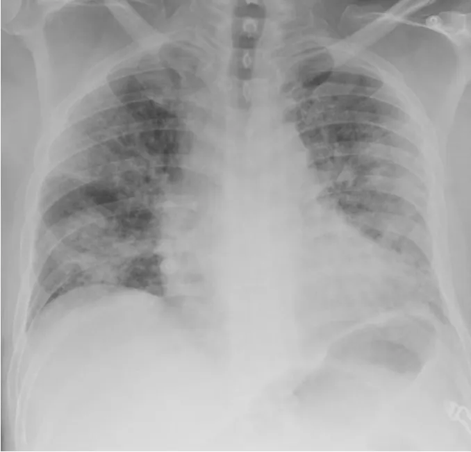
Studies have shown chest radiographs of patients with DM to have increased bilateral airspace consolidation compared to patients without DM[12,13].The severity of chest radiograph changes in patients with DM has indicated a significant correlation with mortality, as evidenced in multivariate analysis by Cellina
[14].Patients with bilateral peripheral alveolar disease (Figure 1) often present at a later stage and have a worse outcome.However, some patients with COVID-19 have preserved lung compliance despite being acutely hypoxaemic, suggesting poorer outcomes result from processes other than alveolar damage[15].
A few years ago, I began to prepare my children for the fact that Christmas that year was going to be a small one. Their response was, “Yeah sure, Mom, we’ve heard that before!” I had lost my credibility because I had told them the same thing the previous year, while going through a divorce. But then I had gone out and charged every credit card to the max. I even found some creative financing techniques to pay for their stocking stuffers. This year was definitely going to be different, but they weren’t buying it.
COVID-19 infection in patients with DM leads to hyperglycaemia, and in some cases leads to DKA and/or HHS[2].It has been shown that there is a positive correlation between daily average blood glucose readings and CXR findings.Similarly, post-admission day-1 hyperglycaemia was found to be the strongest independent predictor for COVID-19 CXR changes.This was a stronger predictor than age, body mass index, and temperature[18].

Then the hero betook himself to the King, who was obliged now, whether he liked it or not, to keep his promise, and hand him over his daughter and half his kingdom
Studies have shown mortality rates to be higher among patients with poorlycontrolled DM and COVID-19 than the general population with COVID-19[22,23].In particular, high HbA1c levels have been linked with inflammation and hypercoagulability, resulting in an increased mortality rate in patients with DM suffering from COVID-19[24].However, the accuracy of these results may be influenced by other comorbidities such as ischaemic heart disease and stroke.No large-scale studies have yet shown an association between worse CT findings and mortality in DM.
The radiological finding of subsegmental or segmental thrombi in peripheral segments of lung affected by acute lung injury and the absence of deep vein thrombosis (DVT) in patients with COVID-19 infection, assumes the theory of immunothrombosis[27].Monfardini
[29] found 76% of patients with a moderatehigh pre-test probability of PE and positive D-dimer level (a fibrin degradation product measured to help diagnose thrombosis), had positive CTPA findings.Nevertheless, only 15% of these patients were associated with ultrasound detected lower limb DVT[29], suggesting the remainder probably represented immunothrombosis.A meta-analysis of twenty-seven studies by Suh
[30] revealed DVT was only found in 42% of patients with PE.
A high incidence of venous and arterial thrombotic complications in critically ill patients with COVID-19 has been reported previously[25].Recent literature based on autopsy studies shows that the origin of thrombotic lesions in COVID-19 is largely unknown.Lung histopathological analysis found multiple thrombi in small to medium pulmonary arteries giving rise to the theory of COVID-19 associated immunothrombosis, contrary to the conventional thromboembolic pathomechanism of PE[26,27].In situ microvascular thrombosis or immunothrombosis occurs due to alveolar injury, inflammatory storm and disruption of the thromboprotective pulmonary vascular endothelium.COVID-19 clinical outcomes are worse in patients with diseases associated with endothelial dysfunction such as systemic hypertension, DM and obesity[28].
A literature search was conducted on PubMed using the keywords of COVID-19 or Coronavirus; CXR or x-ray or radiograph; CT chest; CTPA or pulmonary embolism or PE; and diabetes mellitus or diabetes within the title or abstract.
Patients with poorly-controlled DM are likely to have more severe COVID-19 pneumonia.A recent study by Lu
[21] using a quantitative artificial intelligence algorithm found parameters including the percentage of ground glass volume (PGV) and percentage of consolidation volume (PCV), positively correlated with fasting blood glucose and HbA1c.Unlike semi-quantitative methods, results using this approach were not affected by inter- and intra-observer variability.Raoufi
[20] used a semi-quantitative method to study 117 patients with DM in Iran and found no significant difference in patients with well-controlled (defined as maintaining glycaemic variability between 3.9-10 mmol/L) and poorly-controlled DM.However, the poorly-controlled group contained almost 4 times the number of patients (93
24).Furthermore, the median age of patients in the well-controlled group were older (75
62 years) which may have been a confounding factor for this negative result[20].
CONCLUSlON
DM predisposes to immune deregulation and reduced expression of the ACE2 receptor, leading to severe acute lung injury[5,6].Studies have proven a link between DM and more severe airspace consolidation based on chest x-ray findings[12,13].Furthermore, CXR evidence suggests DM is associated with higher mortality in COVID-19.The exact pathogenesis of this is unclear but may be related to microvascular immunothrombosis[26,28].
There is now quantitative evidence to suggest poorly controlled DM is associated with more severe lung injury on CT[21].However, no large-scale studies have investigated a direct link between CT findings and mortality in DM.Although the incidence of PE is greater in critically ill patients with COVID-19[25], no link has been established between poorly controlled DM and the risk of PE.
When we got home, we talked about what we could do. We decided3 to buy fifty pounds of potatoes and live on them for a month. This would allow us to save twenty dollars of our grocery4 money for the offering. Then we thought that if we kept our electric lights turned out as much as possible and didn’t listen to the radio, we’d save money on that month’s electric bill. Darlene got as many house- and yard-cleaning jobs as possible, and both of us baby-sat for everyone we could. For fifteen cents we could buy enough cotton loops5 to make three potholders to sell for a dollar. We made twenty dollars on potholders. That month was one of the best of our lives.
As new research into COVID-19 is produced and evidence emerges from autopsy studies, the understanding of pathobiology of the disease has evolved.However, there remains scope for future research; particularly whether small pulmonary thromboses represent venous thromboembolism, immunothrombosis, or a combination of both.Furthermore, a direct link between DM and immunothrombosis may help to guide future management strategies.
ACKNOWLEDGEMENT
We will like to thank Kirresh OZ for providing radiographs.
1 CCSE Dashboard [Internet].COVID-19 Dashboard by the Center for Systems Science and Engineering (CSSE) at Johns Hopkins University (JHU).[cited 20 February 2021].Available from: https://www.arcgis.com/apps/opsdashboard/index.html#/bda7594740fd40299423467b48e9ecf6
2 Rafique S, Ahmed FW.A Case of Combined Diabetic Ketoacidosis and Hyperosmolar Hyperglycemic State in a Patient With COVID-19.
2020; 12: e8965 [PMID: 32766007 DOI: 10.7759/cureus.8965]
3 Barron E, Bakhai C, Kar P, Weaver A, Bradley D, Ismail H, Knighton P, Holman N, Khunti K, Sattar N, Wareham NJ, Young B, Valabhji J.Associations of type 1 and type 2 diabetes with COVID-19-related mortality in England: a whole-population study.
2020; 8: 813-822 [PMID: 32798472 DOI: 10.1016/S2213-8587(20)30272-2]
4 Ahmed FW, Kirresh OZ, Robinson AV.A Retrospective Study Assessing the Effect of Diabetes on Mortality in Patients With COVID-19 at a Teaching Hospital in the United Kingdom [Internet].2021.[cited 20 February 2021].Available from: https://www.cureus.com/articles/54241
5 Sartore G, Ragazzi E, Faccin L, Lapolla A.A role of glycation and methylation for SARS-CoV-2 infection in diabetes?
2020; 144: 110247 [PMID: 33254553 DOI: 10.1016/j.mehy.2020.110247]
6 Sardu C, Gargiulo G, Esposito G, Paolisso G, Marfella R.Impact of diabetes mellitus on clinical outcomes in patients affected by Covid-19.
2020; 19: 76 [PMID: 32527257 DOI: 10.1186/s12933-020-01047-y]
7 Cleverley J, Piper J, Jones MM.The role of chest radiography in confirming covid-19 pneumonia.
2020; 370: m2426 [PMID: 32675083 DOI: 10.1136/bmj.m2426]
8 Ai T, Yang Z, Hou H, Zhan C, Chen C, Lv W, Tao Q, Sun Z, Xia L.Correlation of Chest CT and RTPCR Testing for Coronavirus Disease 2019 (COVID-19) in China: A Report of 1014 Cases.
2020; 296: E32-E40 [PMID: 32101510 DOI: 10.1148/radiol.2020200642]
9 Herpe G, Lederlin M, Naudin M, Ohana M, Chaumoitre K, Gregory J, Vilgrain V, Freitag CA, De Margerie-Mellon C, Flory V, Ludwig M, Mondot L, Fitton I, Jacquier ARR, Ardilouze P, Petit I, Gervaise A, Bayle O, Crombe A, Mekuko Sokeng M, Thomas C, Henry G, Bliah V, Le Tat T, Guillot MS, Gendrin P, Garetier M, Bertolle E, Montagne C, Langlet B, Kalaaji A, Kayayan H, Desmots F, Dhaene B, Saulnier PJ, Guillevin R, Bartoli JM, Beregi JP, Tasu JP.Efficacy of Chest CT for COVID-19 Pneumonia Diagnosis in France.
2021; 298: E81-E87 [PMID: 32870139 DOI: 10.1148/radiol.2020202568]
10 Ufuk F, Sava? R.Chest CT features of the novel coronavirus disease (COVID-19).
2020; 50: 664-678 [PMID: 32394687 DOI: 10.3906/sag-2004-331]
11 Ye Z, Zhang Y, Wang Y, Huang Z, Song B.Chest CT manifestations of new coronavirus disease 2019 (COVID-19): a pictorial review.
2020; 30: 4381-4389 [PMID: 32193638 DOI: 10.1007/s00330-020-06801-0]
12 Elemam NM, Hannawi H, Salmi IA, Naeem KB, Alokaily F, Hannawi S.Diabetes mellitus as a comorbidity in COVID-19 infection in the United Arab Emirates.
2021; 42: 170-180 [PMID: 33563736 DOI: 10.15537/smj.2021.2.25700]
13 Bhandari S, Rankawat G, Singh A, Gupta V, Kakkar S.Impact of glycemic control in diabetes mellitus on management of COVID-19 infection.
2020; 1-6 [PMID: 32905072 DOI: 10.1007/s13410-020-00868-7]
14 Cellina M, Gibelli D, Valenti Pittino C, Toluian T, Marino P, Oliva G.Risk Factors of Fatal Outcome in Patients With COVID-19 Pneumonia.
2020; 1-8 [PMID: 32907676 DOI: 10.1017/dmp.2020.346]
15 Gattinoni L, Coppola S, Cressoni M, Busana M, Rossi S, Chiumello D.COVID-19 Does Not Lead to a "Typical" Acute Respiratory Distress Syndrome.
2020; 201: 1299-1300 [PMID: 32228035 DOI: 10.1164/rccm.202003-0817LE]
16 Izzi-Engbeaya C, Distaso W, Amin A, Yang W, Idowu O, Kenkre JS, Shah RJ, Woin E, Shi C, Alavi N, Bedri H, Brady N, Blackburn S, Leczycka M, Patel S, Sokol E, Toke-Bjolgerud E, Qayum A, Abdel-Malek M, Hope DCD, Oliver NS, Bravis V, Misra S, Tan TM, Hill NE, Salem V.Adverse outcomes in COVID-19 and diabetes: a retrospective cohort study from three London teaching hospitals.
2021; 9 [PMID: 33408084 DOI: 10.1136/bmjdrc-2020-001858]
17 Mozzini C, Cicco S, Setti A, Racanelli V, Vacca A, Calciano L, Pesce G, Girelli D.Spotlight on Cardiovascular Scoring Systems in Covid-19: Severity Correlations in Real-world Setting.
2021; 46: 100819 [PMID: 33631706 DOI: 10.1016/j.cpcardiol.2021.100819]
18 Iacobellis G, Penaherrera CA, Bermudez LE, Bernal Mizrachi E.Admission hyperglycemia and radiological findings of SARS-CoV2 in patients with and without diabetes.
2020; 164: 108185 [PMID: 32360710 DOI: 10.1016/j.diabres.2020.108185]
19 Guo W, Li M, Dong Y, Zhou H, Zhang Z, Tian C, Qin R, Wang H, Shen Y, Du K, Zhao L, Fan H, Luo S, Hu D.Diabetes is a risk factor for the progression and prognosis of COVID-19.
2020; e3319 [PMID: 32233013 DOI: 10.1002/dmrr.3319]
20 Raoufi M, Khalili S, Mansouri M, Mahdavi A, Khalili N.Well-controlled
poorly-controlled diabetes in patients with COVID-19: Are there any differences in outcomes and imaging findings?
2020; 166: 108286 [PMID: 32592836 DOI: 10.1016/j.diabres.2020.108286]
21 Lu X, Cui Z, Pan F, Li L, Liang B, Yang L, Zheng C.Glycemic status affects the severity of coronavirus disease 2019 in patients with diabetes mellitus: an observational study of CT radiological manifestations using an artificial intelligence algorithm.
2021; 58: 575-586 [PMID: 33420614 DOI: 10.1007/s00592-020-01654-x]
22 Wu ZH, Tang Y, Cheng Q.Diabetes increases the mortality of patients with COVID-19: a metaanalysis.
2021; 58: 139-144 [PMID: 32583078 DOI: 10.1007/s00592-020-01546-0]
23 Zhu L, She ZG, Cheng X, Qin JJ, Zhang XJ, Cai J, Lei F, Wang H, Xie J, Wang W, Li H, Zhang P, Song X, Chen X, Xiang M, Zhang C, Bai L, Xiang D, Chen MM, Liu Y, Yan Y, Liu M, Mao W, Zou J, Liu L, Chen G, Luo P, Xiao B, Zhang Z, Lu Z, Wang J, Lu H, Xia X, Wang D, Liao X, Peng G, Ye P, Yang J, Yuan Y, Huang X, Guo J, Zhang BH.Association of Blood Glucose Control and Outcomes in Patients with COVID-19 and Pre-existing Type 2 Diabetes.
2020; 31: 1068-1077.e3 [PMID: 32369736 DOI: 10.1016/j.cmet.2020.04.021]
24 Wang Z, Du Z, Zhu F.Glycosylated hemoglobin is associated with systemic inflammation, hypercoagulability, and prognosis of COVID-19 patients.
2020; 164: 108214 [PMID: 32416121 DOI: 10.1016/j.diabres.2020.108214]
25 Klok FA, Kruip MJHA, van der Meer NJM, Arbous MS, Gommers D, Kant KM, Kaptein FHJ, van Paassen J, Stals MAM, Huisman MV, Endeman H.Confirmation of the high cumulative incidence of thrombotic complications in critically ill ICU patients with COVID-19: An updated analysis.
2020; 191: 148-150 [PMID: 32381264 DOI: 10.1016/j.thromres.2020.04.041]
26 Patel BV, Arachchillage DJ, Ridge CA, Bianchi P, Doyle JF, Garfield B, Ledot S, Morgan C, Passariello M, Price S, Singh S, Thakuria L, Trenfield S, Trimlett R, Weaver C, Wort SJ, Xu T, Padley SPG, Devaraj A, Desai SR.Pulmonary Angiopathy in Severe COVID-19: Physiologic, Imaging, and Hematologic Observations.
2020; 202: 690-699 [PMID: 32667207]
27 van Dam LF, Kroft LJM, van der Wal LI, Cannegieter SC, Eikenboom J, de Jonge E, Huisman MV, Klok FA.Clinical and computed tomography characteristics of COVID-19 associated acute pulmonary embolism: A different phenotype of thrombotic disease?
2020; 193: 86-89 [PMID: 32531548]
28 Loo J, Spittle DA, Newnham M.COVID-19, immunothrombosis and venous thromboembolism: biological mechanisms.
2021; 76: 412-420 [PMID: 33408195 DOI: 10.1136/thoraxjnl-2020-216243]
29 Monfardini L, Morassi M, Botti P, Stellini R, Bettari L, Pezzotti S, Alì M, Monaco CG, Magni V, Cozzi A, Schiaffino S, Bnà C.Pulmonary thromboembolism in hospitalised COVID-19 patients at moderate to high risk by Wells score: a report from Lombardy, Italy.
2020; 93: 20200407 [PMID: 32735448 DOI: 10.1259/bjr.20200407]
30 Suh YJ, Hong H, Ohana M, Bompard F, Revel MP, Valle C, Gervaise A, Poissy J, Susen S, Hékimian G, Artifoni M, Periard D, Contou D, Delaloye J, Sanchez B, Fang C, Garzillo G, Robbie H, Yoon SH.Pulmonary Embolism and Deep Vein Thrombosis in COVID-19: A Systematic Review and Meta-Analysis.
2021; 298: E70-E80 [PMID: 33320063 DOI: 10.1148/radiol.2020203557]
31 Kaminetzky M, Moore W, Fansiwala K, Babb JS, Kaminetzky D, Horwitz LI, McGuinness G, Knoll A, Ko JP.Pulmonary Embolism at CT Pulmonary Angiography in Patients with COVID-19.
2020; 2: e200308 [PMID: 33778610 DOI: 10.1148/ryct.2020200308]