Diagnostic value of ultrasound in the spontaneous rupture of renal angiomyolipoma during pregnancy:A case report
Tong Zhang,Shuai Xue,Zheng-Min Wang,Xiu-Mei Duan,Dong-Xuan Wang
Tong Zhang,Dong-Xuan Wang,Department of Ultrasound,The First Hospital of Jilin University,Changchun 130000,Jilin Province,China
Shuai Xue,Zheng-Min Wang,Department of Thyroid Surgery,The First Hospital of Jilin University,Changchun 130000,Jilin Province,China
Xiu-Mei Duan,Department of Pathology,The First Hospital of Jilin University,Changchun 130000,Jilin Province,China
Abstract
Key words:Angiomyolipoma;Kidney;Rupture;Pregnancy;Ultrasonography;Case report
INTRODUCTION
Renal angiomyolipoma (RAML),a common benign tumor,is composed of mature blood vessels,smooth muscle,and fatty tissue.Some scholars believe that RAML consists solely of tumors and may originate from epithelioid cells around the blood vessels[1].Its incidence in the general population is estimated to be 0.13%,and about 80% of affected patients are women[2,3].Spontaneous rupture and hemorrhage is a lifethreatening primary complication of RAML.Pregnancy is a risk factor for increasing the size,leading to rupture of RAML.Because computed tomography (CT) exposes the patient to radiation and magnetic resonance imaging (MRI) is time-consuming,these imaging methods are not always advised for pregnant women.Ultrasound is a quick and convenient method that does not predispose the patient to radiation;therefore,it is preferred for pregnant women.However,because ultrasound doctors lack understanding of the disease,it is more prone to misdiagnosis.
CASE PRESENTATION
Chief complaints
A 38-year-old woman in her 19thwk of pregnancy (G2P1) was referred to our clinic for a sudden persistent pain on the left side of the waist on July 28,2017.
History of present illness
The patient’s physical examination revealed tenderness over the left kidney area.Her blood pressure was 120/85 mmHg,heart rate was 86 beats/min,and body temperature was 36.8 °C.The patient had no significant medical history.She had not undergone any related abdominal examination previously.
History of past illness
The patient had no significant medical history.She had not undergone any related abdominal examination previously.
Physical examination
The patient was hospitalized and given conservative treatment,but her left-side waist pain continued to be intense.Because the size of the tumor was so large,and the fetalheart rate was unstable,the patient decided to undergo left nephrectomy after the induction of labor.
Laboratory examinations
Laboratory tests indicated that the patient’s hemoglobin level was 80 g/L,and the hematocrit was 0.242 L/L.On the 2ndd,hemoglobin was 95 g/L,and the hematocrit was 0.286 L/L.
Imaging examinations
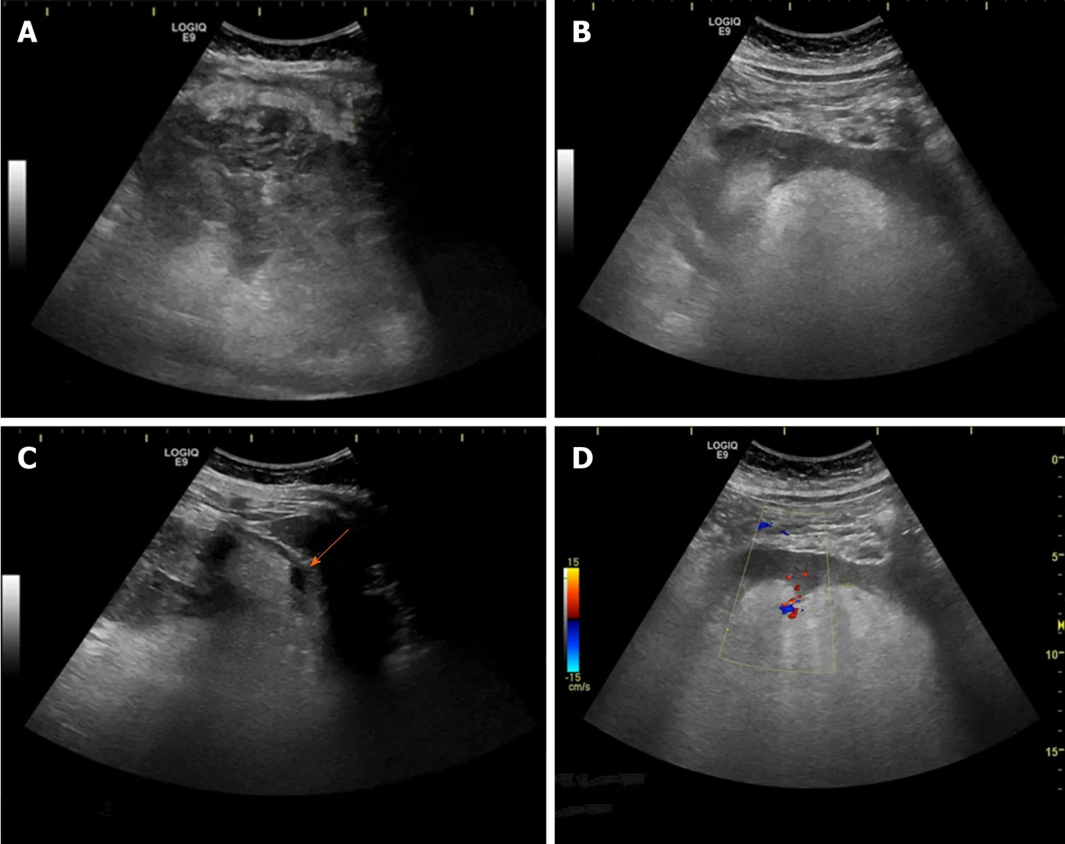
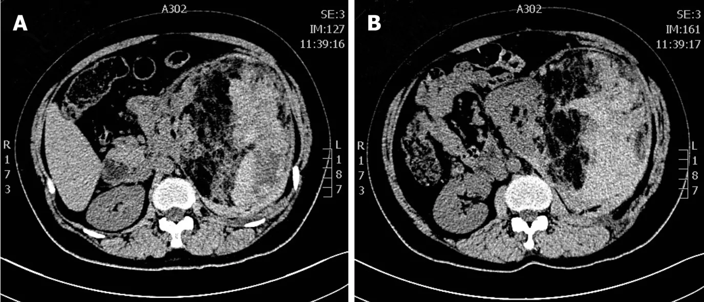
Ultrasound examination of the urinary system (LOGIQ E9,GE) revealed a giant nonhomogenous lump in the left kidney area,which had caused the left kidney to move to the midabdomen.The size of the lump was approximately 159 mm × 100 mm,and the border was faintly visible.The lump showed a “striped sign” in which the outer part was hypoechoic with a strong stripe echo (Figure 1A),and the inner part near the left kidney was hyperechoic (Figure 1B).A stripe-shaped echoless zone was seen around the lump (Figure 1C) (arrow).Color Doppler flow image showed some spot-like blood flow signals around the lump (Figure 1D).A hyperechoic nodule was seen in the right kidney with a size of 30 mm × 25 mm.There was a fetus echo in the uterus.Preoperative CT showed a large,mixed-density mass in the left kidney(Figure 2).The density of the area adjacent to the kidney was low,and the area far from the kidney showed high density.
FINAL DIAGNOSIS
The final diagnosis of the present case was rupture and hemorrhage of the left RAML,right kidney hamartoma,and pregnancy.
TREATMENT
The patient underwent left nephrectomy after the induction of labor.During the operation the renal artery spurted after the left nephrectomy,the blood pressure dropped to 80/50 mmHg,and the blood loss reached 1000 mL.Four units of red blood cell suspension were transfused after emergency hemostasis,and the blood pressure rose to 100/60 mmHg.After the operation,gastrointestinal decompression and intravenous nutrition were given to the patient.The gastric tube was removed on the 2ndd after the patient exhausted.Antibiotics were continuously applied for 5 d,and the patient was discharged on the 6thd after the operation.
OUTCOME AND FOLLOW-UP
The pathological result was rupture and hemorrhage of the vascular leiomyoma lipoma.We instructed the patient to review regularly every 6 mo,but the patient has not reviewed in our hospital.
DISCUSSION
RAML,a common benign tumor,is composed of mature blood vessels,smooth muscle,and fatty tissue.Spontaneous rupture and hemorrhage is one of the main complications of RAML,which may be related to the lack of elastic fibers in the blood vessel wall or the sudden increase in renal intratumor and intravascular pressure.It is generally considered that tumors with a diameter of >4 cm are more likely to rupture.However,there are tumors smaller than 4 cm that can still rupture[4].Thus,an assessment based only on tumor size is not accurate.Aneurysm formation,tuberous sclerosis complex (TSC),pregnancy coagulopathy,trauma,and hormone levels are also closely associated with rupture[2].About 80% of patients with TSC can develop angiomyolipoma[2],and approximately 20% of patients have TSC[5].The TSC-related renal hamartomas tend to be multiple,large,and bilateral,and these lesions are more likely than isolated lesions to rupture[6].
Although rupture and hemorrhage of RAML during pregnancy is rare,thiscondition has received increasing attention because of the difficulty of diagnosis and treatment.The rupture of RAML can occur at various stages of pregnancy.One study reported that the mean gestational age of patients was 27.7 wk,and the average tumor size was 10.1 cm at the time of diagnosis[7].Pregnancy also plays a role in the growth and rupture of RAML,but the mechanism is not clear.Studies have shown that about 25% of patients have estrogen and progesterone receptors in tumor specimens,and estrogen levels increase during pregnancy,which may accelerate tumor growth[8].In addition,the increase in circulating blood volume during pregnancy,which causes an increase in blood pressure,coupled with the rise of intra-abdominal pressure,may contribute to the growth and rupture of RAML.

Figure 1 Ultrasound images of a giant nonhomogenous lump in the left kidney area.A:The part of the lump located in the left kidney area was hypoechoic with a strong stripe echo;B:The lump caused the left kidney to move to the middle abdomen.The inner part of the lump near the left kidney was hyperechoic;C:A stripe-shaped echoless zone was seen around the lump (arrow);D:The color Doppler flow image showed some spot-like blood flow signals around the lump.

Figure 2 Computed tomography exhibited a large,mixed-density mass in the left kidney.The density of the area adjacent to the kidney was low(A),and the area far from the kidney showed high density (B).
In the clinical setting,patients may present with severe pain on one side of the waist,and the huge mass may be palpated or can manifest as gross hematuria.As we all know,ultrasound is a quick and safe examination with no radiation exposure,and it is always preferred for pregnant women.Because a typical RAML is rich in fat,ultrasound examination is sensitive and can show a hyperechoic nodule.For the lowfat RAMLs,ultrasound,which shows various forms of echo mode,is not very sensitive or accurate[9].At this time,an ultrasound must be combined with other imaging methods,such as CT and MRI.
The ultrasound findings of rupture and hemorrhage of RAML show a strong,lamellar heterogeneous echogenic mass,similar to “onion skin” in the kidney area when it occurs under the renal capsule.It should be distinguished from the nonbleeding hamartomas,which also can appear as mixed.If the tumor ruptures and bleeds out of the renal capsule,there is a stripe echoless zone around the hematoma,and the no-echo area may be more reliable for the diagnosis of the rupture.The Color Doppler flow image can measure the blood flow of the tumor and determine the presence of an aneurysm.Contrast-enhanced ultrasound can identify the presence of hematomas and the existence of active bleeding and aneurysm formation[10].
Differential diagnosis includes obstetric factors such as abruptio placentae,placenta previa,uterine rupture,and ruptured ectopic pregnancy.In addition,it also needs to be distinguished from bilateral renal cell carcinoma,perinephric abscess,and primary retroperitoneal tumor.Renal cell carcinoma has general characteristics of cancer.The patient with typical renal cell carcinoma will have intermittent,painless,gross hematuria clinically.Most of them are hypoechoic on ultrasound,and the color blood flow is mostly spherical.For the atypical cases,combining enhanced CT or MRI can improve the accuracy of diagnosis.
In summary,although the gold standard for diagnosis is pathology,ultrasound examination can facilitate a preliminary diagnosis with its characteristics.Combining clinical data (female with TSC,related medical history,multiple bilateral hyperechoic nodules,and clinically with acute low back pain) can increase the accuracy of ultrasound diagnosis.
In the ultrasound images of this case,the inside part of the mass near the left kidney was homogeneously hyperechoic,which highly suggested the diagnosis of hamartoma.The lateral part of the mass was hypoechoic with a strong stripe-like echoless area,which suggested the diagnosis of rupture and hemorrhage.In addition,the patient appeared with a sudden persistent pain on the left waist,and the ultrasound showed a right kidney hamartoma,which was more helpful for the diagnosis.
At present,no consensus has been reached on the optimal treatment for this condition.When an RAML is detected during pregnancy,the patient should be informed of the risk of rupture.If the tumor is large (i.e.>4 cm),active surgery or arterial embolization before pregnancy is recommended.Studies have shown that renal artery embolization with less trauma has better nephron retention than surgery[11],especially for patients with TSC or bilateral multiple hamartomas[12].For patients with ruptured hemorrhage during pregnancy,the most appropriate treatment depends on the gestational week and the condition of the pregnant woman.When RAML causes maternal hemodynamic instability,it should be managed actively with surgical treatment or arterial embolization simultaneously with antishock therapy.If the patient’s blood pressure is stable,and tumor bleeding is steady with no progression,temporary conservative treatment can be considered[13].Alongside close clinical observation,the patient may choose natural birth or pregnancy termination before treating the lesion.After 28 wk of pregnancy,if the fetus’s lungs are mature and the mother’s condition is severe,a cesarean delivery may be required to terminate the pregnancy.There are some successful cases in the literature of conservative treatment[13-15],and there is also a case report of a patient who underwent radical nephrectomy during pregnancy while continuing the pregnancy to full term,which of course,is associated with very great challenges[16].
In addition,for the patients undergoing conservative treatment,ultrasound has an advantage of no fetal radiation damage.For the patients who cannot perform MRI in the emergency department,or who need emergency surgery in critical condition,ultrasound is an important method to make the diagnosis and differential diagnosis.
In this case,the patient’s left-side waist pain remained intense after conservative treatment.Taking into consideration the huge size of the tumor and the patient’s general condition,there was a great risk of continuing the pregnancy,and she chose to terminate the pregnancy and underwent left nephrectomy.
CONCLUSION
Rupture and hemorrhage of RAML during pregnancy is compared with a “bombexplosion,” which is life-threatening.Ultrasound has many advantages,such as rapid diagnosis and no radiation.Although ultrasound examination lacks specificity,it can lead to a preliminary disease diagnosis,facilitating a relevant differential diagnosis to reduce misdiagnosis,which can prompt further treatment.Therefore,ultrasound examination played an important role in the diagnosis of the condition during pregnancy.Depending on the patient’s condition,treatment should be individualized.
 World Journal of Clinical Cases2020年17期
- World Journal of Clinical Cases的其它文章
- Diagnosis and treatment of an elderly patient with 2019-nCoV pneumonia and acute exacerbation of chronic obstructive pulmonary disease in Gansu Province:A case report
- Active surveillance in metastatic pancreatic neuroendocrine tumors:A 20-year single-institutional experience
- Shear wave elastography may be sensitive and more precise than transient elastography in predicting significant fibrosis
- Diagnosis and treatment of mixed infection of hepatic cystic and alveolar echinococcosis:Four case reports
- Surgical strategy used in multilevel cervical disc replacement and cervical hybrid surgery:Four case reports
- Gallbladder sarcomatoid carcinoma:Seven case reports
