What is the cause of the neck hematoma? A rare complication of percutaneous coronary intervention of acute coronary syndrome: a case report
Hua WANG, Zhao-Jun XU, Xiao-Yang ZHOU
HwaMei Hospital, University of Chinese Academy of Sciences, Ningbo, China
Keywords: Bleeding; Percutaneous coronary intervention; Neck hematoma
As the “gold standard” for diagnosis and treatment of coronary heart disease, percutaneous coronary intervention(PCI) has become one of the most common interventional methods in clinical practice. Besides the cardiac complication, one of the most frequent complications is post PCI bleeding. It was found that the median bleeding rates in various hospitals varied between approximately 2% and 10% between hospitals in the 5thand 95thpercentile.[1]
A 67-year-old man presented with a history of hypertension with the dosage of amlodipine 5 mg daily. With more than 20 days of chest distress and pain, his computed tomographic angiography (CTA) showed severe proximal narrowing of the left coronary artery. His dynamic electrocardiogram showed: (1) sinus rhythm; and (2) single atrial premature beats 37 times, once in pairs, and short-time atrial tachycardia two times. Eight months before, he survived the same chest pain and CTA showed the same condition, he took the dosage of medicine (aspirin enteric-coated tablets 100 mg daily, betaclock 25 mg daily and atorvastatin calcium 20 mg daily) to control the symptoms. After discussion with the patient, the interventional plan was to attempt a selective percutaneous coronary intervention (PCI).
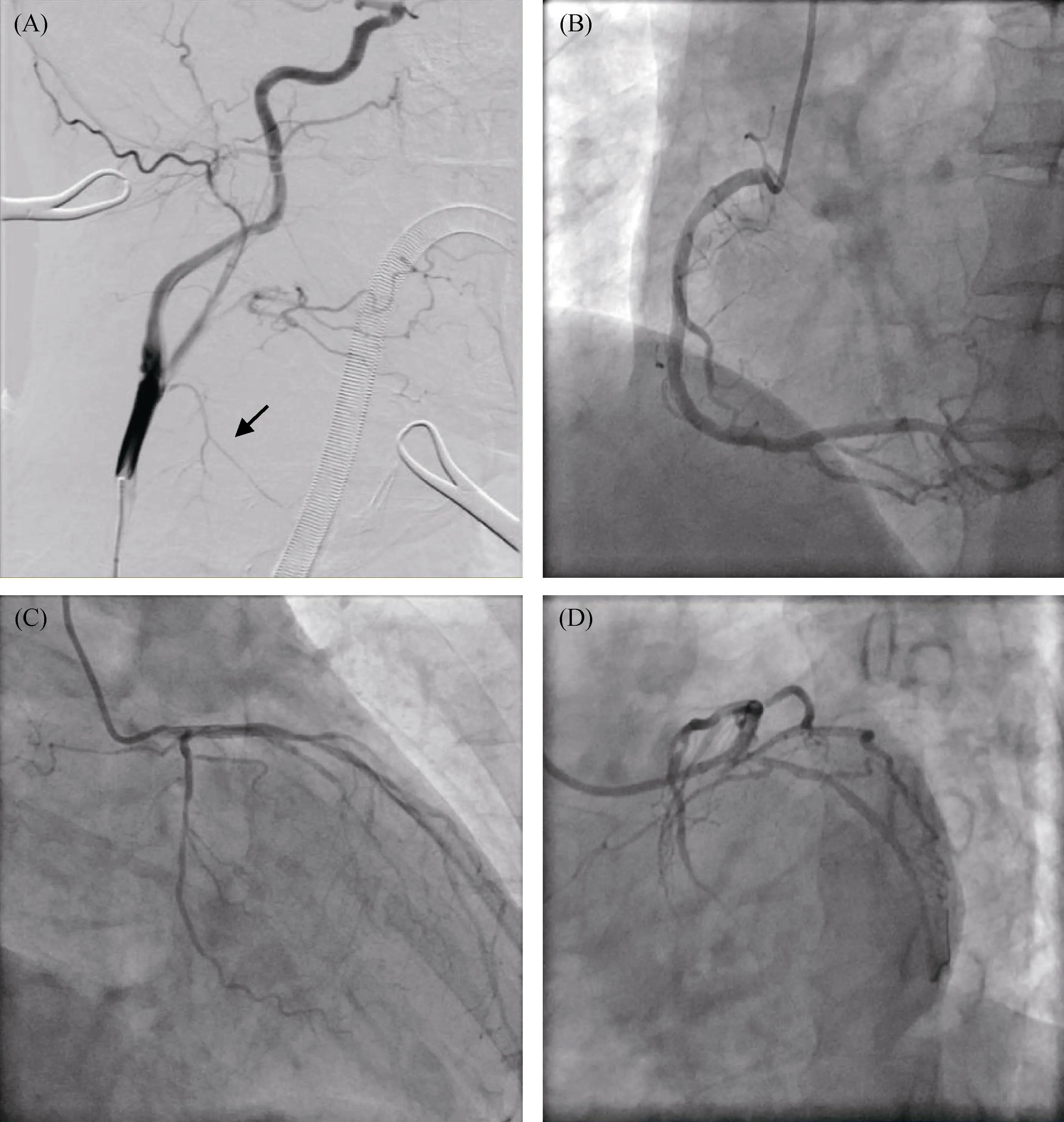
The procedure was carried out to use a right radial 6Fr access. The radial angiography catheter was inserted under the guidance of the loach guide wire, from the radial artery to the brachial artery, the ascending aorta, and the root of the aorta. After it was grafted to the left coronary artery, the radiography was performed under multiple orientations. The left anterior descending artery had a 70%–80% mid-vessel stenosis and the left circumflex had an 80% mid stenosis.After being grafted to the right coronary ostium, the angiography was performed under multi-directional projection,and it was found that the right coronary had a 30% stenosis and diffuse multi-segment disease. The left and right coronary arteries were right-handed (Figure 1).
Discussing the condition with the patient and his family members, he had been proposed an anterior descending branch and circumflex branch PCI. 7000 IU of heparin was injected into the arterial sheath. The 6F-EBU large-lumen catheter was used, and it was sent to the left coronary artery under the guidance of 0.035 loach guide wire.
The Runthough guide wire was pushed to the distal end of the circumflex branch, and a 2.0 × 20 mm balloon was expanded at the lesion in the middle segment of the circumflex branch. An angiography showed a residual stenosis of 30% in the middle section, and a 2.5 × 20 mm stent was placed in the narrow section of the circumflex branch. Later,it was expanded twice at 8 ATM × 8 s, and the residual stenosis was 0 on angiography. After the operation, the tube was withdrawn and the sheath was manually compressed to stop the bleeding. The sterile dressing was pressurized and bandaged. The patient had no discomfort during the operation and returned to the ward. 7000 IU of heparin was shared during the operation, and the contrast agent iodixanol was 160 mL during the operation.
An hour later, the patient found swelling and painful in the neck. The bedside ultrasonography of carotid artery indicated a large hematoma (76 × 43 mm) which commuted with the primary subclavian artery. The vascular surgeon was called to have an angiography in the Cath lab immediately. Unfortunately, they couldn’t find the bleeding blood vessel. Under direct vision of surgery, they found the cause of neck hematoma: a subcutaneous mass in the right neck was cut to see the inside of the carotid artery, and there was a large amount of hematoma around the thyroid. After the hematoma was cleared, the common carotid artery was explored. Behind the internal carotid artery, the proximal and distal of superior thyroid artery was rupture and bleeding.The surgeon sutured it and washed around the carotid artery.After irrigation, a drainage piece was placed locally, and the neck incision was sutured layer by layer after hemostasis.Then the patient was transferred to the ICU to continue treatment after local pressure bandaging.

Figure 1. Angiography images. (A): Carotid angiography; (B): right coronary artery; (C): left coronary system; and (D): diagonal and circumflex branch.
At the time, the cardiologist remembered that he had passed the loach guide wire without direct imaging, and felt resistance, then returned, and performed the surgical operation again under direct imaging. The guide wire traveled from the radial artery to the brachial artery, ascending aorta, and aortic arch, up to the brachiocephalic trunk, the common carotid artery, and reaches the superior thyroid artery, damaging it and causing neck hematoma formation in a short time.
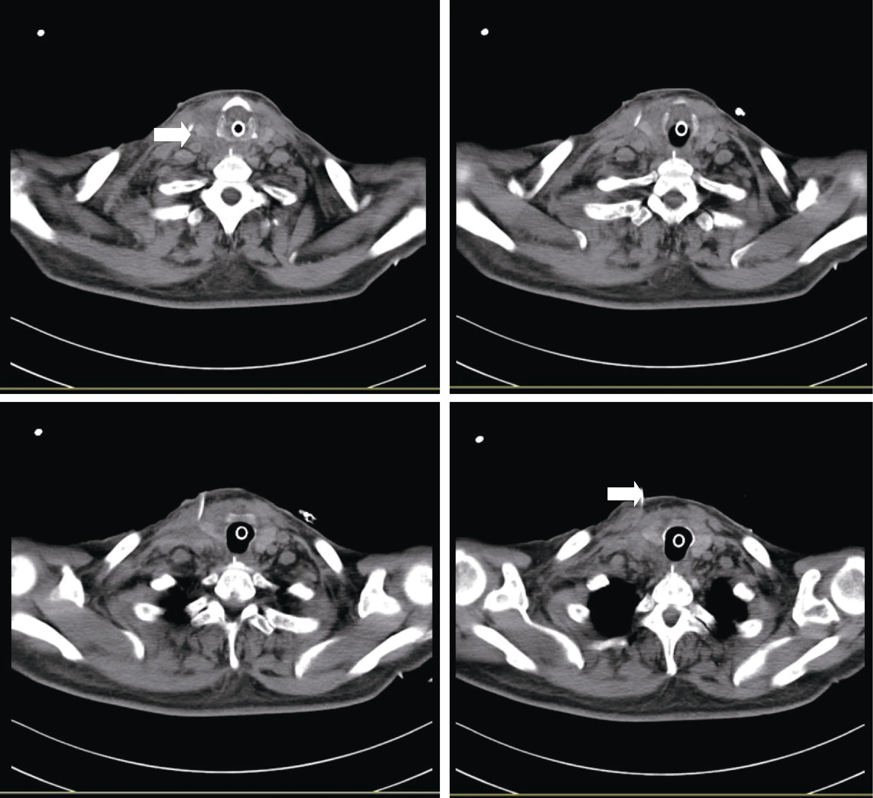
Three days later, the CT showed the right neck had a dense streaking shadow and a drainage piece (Figure 2).Due to the neck hematoma, he stayed in ICU for 4 days and suffered high fever and pneumonia. He was eventually discharge from hospital for 31 days.
Post PCI bleeding is one of the most common complications. Hemorrhage and subcutaneous hematoma are the most common vascular complications, most of which are in the puncture site, and occasionally in the gastrointestinal tract or retroperitoneal space. The amount of bleeding is not necessarily proportional to the size of the hematoma. The blood of the thin patients is easy to penetrate the subcutaneous tissue, and the blood of the obese patients is easy to seep from the puncture, especially within 3–4 h after surgery. Many effective strategies were done to decrease this complication, including anticoagulation with bivalirudin,radial access procedures and vascular closure devices.[2]In our case, the cardiologist was too confident to allow the guide wire to enter the superior thyroid artery without direct vision of the radiography. It caused a neck hematoma and may cause progressive dyspnea over time. The emergency surgery to clear the hematoma made the patient safe. If the guide wire went up along the right internal carotid artery, it may cause a more serious complication which called cerebral hemorrhage. PCI of chronic total occlusions has certain blindness. Hard guide wire is easy to cause a series of possible complications. Intramural hematoma is a rare and potentially fatal complication of percutaneous procedures as it may cause compression of cardiac chambers with similar haemodynamic effects as a proper pericardial tamponade,[3]but with no evidence of significant pericardial effusion. It requires the cardiologist to quickly identify and handle it correctly.

Figure 2. Neck computerized tomography and the drainage piece.
Bleeding following PCI is associated with elder patients because the prevalence of coronary artery disease increase with age, reaching 30.6% and 21.7% in male and female patients over the age of 80 respectively.[4]It will cause a prolonged hospital stay[5,6]and an increasing in-hospital mortality.[7]In order to reduce the cost of hospitalization and obtain a higher success rate, we need to fully evaluate patients before PCI, operate carefully during the procedure,and effectively prevent various complications to improve the survival rate of the patients.
Acknowledgements
This work was supported by Zhejiang Provincial Medicine and Health Technology Plan 2019 (No. 2019RC264) to Wang H. The authors have no conflicts of interest to declare.
 Journal of Geriatric Cardiology2020年5期
- Journal of Geriatric Cardiology的其它文章
- Midventricular Takotsubo syndrome
- Ischemia/hypoxia inhibits cardiomyocyte autophagy and promotes apoptosis via the Egr-1/Bim/Beclin-1 pathway
- Sagittal abdominal diameter as a marker of visceral obesity in older primary care patients
- Association of frailty with all-cause mortality and bleeding among elderly patients with acute myocardial infarction: a systematic review and meta-analysis
- Association between serum uric acid level and endothelial dysfunction in elderly individuals with untreated mild hypertension
- Relationship between high sensitivity C-reactive protein and angiographic severity of coronary artery disease
