Delayed cardiac tamponade after simultaneous transcatheter atrial septal defect closure and left atrial appendage closure device implantation: a particular case report
Jian-Ming WANG, Qi-Guang WANG, Xian-Yang ZHU
Department of Congenital Heart Disease, General Hospital of Northern Theater Command, Shenyang, China
Keywords: Atrial fibrillation; Atrial septal defect; Cardiac catheterization; Cardiac tamponade; Left atrial appendage
Percutaneous left atrial appendage (LAA) occlusion evolved as an alternative treatment to the patients who are contraindicated or cannot tolerate oral anticoagulants with nonvalvular atrial fibrillation (AF) at risk of stroke or systemic embolism.[1]Abnormal hemodynamic changes in elder atrial septal defect (ASD) patients cause remodeling of the left atrium, which eventually leads to right heart failure.[2]As the ASDs elderly are associated with a higher incidence of AF, simultaneous transcatheter ASD and LAA closure has become a new effective therapeutic strategy. However, only a limited number of articles involving cardiac tamponade complications have been published in the literature. What’s more, previous studies involving early hemodynamically irrelevant pericardial effusion after the procedure attribute to multiple repositioning attempts of LAA occluder or delivery sheath injured the atrial wall. We hereby describe a particular case of simultaneous closure of LAA and ASD, who was transferred to surgery for unacceptable delayed cardiac tamponade and may suggest that abrasion effect of LAA occluder disks might be potential risk of delayed cardiac tamponade after LAA closure.
A 61-year-old man who complained of palpitation and shortness of breath after activity for two months was diagnosed as a secundum ASD (36 mm under transthoracic echocardiography, TTE), and he had suffered from AF more than 12 months with left atrium dilation (49 mm under TTE). He had a history of transient ischemic attack (CHA2DS2VAS score was 4). He remained at high risk of bleeding with anticoagulation, as he had a repeated epistaxis and gingival hemorrhage (HASBLED score was 3). He was referred for consideration of simultaneous transcatheter ASD closure and LAA closure device implantation.
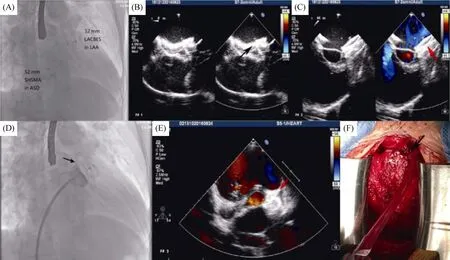
Transesophageal echocardiography (TEE) was used for preoperative evaluation of cardiac structure and function. A large central ASD at bi-atrial view, wind bag shape of LAA without thrombus and mild mitral valve regurgitation at left-sagittal view were demonstrated (Figure 1). After general anesthesia, the right radial artery and right femoral vein were punctured, then the internal and external sheath were placed. The coronary angiography was performed through the right radial artery, and no stenosis was found in the left main trunk, the anterior descending branch, the circumflex branch and the right coronary artery. The pressure of the pulmonary artery and right ventricle was measured through the femoral vein by end-hole catheter (40/16 mmHg and 40/0 mmHg respectively). Thereafter, the 0.035" × 260 cm stiff guide wire was inserted through the atrial septal defect, from the left atrium to the left superior pulmonary vein. Along the guide wire, a 12 Fr sheath (Push Medical, Shanghai, China) was transferred into the left superior pulmonary vein, then the 6 F pigtail catheter was sent into the sheath, which exited from the left superior vein to the left atrium, landing in the LAA. The LAA angiography was performed under the right anterior oblique 30° + the head 20° and the right anterior oblique 30° + the foot 20° respectively. The angiography demonstrated that the shape of the LAA was wind bag type, and the maximum diameter of its neck ostium, landing zone and depth were 31 mm, 28 mm and 5 mm respectively. A 32-mm domestic LAA occluder[3](LACBES?, Push Medical, Shanghai, China) was introduced in the 12 Fr delivery sheath and placed into the LAA. The device was successfully deployed. Before the occluder released, a stable position and favorable shape of discs of the occluder without residual leak was confirmed angiographically by a tug test (Figure 2A). Subsequently, the maximum diameter of ASD was measured (39 mm) under TEE. A 52-mm domestic ASD occluder (SHSMA, Lepu Medical, Beijing, China) was selected to close the ASD through a 14 Fr delivery sheath (Lepu Medical, Beijing, China). The blood flow of the left superior pulmonary vein, mitral valve, aortic valve, and coronary sinus were normal and no pericardial effusion was found during the procedure under TEE.

Figure 1. TEE preoperative evaluation. (A): 36 mm ASD (black arrow) at bi-atrial view; (B): wind bag shape of LAA without thrombus (red arrow) was demonstrated; and (C): left atrium dilation with mild mitral valve regurgitation (*) at left-sagittal view. ASD: atrial septal defect; LA: left atrium; LAA: left atrial appendage; LV: left ventricle; RA: right atrium; RV: right ventricle; TEE: transesophageal echocardiography.

Figure 2. The angiography video and echocardiography review. (A): LAA occluder in favorable shape and position after released under fluoroscopic; (B): ASD occluder in optimal position without residual leak under TTE (black arrow); (C): LAA occluder without dislodgement under TTE in 24 h postimplantation assessment (red arrow); (D): after contrast injections, the upper left sealing disc of LAA occluder localized filling (black arrow) by review of angiography video; (E): the upper left sealing disc of LAA occluder (red arrow) might have been up against the LAA root under TTE assessment; and (F): intraoperative visulization of the left atrium and the left upper pulmonary vein ruptured (black arrow) and bleeding. ASD: atrial septal defect; LAA: left atrial appendage; TTE: transthoracic echocardiography.
The optimal position of the ASD and LAA occluders without residual leak, dislodgement or mitral apparatus damage were confirmed by first postoperative 24 hours TTE examination (Figure 2B and 2C). The syndrome of nausea, vomiting, chest tightness, palpitation and cold sweat were suddenly appeared 30 hours after the procedure. A large amount of pericardial effusion was observed under TTE, and cardiac tamponade was diagnosed and immediately the drainage tube followed with pericardientesis was placed under local anesthesia. In the following days, dynamic observation of echocardiography showed that the patient still had slow infiltration of the pericardial effusion and intermittent exudation of the pericardial effusion, especially 300 mL hemorrhagic effusion on the forth postoperative day. The angiography video and echocardiography during the procedure were reviewed carefully. We assumed that the abrasion effect of the upper left sealing disc of LAA occluder against the LAA root might lead to delayed cardiac tamponade (Figure 2D and 2E). Considering the existence of active bleeding in the heart, we urgently requested cardiac surgery consultation. On the fifth postoperative day, emergency surgeries including the removal of ASD and LAA occluders, repair of ASD, and hemostasis suture were successfully performed. The left atrium and the left upper pulmonary vein ruptured and bleeding were demonstrated during the surgery (Figure 2F). This patient was discharged on the ninth day after surgery uneventfully.
As abnormal blood volume of the left atrium, and interstitial tissue fibrosis of atrial wall in ASD patients, which leads to electrical anatomical remodeling of the atrium, the AF is more likely to occur. AF detection rate was 52% in ASD patients aged 60 years or older.[4]Intratrial communication is an independent risk factor for stroke, and the presence of intratrial communication in patients with nonvalvular AF will significantly increase the risk of stroke. In addition, percutaneous LAA occlusion evolved as an alternative treatment to oral anticoagulationin patients with nonvalvular AF at risk of stroke or systemic embolism. For ASD patients with nonvalvular AF who cannot tolerate anticoagulation therapy, simultaneous closure of LAA and ASD is an effective therapeutic strategy to prevent stroke and correct abnormal hemodynamics.[5,6]It also can effectively avoid the complications associated with atrial septal puncture and shorten the operation time. However, simultaneous closure through intratrial communication might complicate the procedure. For some large ASDs, the delivery sheath is difficult to be fixed, resulting in repeated operations.
Major procedural complications including pericardial tamponade, device embolization, major bleeding, stroke, transient ischemic attack, cardiac dysfunction,etc; in which pericardial tamponade is more frequent.[7]what’s more, procedural complications especially hemodynamically irrelevant pericardial effusion was reported immediately after simultaneous transcatheter ASD and LAA closure, they were all treated conservatively. In addition, these patients with procedural tamponade were successfully treated with pericardiocentesis.[7]Dezsoe,et al.[8]reported two pericardial tamponade cases for several unsuccessful attempts with different sizes LAA occluders and recrossing the intratrial communication with the delivery sheath resulted in atrial wall injury, and both patients with procedural tamponade were successfully treated with pericardiocentesis and discharged the following day. However, in this case, the patient was found delayed cardiac tamponade 30 hours after procedure due to left atrium and the left upper pulmonary vein ruptured and bleeding caused by abrasion effect of LAA occluder disks, except for repeated attempts or sheath injury.
To our knowledge, this is the first case of such patient with large left atrium, simultaneous closure of LAA and ASD, who was transferred to surgery for delayed unacceptable pericardial effusion. So we come up with a hypothesis that, the ASDs elderly are associated with a higher incidence of AF, more likely leading to large fibrotic fragile left atrium as abnormal hemodynamic changes,[9]which further increases the potential damage to the left atrium or LAA wall caused by the direct incision effect from the rim of the disc of LAA occluder. It’s debatable for the expansion of clinical indications as Shingo,et al.[10]had reported that the LAA closure for “primary primary” prevention during percutaneous LAA closure of ASDs in patients with large atrium but no AF had been performed.
In conclusion, simultaneous closure of LAA and ASD is a viable option for patients with nonvalvular AF at risk of stroke or systemic embolism but there are technically difficult considerations to prevent the cardiac tamponade. Carefully observing the shape of discs of LAA occluder, and assess the position and its surroundings intraprocedural angiocardiography and TEE in different projections can contribute to procedural success. In the case of our elderly patient, it was suggested that abrasion effect of LAA occluder might be potential risk of delayed cardiac tamponade after LAA closure.
Acknowledgments
This study was supported by the Ph.D. Launch Programs Foundation of Liaoning Province (2019-BS-266). All authors had no conflicts of interest to disclose.
 Journal of Geriatric Cardiology2019年12期
- Journal of Geriatric Cardiology的其它文章
- Quantitative flow ratio and intravascular ultrasound guided percutaneous coronary intervention of left anterior descending lesion concomitant with severe coronary myocardial bridge
- Coarctation of the aorta in twins with severe hypertension
- Risks of incident heart failure with preserved ejection fraction in Chinese patients hospitalized for cardiovascular diseases
- Pacemaker therapy in very elderly patients: survival and prognostic parameters of single center experience
- Irregular surface of carotid atherosclerotic plaque is associated with ischemic stroke: a magnetic resonance imaging study
- Hypoxia promotes pulmonary vascular remodeling via HIF-1α to regulate mitochondrial dynamics
