Next generation sequencing reveals co-existence of hereditary spherocytosis and Dubin-Johnson syndrome in a Chinese gril:A case report
Yuan Li,Yang Li,Yang Yang,Wen-Rui Yang,Jian-Ping Li,Guang-Xin Peng,Lin Song,Hui-Hui Fan,Lei Ye,You-Zhen Xiong,Zhi-Jie Wu,Kang Zhou,Xin Zhao,Li-Ping Jing,Feng-Kui Zhang,Li Zhang
Yuan Li,Yang Li,Yang Yang,Wen-Rui Yang,Jian-Ping Li,Guang-Xin Peng,Lin Song,Hui-Hui Fan,Lei Ye,You-Zhen Xiong,Zhi-Jie Wu,Kang Zhou,Xin Zhao,Li-Ping Jing,Feng-Kui Zhang,Li Zhang,Anemia Therapeutic Center,Institute of Hematology and Blood Diseases Hospital,CAMS and PUMC,Tianjin 300020,China
Abstract
Key words:Hereditary spherocytosis;Dubin-Johnson syndrome;Hemolytic anemia;Jaundice;Next generation sequencing;ABCC2;SPTB;Case report
INTRODUCTION
Hereditary spherocytosis(HS)is a hereditary disease of hemolytic anemia that occurs due to the erythrocyte membrane defects caused by the gene mutation in the erythrocyte membrane protein,and its common clinical manifestations include hemolysis,anemia,jaundice,splenomegaly,etc.Dubin-Johnson syndrome(DJS),which commonly results in jaundice,is a benign hereditary disorder of bilirubin clearance that occurs only rarely.The co-occurrence of HS and DJS is extremely rare[1]and leads to severe jaundice.We recently diagnosed and treated a case of co-occurring HS and DJS in a patient who had severe jaundice and moderate anemia intensified by the infection of the upper respiratory tract.We applied next generation sequencing(NGS)to arrive at a definite diagnosis.The case is reported as follows.
CASE PRESENTATION
Chief complaints
A 21-year-old female patient presented to our department on November 10,2017 because of severe jaundice,severe splenomegaly,and mild anemia since birth(Table 1).
History of past illness
She did not undergo irradiation treatment.When she was two years old,the yellow staining was intensified in her skin and sclera and she started having dark brown urine.Other clinical manifestations included anemia,splenomegaly,and elevated bilirubin,but her transaminases were normal.All later clinicians that she visited could not clarify her situation any further except for giving the diagnosis of “hemolytic anemia” and did not advise any treatment either.Her history of past illness was negative.She did not receive blood transfusion or surgery.Her regular blood routine testing showed the following:WBC 8.4-10.8 × 10⁹/L(normal:4-10 × 109/L),hemoglobin(HGB)106-111 g/L(110-150 g/L),platelets(PLT)186-241 × 109/L(100-300 × 109/L),percentage of neutrophils(NE%)56.9%-60.5%(normal:50%-70%),percentage of lymphocytes(LY%)33.1%-34.7%(normal:20%-40%),and percentage of reticulocytes(RET%)7.8%-11.3%(normal:0.5%-1.5%).Her liver function results were:Total bilirubin(TBIL)132-162.8 μmol/L(normal:5.0-21.0 μmol/L),direct bilirubin(DBIL)34.4-40 μmol/L(normal:0-3.4 μmol/L),and indirect bilirubin(IBIL)98.6-137 μmol/L(normal:0-13.6 μmol/L).She experienced intensified anemia and jaundice upon fatigue or infection.She sought further diagnosis of her situation in her current visit to our clinic.
Personal and family history
She was the only child of her family and her parents had no similar clinical manifestations including anemia,splenomegaly,and elevated bilirubin.
Physical examination
On physical examination,the patient demonstrated obvious anemic appearance and intense yellowing of the skin,without any edema.The liver was not palpable,but the spleen was palpable at 6 cm below the rib margin.
Laboratory examinations
Blood tests gave the following results:WBC 4.58 × 10⁹/L(normal:4-10 × 109/L),absolute neutrophil count(ANC)3.22 × 10⁹/L(normal:2-7 × 109/L),red blood cells(RBC)2.64 × 10¹²/L(normal:3.5-5 × 1012/L),HGB 85 g/L(normal:110-150 g/L),mean corpuscular volume 87.9 fL(normal:80-100 fL),mean corpuscular hemoglobin 32.2 pg(normal:27-34 pg),mean corpuscular hemoglobin concentration 366 g/L(normal:320-360 g/L),PLTs 170 × 109/L(normal:100-300 × 109/L),RET% 7.89%(normal:0.5%-1.5%),and ARC 0.2083 × 10¹²/L(normal:0.024-0.084 × 1012/L).Theurine test gave all normal results except for elevated urobilinogen(+).The liver and kidney function tests showed:total protein 69.4 g/L(normal:66-83 g/L),albumin 42.2 g/L(normal:35-52 g/L),globulin 27.2 g/L(normal:20-35 g/L),alanine aminotransferase 9.7 U/L(normal:0-35 U/L),aspartate aminotransferase 11.6 U/L(normal:0-35 U/L),alkaline phosphatase 50.4 U/L(normal:30-120 U/L),γ-glutamyl transpeptidase 9.5 U/L(normal:8-57 U/L),TBIL 111.8 μmol/L(normal:5.0-21.0 μmol/L),DBIL 35.4 μmol/L(normal:0-3.4 μmol/L),DBIL/TBIL ratio 31.7%(normal:20%),IBIL 76.4 μmol/L(normal:0-13.6 μmol/L),blood urea nitrogen 2.54 mmol/L(normal:2.8-7.6 mmol/L),creatinine 58.3 μmol/L(normal:49-90 μmol/L),uric acid 362 μmol/L(normal:154.7-357 μmol/L),and lactate dehydrogenase 189.6 U/L(normal:0-248U/L).Hemolysis test showed reduced plasma haptoglobin(0.375 g/L;normal:0.5-2.0 g/L),and plasma-free hemoglobin was 37.1 mg/L(normal:0-40 mg/L).Eosin-5’-maleimide(EMA)flow cytometry showed that the mean fluorescence intensity attenuation of the RBC EMA was 23.41%(normal:<16%).The RBC osmotic fragility(EOF)test showed that hemolysis started at 0.6%(normal:0.44%)and completed at 0.36%(normal:0.32%).The acidified glycerol lysis test(AGLT50)gave a result of 60 s(normal:>290 s).

Table 1 The charactistics of the patient
The patient was found negative in the hemoglobin A2 test,anti-alkaline hemoglobin test,heat instability test,hemoglobin acetate membrane electrophoresis,direct Coombs test,cold agglutinin test,denatured globin corpuscle test,isopropanol test,methemoglobin reduction test,and acid hemolysis test.The patient had normal activities of erythrocyte pyruvate kinase,erythrocyte pyrimidine 5’-nucleotidase,6-phosphate glucose dehydrogenase,and erythrocyte glucose phosphate isomerase,and there was no anomaly in immunoglobulin quantification,antinuclear antibody,etc.Iliac marrow smear showed obviously active hyperplasia,with 44%(normal:40%-60%)of granulocytes and 48.5%(normal:15%-25%)of erythrocytes.The peripheral blood smear was rich in small spherical RBC,which accounted for 70% of the mature RBC.Bone marrow autopsy showed relatively normal(50%)myeloproliferation based on HE and PAS staining,as well as escalated erythrocytes/granulocytes ratio.The reticular fiber dyeing result was MF-0.The karyotype was 46.An abdominal CT scan showed enlarged spleen with minor effusion.The liver had normal size,proportionate lobes,and parenchyma of uniform density.The intrahepatic and extrahepatic bile ducts showed no sign of dilation,and the hepatic portal had a clear structure.No anomaly was observed for the gallbladder,pancreas,kidneys,or abdominal cavity.Abdominal ultrasound examination showed hyperechogenic liver parenchyma,moderately enlarged spleen,as well as normal gallbladder and pancreas.Hence,as a result of the preceding clinical findings,the patient was diagnosed with HS.
Imaging examinations
However,because of the extraordinary jaundice of the patient,whole exome sequencing was carried out additionally after the diagnosis of HS.
DNA for NGS was extracted from peripheral blood of the patient and her parents.Agarose electrophoresis,Qubit 2.0 fluorometer dsDNA HS Assay(Thermo Fisher Scientific),and 2100 Bioanalyzer and Herculase II Fusion DNA Polymerase(Agilent)were used for DNA library preparation according to the instructions.Targeted fragments were captured with exome capture probes(Aligent)and sequenced on the Illumina HiSeq X platform following Illumina-provided protocols.The sequencing quality was determined with FastQC software.After data filtration,the clean reads were mapped to human reference genome(hg19)using SentieonBWA software.Then,the mapped reads were used to detect SNV and InDel with Sentieon(the same algorithm with GATK),and annotated using ANNOVAR/VEP software.All the variants were annotated with VEP software and the pathogenic variants wre screened by ClinVar,OMIM,and HGMD.Function prediction of missense mutations and annotation of non-coding sequences were performed with PolyPhen-2 and Sorting Intolerant from Tolerant(SIFT).
Blood(2 mL)was drawn for Sanger sequencing from the patient and her healthy parents.All the suspicious pathogenic variants were validated in the patient and her parents using Sanger sequencing.Primers were designed with Primer 3 software,and BLAST in NCBI database was used to confirm specificity.PCR amplification product was sequenced with an ABI 3500D x DNA Analyser(Applied Biosystems,Foster City,CA,United States)and analyzed with sequencing analysis software(Thermo Fisher).
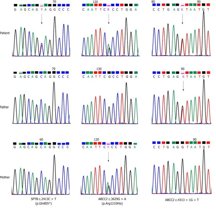
The sequencing results revealed ade novoheterozygous mutation of theSPTBgene(NM_000347.5),c.2413C > T(p.Gln805*),as well as two inherited novel heterozygous mutations of theABCC2gene(NM 000392.4),c.4313+1 G > T from the father and c.3629G > A(R1210H)from the mother(Figure 1).Neither of these mutations had been observed in the Clin Var,OMIM,and HGMD databases,indicating that these variants are very rare.All three mutations were predicted to be harmful and pathogenic with PolyPhen-2 and SIFT.These mutations were identified as pathogenic variants following the 2013 ACMG guidelines[2].
FINAL DIAGNOSIS
The final diagnosis was co-existence of HS and DJS.
TREATMENT
The patient was recommended to receive oral doses of ursodeoxycholic acid and phenobarbital in addition to splenectomy.She refused splenectomy and was then discharged.
OUTCOME AND FOLLOW-UP
The patient first took ursodeoxycholic acid in combination with phenobarbital for three months.The treatment successfully increased HGB to 114 g/L(normal:110-150 g/L)and reduced TBIL to 88.69 μmol/L(normal:5.0-21.0 μmol/L),DBIL to 29.9 μmol/L(normal:0-3.4 μmol/L),DBIL/TBIL to 33.7%(normal:20%),and IBIL to 58.79 μmol/L(normal:0-13.6 μmol/L).
DISCUSSION
The diagnosis of HS was straightforward.Hemolytic anemia could be readily inferred since the patient experienced the symptoms since birth.Anemia,jaundice,enlarged spleen,increased reticulocytes in peripheral blood,and marrow morphology all pointed to proliferative anemia.The key evidence of HS included notable increase in the small spherical mature RBC in the peripheral blood smear,escalated RBC osmotic fragility in the EOF and AGLT50 tests,and notable defect of erythrocyte membrane protein in the EMA test.The genome sequencing revealed heterozygous mutation of theSPTBgene and thus verified the diagnosis of HS.
Although the patient was clearly diagnosed with moderate HS,this could not convincingly account for the severe jaundice and the significantly elevated levels of TBIL,DBIL,and IBIL.Since the imaging results clearly excluded any obstruction of intrahepatic and extrahepatic bile ducts,and the activities of all liver enzymes werefound normal,the observed degree of hemolysis did not suffice to reasonably explain the severity of jaundice.Consequently,we suspected that another disease aside from HS was likely,and we naturally turned out attention to two hereditary disorders of bilirubin clearance,i.e.,DJS and the Rotor syndrome(RS),because the patient had experienced elevated DBIL since birth and showed normal liver enzymes without abnormality in the liver and bile duct images.

Figure 1 Sanger sequencing confirmed a de novo heterozygous mutation of the SPTB gene(NM_000347.5).
DJS,first reported in 1954[3],is a rare,chronic,benign,autosomal recessive disorder characterized by elevated DBIL along with normal liver function.Its pathogenesis has been largely clarified and could be accounted by the mutation of theABCC2gene.
The humanABCC2gene is located at chromosome 10q24,and its introns and exons have been well sequenced.TheABCC2gene has 32 exons that encode theABCC2transporter,i.e.,the multidrug resistance-associated protein 2.TheABCC2transporter is an ATP-binding cassette transporter at the apical membrane of hepatocytes consisting of 1545 amino acids.It is the major carrier that is responsible for transporting bilirubin into bile,and it becomes activated by the ATP hydrolysis of two ATP-binding regions to expel the substrate outside the cellviaan energy dissipation process.Mutation of theABCC2gene in the DJS patients leads to defectedABCC2transporter and thus impaired ability to transport DBIL into the bile,and the high serum DBIL in turn results in jaundice.
To date,34 kinds ofABCC2mutations have been found in DJS patients[4-17],including 6 kinds of nonsense mutations(17.4%),14 kinds of missense mutations(41.2%),4 kinds of small deletions(11.8%),4 kinds of splice mutations(11.8%),and 6 kinds of large deletions(17.4%).At least 23 mutations(67.6%)of the total 34 are predicted to be harmful.The mutations may result in the maturity disorder or positioning error of theABCC2transporter and reduce the proteinactivity[18].Compound heterozygous mutations were found in theABCC2gene of this patient,which were all previously unreported missense mutations.
The c.4313+1 G > T mutation from the father was located at the boundary between the 30thexon and the 30thintron of theABCC2gene(i.e.,the donor point).It could result in exon skipping and was predicted to be pathogenic.The c.3629 G > A(R1210H)mutation from the mother was located at the 26thexon of theABCC2gene,which is responsible for expressing the cytoplasmic side of the membrane-spanning domain 2 of theABCC2transporter[5].This mutation was also predicted to be pathogenic and could be involved in this disease.Therefore,both parents of the patient carriedABCC2mutations but had a normal phenotype,which is in agreement with the genetics of the DJS.Therefore,the diagnosis of DJS could be established unequivocally.More data on theABCC2transporter and DJS patients are needed to clarify the relationship between the genotype and phenotype of DJS patients,since mutations occur across the entireABCC2gene and no hotspot mutation has been discovered to date.
NGS is not yet a regular examinaiton in the diagnosis of DJS.Current clinical diagnosis of DJS generally requires liver function tests,urinary coproporphyrin excretion,liver histopathology(sampled by liver puncture),and sodium bromosulfonate(BSP)test.Diagnosis can be made when the following evidence is gathered:The liver function tests show normal enzyme activities and elevated DBIL(usually 34.2-85.5 μmol/L);urinary coproporphyrin excretion show normal results and 80% of the discharged porphyrin is type I coproporphyrin;the liver histopathology showed “black liver” due to the diffusive deposition of lipofuscinmelanin complex in the hepatocytes[19].Although sodium bromosulfonate test is no longer used in clinical practice and has been a redundant test over many years,the retention of BSP is normal or slightly elevated at 45 min,and is further increased at 90 and 120 min,indicating that upon the maximum excretion of BSP,the transportation of BSP by the liver declines notably and the storage of BSP decreases only slightly[19],which helps in understanding the dynamics of bilirubin metabolism.
Despite the additional cost required by NGS,we recommend adopting NGS as a regular examination for the diagnosis of DJS because of the following advantages.First,NGS is very simple to operate and circumvents the tedious procedures required in the BSP test and urinary coproporphyrin excretion.In addition,NGS commonly only requires sampling peripheral blood and is thus much safer and less invasive than the liver puncture required by histopathology examination,therefore avoiding the risk of serious complications such as peritonitis.More importantly,the high accuracy of NGS allows the direct identification of the pathogenic gene,simplifies the diagnosis procedures,and improves the efficiency,which is particularly advantageous in distinguishing DJS and RS.
RS is rare autosomal recessive disorder of bilirubin clearance.It is characterized by the disorder of hepatocytes in the storage and uptake of bilirubin due to the mutation ofSLCO1B1/OATP1B1 orSLCO1B3/OATP1B3 genes,and it is clinically manifested as jaundice[20].The phenotypes of DJS and RS cannot be differentiated clinically,although RS is also diagnosed after liver function tests,urinary coproporphyrin excretion,liver histopathology(sampled by liver puncture),and BSP test.Since the current patient had elevated serum bilirubin due to HS,it was highly difficult(if possible)to determine by conventional tests if DJS or RS was occurring.However,this problem was easily solved by NGS,since the heterozygous mutation ofABCC2was detected while the mutations ofLCO1B1/OATP1B1 andSLCO1B3/OATP1B3 were excluded.Therefore,RS was ruled out for the patient,and NGS greatly simplified the diagnosis by obviating the standard cumbersome tests.
Splenectomy is the classical treatment for HS.The patient showed the indications for splenectomy since she already had moderate HS,although she declined surgery.Since DJS normally has a good prognosis,no special treatment is necessary and DJS patients only need to avoid situations that may aggravate jaundice such as fatigue and infection.Nevertheless,because the combination of DJS and HS posed a significant risk for cholelithiasis in this patient,ursodeoxycholic acid and phenobarbital are the recommended medications[12].This patient showed improvements after taking these two drugs.
CONCLUSION
In summary,to the best of our knowledge,the current case is probably the first case that was definitively diagnosed with the co-occurrence of HS and DJS by NGS.We suggest that inherited disorders of bilirubin clearance should be investigated if patients with inherited hemolytic anemia present with severe hyperbilirubinemia.Theroutine application of NGS is also recommend to efficiently render a definite diagnosis when inherited disorders are suspected.
 World Journal of Clinical Cases2019年20期
- World Journal of Clinical Cases的其它文章
- Clinical use of low-dose aspirin for elders and sensitive subjects
- Distribution and drug resistance of pathogenic bacteria in emergency patients
- Comparative analysis of robotic vs laparoscopic radical hysterectomy for cervical cancer
- Feasibility of laparoscopic isolated caudate lobe resection for rare hepatic mesenchymal neoplasms
- Soft tissue release combined with joint-sparing osteotomy for treatment of cavovarus foot deformity in older children:Analysis of 21 cases
- Clinical characteristics of sentinel polyps and their correlation with proximal colon cancer:A retrospective observational study
