Primary malignant melanoma of the biliary tract:A case report and literature review
Soledad Cameselle-García, José Luis Fírvida Pérez, María C Areses, Jesús Daniel Fernández de Castro,Juan Mosquera-Reboredo, Jesús García-Mata
Abstract
Key words: Biliary tract; Case report; Immunotherapy; Malignant melanoma
INTRODUCTION
Melanoma is a malignant tumour arising from melanocytes whose origin is the neural crest[1].This is a very aggressive neoplasia with a high capacity for remote dissemination[2].In the last few years there has been a major increase in the incidence of this neoplasia[3,4]due to early diagnosis and overdiagnosis of benign lesions[5,6],whereby mortality remains virtually stable[4,7,8].Its location in the mucosa is less common and associated with a delay in diagnosis2.Location in the biliary tract in the form of primary tumour is exceptional with only 13 cases reported in the literature[9-20].Increased knowledge of molecular biology has enabled more development of treatments for this disease[2,4].This paper reviews the most important features of primary malignant melanoma of the biliary tract (MBT) cases using the PubMed database (up to January 2019) and reports the first patient treated with immunotherapy.
CASE PRESENTATION
Chief complaints
A 51-year-old man with a personal history of smoking (one pack/d) and high blood pressure.The patient was admitted to the gastrointestinal department to study obstructive jaundice of a 1 wk clinical course.
Laboratory examinations
Initial analysis revealed:total bilirubin 13.9 mg/dL, GGT 900 U/L, and AF 324 U/L.
Imaging examinations and treatment
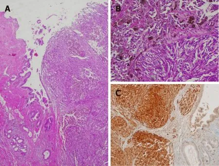
Magnetic resonance cholangiopancreatography (MRC) revealed dilatation of the intrahepatic biliary tract and stenosis of the common hepatic duct.Cytological samples obtained by endoscopic retrograde cholangiopancreatography were negative for malignancy (non-representative samples).Abdominal computed tomography (CT)confirmed the findings of the MRC and was negative for distant metastasis.Given the suspicion of biliary tract neoplasia, cholecystectomy and resection of the common hepatic conduct were performed with hepatic jejunostomy free of complications.Anatomopathological examination, including the immunohistochemical study,revealed a malignant melanoma measuring 1 cm in diameter (Figure 1).A total of two adenopathies were analysed on a perichole-cystic and peri-portal level; these did not reveal metastasis.
After intervention, the patient was referred to the Department of Medical Oncology, where primary origin was excluded in the skin, mucosa, and eyes.This confirmed diagnosis of primary biliary tract melanoma.
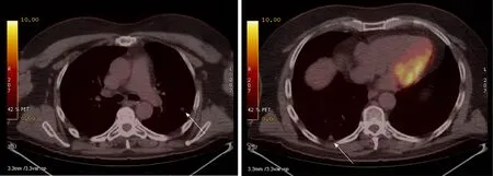
CT performed 12 mo after the procedure revealed several subcentimetric lung nodules in the posterior and apical segments of the left upper lobe (LUL), in the anterior and superior segments of the left lower lobe (LLL), and in the posterior-basal region of the right lower lobe (RLL).These nodules revealed pathological increased uptake on positron emission tomography-CT (Figure 2).Wedge resection was performed on the LUL and LLL free of complications.Second, atypical segmentectomy was performed in the RLL.Anatomopathological study confirmed the diagnosis of lung metastasis from melanoma (positive for HMB45, vimentin, and S100 protein but negative for cytokeratin in the immunohistochemical studies).BRAFgene mutation in tumour tissue was studied by real-time polymerase chain reaction(Cobas 4800 BRAF V600 Mutation Test; Roche Diagnostics, Indianapolis, IN, United States); this proved to be negative.
FINAL DIAGNOSIS
Pulmonary metastasis from primary melanoma of the biliary tract.
TREATMENT
After confirming the diagnosis of pulmonary metastasis from primary malignant melanoma of the biliary tract, the patient started treatment with intravenous nivolumab at a dose of 3 mg/kg every 2 wk.
OUTCOME AND FOLLOW-UP
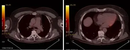
Positron emission tomography-CT studies performed 4 mo from starting nivolumab,subsequently every 6 mo and recently at 36 mo from pulmonary surgery, revealed a prolonged complete response (Figure 3).Tolerance to treatment was excellent; no immunomediated toxicity presented.
DISCUSSION
Melanoma is considered a multifactorial disease that arises from interactions between genetic susceptibility and environmental exposure[2,4].Ultraviolet radiation (UVR) is the most significant modifiable risk factor.Both UVA-A and UVR-B have genotoxic effects either because of generation of oxygen radicals or abnormality in the nucleotide sequence, among others[21].Melanomas are mainly classified according to their location and relationship to solar exposure[2].Types of melanoma that arise on the skin exposed to sun are:Melanoma with superficial extension/melanoma with low cumulative solar damage, lentigo-malignant melanoma/melanoma with significant solar damage, and desmoplastic melanoma[2].Melanomas that arise on skin protected from the sun or free of aetiological association UVR are:spitz melanoma,acral melanoma, melanoma of mucosa, melanoma that arises in congenital naevi,melanoma that arises in blue naevus, and uveal melanoma[2].The four most common histological subtypes of melanoma are superficial spreading melanoma, lentigo maligna melanoma, acrallentiginous melanoma, and nodular melanoma[2].
Although these are very useful classifications in clinical practice, new classifications are being developed based on genetic abnormalities, which have therapeutic indications.In general, melanoma is a tumour with a high mutational load[2,22].Mutations are common indrivergenes such asBRAFandRAS[2,23].Those melanomas that appear in exposed areas have more abnormalities in the genesBRAF,N-RAS, andPTEN.However, it appears that those not exposed to UVR (melanoma of mucosa and acral melanoma) have a higher number of chromosomal abnormalities (amplifications and deletions) and gene amplification, essentially of CDK4 and CCND1[2,23].Therefore,it appears that melanomas that arise in areas not exposed to radiation would have a higher mutational load, and therefore, predict a greater response to immunotherapy[22].

Figure 1 Primary melanoma of the biliary tract.
The most common melanoma location is on the skin, mainly the trunk (43.5%) and the limbs (33.9%)[2,7].Melanoma is a highly aggressive tumour that can disseminate to any part of the body, although metastases are more common in the lymph nodes,liver, lungs, and brain.Only 2% to 4% of patients present metastasis in the gastrointestinal system, mainly in the bowel, colon, and stomach[24].There are unusual cases of solitary metastasis of melanoma to the gallbladder[25]and asymptomatic metastasis has been reported in the gallbladder and biliary tract in up to 4% to 20% of autopsies performed on patients with metastatic melanoma[26,27].Therefore, another primary origin in the event of existence of melanoma in the biliary tract should be ruled out.Along these lines, Ricciet al[28]proposed five criteria that could provide guidance around diagnosis of primary melanoma in the biliary tract:(1) Absence of previous melanoma; (2) Absence of melanoma in other locations; (3) Single solitary lesion; (4) Polyploid or papillary form; and (5) Existence of biliary obstructive clinical symptoms.
After reviewing the literature and including this case, there are only 14 cases of MBT reported to date (Table 1).MBT is a very rare entity that mainly affects men(men/women ratio 12:2) and has an average age of presentation of 47 years (range:26-67).The most common form of presentation is abdominal pain together with jaundice and at times a general syndrome.In cases with macroscopic data lesions were black, polyploid, and with endoluminal growth, which conditioned obstructive clinical symptoms associated with clinical presentation.Although only three patients were revealed to have metastatic disease at diagnosis, up to seven revealed remote course after the procedure.The most common site of metastatic dissemination documented was the liver, although other locations were the brain, lung, mesentery,and pelvis.In all cases of localised disease, the treatment of choice was surgery.Radical surgery was performed in those with good “performance status”;duodenopancreatectomy was the most common intervention.Only two patients were not candidates for surgery due to their general condition and/or disease extension at the time of diagnosis.
In the last few years, immunotherapy has changed the paradigms for treatment of metastatic melanoma.Currently, first-line treatment of metastatic melanoma or nonresectable wild-typeBRAFis considered, which demonstrates benefits on overall survival, progression-free survival, response rates, and duration of response[29].While most immunotherapy clinical trials only include malignant melanomas of the skin,this case exemplifies that the excellent response of malignant melanoma to this therapy is not limited exclusively to primary malignant melanomas of the skin.
CONCLUSION
Until a few years ago, malignant melanoma was considered one of the most aggressive tumours and there were hardly any effective systemic treatment options.

Figure 2 Thoracic positron emission tomography/computed tomography scan showing metastatic nodules (arrows).
The overall survival of a patient with metastatic melanoma was less than 1 year.However, knowledge of melanoma’s molecular biology has meant a spectacular change in regard to treatment and survival of melanoma patients.Contrary to what was believed, a greater mutational load has been revealed in those melanomas that appear in sites not exposed to UV[2].This has meant a greater efficacy of immunotherapy[30].Here, for the first time, we describe a case of primary biliary malignant melanoma with pulmonary metastases, successfully treated with immunotherapy.

Table 1 Cases of patients with primary melanoma of common bile duct

Figure 3 Thoracic positron emission tomography/computed tomography scan with no evidence of metastases.
 World Journal of Clinical Cases2019年16期
- World Journal of Clinical Cases的其它文章
- Role of infrapatellar fat pad in pathological process of knee osteoarthritis:Future applications in treatment
- Application of Newcastle disease virus in the treatment of colorectal cancer
- Reduced microRNA-451 expression in eutopic endometrium contributes to the pathogenesis of endometriosis
- Application of self-care based on full-course individualized health education in patients with chronic heart failure and its influencing factors
- Predicting surgical site infections using a novel nomogram in patients with hepatocelluar carcinoma undergoing hepatectomy
- Serological investigation of lgG and lgE antibodies against food antigens in patients with inflammatory bowel disease
