Total segmental aplasia of uterus body in bitch
Leonardo Martins Leal, Fábio Rodrigo Castro Bastos, Tais Harumi de Castro Sasahara, Paola Castro Moraes,Márcia Rita Fernandes Machado
1Centro Universitário Ingá - UNINGá, Maringá, PR, Brazil
2Universidade de S?o Paulo – USP, S?o Paulo, SP, Brazil
3Universidade Estadual Paulista- Unesp, Jaboticabal, SP, Brazil
Keywords:Segmental aplasia Surgery Ovariohysterectomy Reproduction
ABSTRACT Congenital anomalies of the female tubular genital tract are rare and known as segmental aplasia, resulting from incomplete development of paramesonephric ducts during embryonic stage, which originate from the cranial portion of vagina, cervix, uterus and oviducts. In the literature consulted, several reports of uterine horn aplasia are described in bitches,however there is only one report described in the world literature on total aplasia of the body of the uterus. This study aimed to describe the case of a two years old bitch, no breed defined, with 12 kg of weight, with segmental aplasia of uterus body diagnosed during ovariohysterectomy. During surgery it was noted that the bitch had the ovaries and uterine horns in their anatomical position, however an aplasia was noted in the body of the uterus.It was also found that the uterine horns had liquid inside, suggesting hydrometra. It was concluded in this case that although segmental aplasia of the body of the uterus is rare and difficult to diagnose by maintaining normal cyclicity of the female, it should be considered in dogs that do not have bloody vaginal discharge in proestrus and can not become pregnant after natural coverage or artificial insemination.
1. Introduction
Congenital anomalies of the female tubular genital tract are known as segmental aplasia and they result from incomplete development of paramesonephric ducts during embryonic stage, which originates from the cranial portion of the vagina, cervix, uterus and oviducts[1].
Uterine horn aplasia is rare and may be followed by ipsilateral renal agenesis, possibly due to the relationship of the embryonic origin of these structures. During embryonic development, paramesonephric ducts, which are the origin of uterine horns, migrate caudally along the wall of the mesonephric ducts forming the kidneys[2].
The presence of the ovaries does not interfere with normal cyclicity after puberty and for this, it hampers the suspicion of the defect[3]. In unilateral cases, few clinical signs develop even in older animals[4],however, this type of malformation always causes a significant reduction in fertility because it affects the number of offspring,although pregnancy in the contralateral horn is possible[5].
Total segmental aplasia of uterus body is even rarer and to date there has been only one report of this condition in the world literature[6]. Almeidaet al[7] reported a second case aplasia of uterus body; however, it was not a complete aplasia.
Given the scarce studies in the veterinary literature consulted and the rarity of the disease reported, it is important to demonstrate the need for knowledge of its occurrence. So, the objective is to describe the case of a bitch with segmental aplasia of uterine body diagnosed during elective ovariohysterectomy (OH).
2. Case history
A two years old bitch with no breed defined, and with a body weight of 12 kg was brought to neutering campaign to be submitted to an OH, and a twelve-hour fasting of water and food was performed before surgery. The owner related that the bitch never had blood presence prior to estrus, noticed by swelling of the vulva,and that it was nulliparous.
Upon physical examination, all parameters usually assessed in veterinary medicine were within normal limits. No preoperative laboratory exam was performed. The bitch was submitted to general anesthesia with tiletamine/zolazepam.
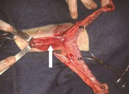
The bitch was positioned dorsally, submitted to trichotomy and previous antisepsis with povidone iodine 2% solution across all abdominal area. A retroumbilical incision was performed using a specific hook for OH, the right uterine horn was pulled out of the abdominal cavity, which was enlarged and had fluid inside,thereafter, the ovaries and left uterine horn was identified. After ovarian pedicle ligation with chrome catgut number 2 and release of the broad uterus ligament, the ligation of the uterine body near the cervix was performed. In this moment, it was observed that the bitch had total aplasia of uterus body (Figure 1), so the uterine vessels were ligated separately with chrome catgut number 2 and sectioned. The suture of the muscular part was made with nylon number 2-0 in a simple continuous pattern, the subcutaneous tissue was approximated with nylon number 3-0 in zigzag and finally the skin was sutured with nylon number 3-0 in “U” type.
After the surgery, the uterus was inspected and it was observed a clear liquid without pus, suggesting hydrometra.

Figure 1. Structures of the two years old bitch submitted to OH.
3. Discussion
Uterus congenital anomalies are rare in bitches. According to McIntyreet al[3], segmental aplasia was responsible for 20% of uterus congenital anomalies in bitches. Frequently observed in uterine horns, especially unilaterally, the incidence of uterine horn aplasia is 1:5 000 to 1:10 000 in necropsied bitches[2]. However, in the case in question, the region of aplasia was observed in the uterus body and in the world literature consulted, there is only one report of this affection[6].
In aplasia of uterine horn, if the ovary and one of the horns are preserved, normal estrous cycles are noted with bleeding in proestrus and pregnancy can be possible[4]. In this report, the absence of a segment in the body of the uterus cranially to the cervix justified the nulliparous condition of the bitch due to the inability of the ejaculate penetration and subsequent fertilization. The absence of the blood in the proestrus observed by the owner can also justified; once the blood discharge, product of erythrocyte diapedesis and rupture of subepithelial capillaries[8] could not be overflowed from the uterus.Although the normal cyclicity hampers the suspicion of uterine segmental defect[3], hydrometra and pyometra could be associated[6,9].In this case, it was observed that the presence of a mild hydrometra and no infection in uterine horns were related. Although the cause of pyometra may have hematogenous origin, the main way of uterus contamination is through the vagina upwardly[5] as aplasia was observed in the uterus body region, and the vagina/uterine horns communication did not exist.
Accidentally ovarian or uterine congenital anomalies are detected during the practice of OH or during exploratory laparotomy[10].Unicorn uterus complicates the OH routine, because the absence of one uterine horn implies modification of surgical anatomy[2]especially in hook technique, which initially identifies the uterine horns to subsequently find the body of the uterus and ovaries. In this case, because aplasia was located in the body of the uterus, the surgical technique was not hampered, and the OH with the hook technique was conducted as a routine.
Although histological analyses are important in identifying structures in microscopic form[6,7], in the present report they were not made, because the case came from a neutering campaign, in which products for fixation and conservation of the material were not available, and thus, only the macroscopic evaluation was performed.It can be concluded in this report that although total segmental aplasia of uterus body is rare and difficult to diagnose by maintaining female normal cyclicity, it should be considered in dogs that do not have bloody vaginal discharge in proestrus and can not become pregnant after natural or artificial insemination.
Conflict of interest statement
The authors declare that there is no conflict of interest.
 Asian Pacific Journal of Reproduction2019年1期
- Asian Pacific Journal of Reproduction的其它文章
- Infertility in China: Culture, society and a need for fertility counselling
- Chronic atrophic endometritis and pyometra in a ferret: A case report
- Comparison of transvaginal cervical length and modified Bishop’s score as predictors for labor induction in nulliparous women
- Value of α-fetoprotein,β-HCG, inhibin A, and UE3 at second trimester for early screening of preeclampsia
- Blood indicators of dry cows before and after administration of a drug STEMB
- Effect of butylated hydroxytoluene on quality of pre-frozen and frozen buffalo semen
