Non-invasive electrical brain stimulation: from acute to late-stage treatment of central nervous system damage
Petra Jenrich-Noack, Elena G. Sergeeva, Bernhard A. Sabel
1 Institute of Medical Psychology, Otto-von-Guericke University Magdeburg, Germany
2 Department of Emergency Medicine, Emory University, Atlanta, GA, USA
How to cite this article: Henrich-Noack P, Sergeeva EG, Sabel BA (2017) Non-invasive electrical brain stimulation: from acute to late-stage treatment of central nervous system damage. Neural Regen Res 12(10):1590-1594.
Non-invasive electrical brain stimulation: from acute to late-stage treatment of central nervous system damage
Petra Jenrich-Noack1,*, Elena G. Sergeeva2, Bernhard A. Sabel1
1 Institute of Medical Psychology, Otto-von-Guericke University Magdeburg, Germany
2 Department of Emergency Medicine, Emory University, Atlanta, GA, USA
How to cite this article: Henrich-Noack P, Sergeeva EG, Sabel BA (2017) Non-invasive electrical brain stimulation: from acute to late-stage treatment of central nervous system damage. Neural Regen Res 12(10):1590-1594.
Non-invasive brain current stimulation (NIBS) is a promising and versatile tool for inducing neuroplasticity, protection and functional rehabilitation of damaged neuronal systems. It is technically simple, requires no surgery, and has significant beneficial effects. However, there are various technical approaches for NIBS which influence neuronal networks in significantly different ways. Transcranial direct current stimulation(tDCS), alternating current stimulation (ACS) and repetitive transcranial magnetic stimulation (rTMS) all have been applied to modulate brain activity in animal experiments under normal and pathological conditions. Also clinical trials have shown that tDCS, rTMS and ACS induce significant behavioural effects and can – depending on the parameters chosen – enhance or decrease brain excitability and influence performance and learning as well as rehabilitation and protective mechanisms.e diverse phaenomena and partially opposing effects of NIBS are not yet fully understood and mechanisms of action need to be explored further in order to select appropriate parameters for a given task, such as current type and strength,timing, distribution of current densities and electrode position. In this review, we will discuss the various parameters which need to be considered when designing a NIBS protocol and will put them into context with the envisaged applications in experimental neurobiology and medicine such as vision restoration,motor rehabilitation and cognitive enhancement.
non-invasive brain stimulation; transcranial direct current stimulation; transcranial magnetic stimulation; transorbital alternating current stimulation; stroke; trauma; neuroprotection; restoration of function
Introduction
Non-invasive electrical brain stimulation (NIBS) has increasingly been used during the last decade to modulate excitability with many beneficial effects ranging from enhanced performance to neuroprotection and rehabilitation “aer-effects”(Wagner et al., 2007; Sehic et al., 2016;ut et al., 2017). For example, application of transcranial direct current stimulation (tDCS) over the dorsolateral prefrontal cortex in humans may reduce depression; however, enhancement of fear memory has been described as well. In stroke patients, tDCS was applied to influence maladaptive post-lesional plasticity with a significant amelioration of hand motor function. Delayed treatment with transcorneal alternating current stimulation(ACS) after optic nerve damage significantly improved the visual field of the patients and an acute ACS of rats with corneal electrodes aer optic nerve crush increased the number of surviving neurons (Morimoto et al., 2005; Henrich-Noack et al., 2013; Abd Hamid et al., 2015; Woods et al., 2016) and induced vision recovery in patients with optic nerve damage(Gall et al., 2016). However, although such significant NIBS effects have been demonstrated many times (Yavari et al.,2017), the literature of thousands of reports by now resembles a confusing patchwork when it comes to the details of the stimulation paradigms and experimental treatment designs.ere is no unifying hypothesis about the underlying mechanisms and a wide variety of protocols with different timing,locations and parameters of stimulation have been published.erefore, this perspective suggests a categorization of NIBS techniques and their respective mechanisms with the goal to develop a system which will allow better predictions as to which kind of stimulation may be suitable for acute versus chronic pathophysiological conditions.
Electrical Brain Stimulation – Effects and Mechanisms
Developmental biology has established that sensory stimulation and neuronal activity are necessary for cellular survival and growth. Electro-chemical currents are long known to be the underlying processes of such brain activity. It can be therefore assumed that excitation or inhibition of neuronal networks with extrinsic/induced electrical stimulation may modify mechanisms of plasticity. One way that both mechanisms of development and induced electrical activation may promote and support neuronal plasticity is achieved by stimulating cellular pathways that mediate the synthesis and release of neuronal growth factors. For example, it has been established that during development, growth factors are essential to prevent programmed cell death and they play an important role in developmental plasticity of neurons.ese mechanisms have been a long-studied subject across the whole life span. Not surprisingly therefore, experiments investigating NIBS’s mode of action demonstrated an increase in growth factors, like, for example, insulin-like growth factor(IGF), vascular endothelial growth factor (VEGF) and activity-dependent neurotrophic factor peptide (ADNF). In case of optic nerve damage, for example, the results indicate that the induction of growth factors by extrinsic electrical stimulation is causal for neuronal survival (Morimoto et al., 2005).
In the area of learning and memory, the concept of Hebbian plasticity is widely discussed.us, in the studies of synaptic plasticity, an intensive high frequency stimulation (about 200 Hz), i.e., “tetanization” is applied to induce long-term potentiation (LTP), whereas low frequencies (around 1 Hz) induce long-term depression (LTD). Studies investigating the NIBS’mode of action aer brain damage and diseases indicate that also here processes of Hebbian plasticity may apply. Although most NIBS protocols use frequencies typically corresponding to alpha and beta bands of the human EEG, i.e., 7.5–30 Hz,frequency range to induce Hebbian plasticity are much higher, i.e., most typically 100–200 Hz for LTP. Yet stimulation with 7–13 Hz causes LTP-like “aer-effects” that outlast the time of stimulation (Sergeeva et al., 2012) and those aer-effects can be associated with increased intracellular free calcium in the retinal cells (Prilloff et al., 2007; Henrich-Noack et al., 2013,2017). Calcium can regulate many different cellular processes like signal transduction, gene transcription and cell proliferation.is depends on the temporal/spatial pattern of calcium transients in neurons, which can activate various intracellular components, like, for example Ca2+/calmodulin (CaM)-dependent kinase II, neuronal nitric oxide synthase, scaffolding proteins (like postsynaptic protein-95 (PSD95)) and syntaxin for exocytotic release of synaptic vesicles (Berridge et al., 2003).
In addition to Hebbian plasticity NIBS protocols, specifically direct current may also be tailored to manipulate homeostatic plasticity, i.e., adjust neuronal excitability through mechanisms of synaptic scaling in order to counteract excessive excitation or inhibition (Siebner et al., 2004; Karabanov et al., 2015).
Electric stimulation-induced mechanisms of neuroplasticity may also take place on a more global, brain network level,where oscillation entrainment and altered connectivity may represent system-wide modulation effects (Bola et al., 2014).Moreover, there are many other effects of NIBS on the molecular and cellular level including anti-inflammatory effects,blood-brain barrier recovery, and/or increase in blood flow(Kurimoto et al., 2011; Yang et al., 2012; Bonaz et al., 2016),to name but a few. Glial cells are affected by NIBS as well.Glia can align their processes in an electric field and a special kind of glia in the retina – Mueller glia cells – may be one source of electrical stimulation-induced increase in growth factors (Borgens et al., 1994; Sato et al., 2008a, b; Gellner et al., 2016). Astrocytes are also indirectly affected by NIBS via the neuronal activity, for example reacting to changes in extracellular transmitter concentrations. Regarding microglia cells, it has been demonstrated that morphological changes and activation are induced with current intensities below the threshold of neurodegeneration (Gellner et al., 2016). However, under pathological conditions, NIBS may also reduce microglia activation (Yin et al., 2016).
All these effects may be an indirect consequence of the above-mentioned widely discussed mechanisms of (post-lesional) neuronal plasticity but they may also well be independent mechanisms associated with protection and/or with regeneration (Figure 1). From these considerations, we can conclude that various mechanisms based on concepts of neuroplasticity, protection and/or regeneration may be induced by NIBS. It can be hypothesized that the individual stimulation protocols determine which mechanisms prevail.erefore, considering which kind of NIBS protocol to apply is essential for the outcome and the understanding of the treatment mode.
Different Techniques of NIBS
Three different approaches are used to stimulate the brain non-invasively: the widely used (i) tDCS and (ii) transcranial magnetic stimulation (TMS) as well as (iii) alternating current stimulation which can be sub-divided into a transcranial and a transorbital application technique (i.e., via the eyes).In tDCS, direct currents applied through surface electrodes on the scalp modulate neuronal membrane potentials in the cortical layers beneath the electrodes.e effects are defined by the current polarity; while anodal stimulation depolarizes neurons and increases neuronal excitability, cathodal currents induce hyperpolarization. Accordingly, tDCS can be used as a“two-in-one” treatment, i.e., anodal stimulation re-activating brain areas which suffer from low excitability or de-synchronization aer an incident and cathodal stimulation inhibiting over-activated areas. However, since tDCS currents are direct,i.e., do not have a frequency variable, tDCS can only polarize resting membrane potential to either facilitate or attenuate induction of action potentials and cannot induce spike activity or entrain brain oscillations.is is in contrast with protocols that induce LTD/LTP (Nitsche and Paulus, 2011; Stagg and Nitsche, 2011; Brunoni et al., 2012).
Unlike tDCS, TMS is based on alternating magnetic fields and in addition to the parameters of intensity and polarity, the frequency of alterations can be manipulated.erefore, in addition to polarization effects, TMS also can be used to induce neuronal firing and to interfere with neuronal activity and brain oscillations.ough precise mechanisms still have to be elucidated, TMS is widely known to change synaptic efficacy by inducing LTP-like or LTD-like effects. In reports about therapeutic TMS applications usually the aim is to increase excitability, and this has been used, for example, for the treatment of depression (Fregni and Pascual-Leone, 2007; Bashir et al., 2010; Oberman et al., 2011; Perera et al., 2016).
Similar to TMS, transcranial ACS can induce neuronal firing.e effects are spatially limited to the upper layer of the cortex, although influences on network oscillations are also possible (Abd Hamid et al., 2015). However, notably, in the transorbital ACS method, the current can stimulate neurons and axons along the visual pathway. As the eye is highly conductive due to the water-like vitreous, the current is guided to the retina - where the neurons are excited – and travels even into the brain through the optic nerve foramen. With this method the neuro-activating character of the stimulation can be demonstrated in human studies: when adjusting frequency and current intensity, subjects see phosphenes, a clear indication that retinal ganglion cells are firing (Foik et al., 2015; Gall et al., 2015, 2016).

Figure 1 Concepts and categories of brain damage and diseases

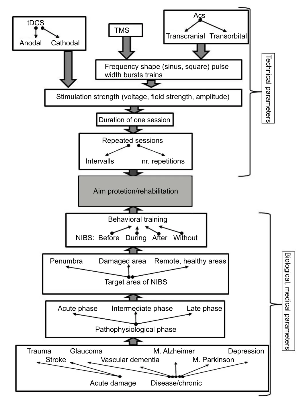
Figure 2 Flow chart for designing non-invasive electrical brain stimulation (NIBS) protocols
Taken together, NIBS therapy and physiological studies of tDCS, TMS and ACS employ neuro-activating and de-activating protocols. Regarding the affected brain regions, usually specific cortical areas are influenced by NIBS, with the exception of transcorneal ACS which allows stimulation of the retina and even different other brain regions. It is also of importance that local changes induced by NIBS can affect global network oscillations.is applies mainly to NIBS methods where alternating current pulses of different frequencies play a role, i.e., in TMS and ACS. In addition to this modulation of local and global network excitability also other, long-term effects like induction of stem cells and growth factors may take place aer NIBS (Corredor and Goldberg, 2009; Rueger et al., 2012; Huang et al., 2015).
NIBS Treatment for Brain Lesions – Basic Concepts
As discussed above, the stimulation location and the stimulation method determine the effects of NIBS. For example, in studies on healthy volunteers, modifications of motor performance can be induced by applying electrodes or stimulating coils on the scalp over the motor cortex. However, in case of brain injury, the situation is more complex because different plasticity mechanisms can be involved and the location of cell death, the post-injury state and the time of treatment have to be considered.
NIBS for Acute Disease State
Not much is known from human studies about NIBS for acute treatment aer brain insults. Patients in acute conditions are highly vulnerable and the risk of unwanted NIBS-side effects cannot be excluded, especially pertaining to the risks of post-lesion epilepsy. However, NIBS applied during the first hours and days post-injury was tested in animal studies (Ni et al., 2009; Morimoto et al., 2010, 2012; Henrich-Noack et al., 2013). Counterintuitively, stimulation protocols which are used for neuronal activation have resulted in neuroprotection.This is surprising as in general over-excitation is one main cause of neuronal death aer an insult and adding even more stimulation input does not seem to be the right strategy in this situation. Yet, this kind of stimulation may have a dual beneficial influence: a vascular and a neuronal one.e interaction of NIBS with the vascular system needs to be studied in more detail; however, as underlying mechanism improved blood flow has been suggested (Kurimoto et al., 2010).
On the neuronal level of analysis, NIBS applied during the acute excitotoxic phase can lead to fast and complete dendritic stripping of neurons and result in input isolation from connected cells. This may interrupt the excitotoxic cascade and help cells survive (Henrich-Noack et al., 2017).Obviously, this rescue comes at a cost as the neurons will be disconnected and not functional.ey seem to change into a mode of functional “silence”. However, survival of such stripped neurons might allow them to later reconnect or regenerate, opening the door for their re-activation. Only through future research may we find ways how such transiently silent survivors might be integrated back again into the brain’s functional connectivity network.
Intermediate Disease State: Post-Lesional Plasticity and Cellular Regeneration by NIBS
Spontaneous recovery by reorganization and neural regeneration, e.g., formation of new synapses, is possible to a limited extent only in a certain time-window after an incidence, i.e.,within weeks aer an insult (Caleo, 2015). Cells at risk which are typically located in the penumbra zone, i.e., the area surrounding an ischemic core, are endangered to either progress slowly towards delayed cell death or be inactivated.ere are different means to help them survive, for example, by electric current stimulation protocols that lead to increased blood flow,induction of growth factors and growth associated proteins,induction of anti-apoptotic mechanisms or anti-inflammatory effects (Corredor and Goldberg, 2009). Besides neurons located inside or in the immediate vicinity of the damaged zone,neurons located in areas far remote from the damage may appear morphologically normal but can still become functionally inactive (“silent”). Here re-activation by LTP-inducing electrical stimulation (tetanization) is possible (Henrich-Noack et al.,2005).erefore, it seems that for the treatment during an intermediate disease state, an activating NIBs protocol should be applied. Indeed, the work of Alber et al. (2017) demonstrated that anodal tDCS over visual cortex in early post-stroke when rehabilitation training is taking place improved the outcome to a greater extent than training alone.
Facilitating Compensation at a Late, Chronic Stage
Another concept of NIBS involves the restoration of function mainly by compensation and activation of residual structures.It can be applied even when the initial damage occurred long ago. At this stage the pathophysiological processes of degeneration and re-organization in lesioned areas are complete. Scars or cavitations have been formed where the damage occurred(Henrich-Noack et al., 2008) and cellular regeneration is not possible anymore.erefore, any therapeutic effort should focus on activating remaining brain structures in other, remote areas or in the damaged area itself (“within-system-compensation”). Regarding the latter, NIBS can improve function of residual structures through synchronization of neuronal firing patterns and modulation of cortical excitability.e protocols to entrain cortical oscillations with a frequency range known to be associated with a certain function were suggested to treat functional loss. For example, it was demonstrated that stimulation in the alpha range (7.5–12.5 Hz) is beneficial for visual restoration (Sabel et al., 2011; Bola et al., 2014). Reduced inhibition of affected local networks can be achieved, for example,by cathodal tDCS stimulation in areas distant from the impaired brain structure (Brunoni et al., 2012; Gall et al., 2015).Moreover, NIBS can improve cell metabolism, oxygenation and glucose supply through increased blood flow in the affected areas (Kurimoto et al., 2010).
In addition, sensory functions other than those lost can be trained to facilitate cross-modal plasticity and compensate for lost abilities (“extra-system-compensation”; e.g., enhanced hearing after vision loss). This requires mechanisms of normal learning, like training to write with the left hand for right-handed people or learning to read Braille aer vision loss(Bedny et al., 2015; Siuda-Krzywicka et al., 2016). In such cases it can be assumed that activating the compensating neuronal network area with electrical stimulation may be beneficial. In addition, anti-depressant effects or mood-improvements induced by NIBS may be an indirect mechanism whereby learning of compensatory skills is facilitated based on increased motivation during training (Dundon et al., 2015).
Summary
Different stages of brain damage and degeneration require adaptation of NIBS protocols depending on whether the aim is compensation, regeneration or protection (for a detailed overview of the relevant parameters seeFigure 2). While significant knowledge has accumulated for intermediate and late diseases stages (Otal et al., 2016), the possible benefits and risks of NIBS for an acute treatment are not yet investigated much and await further research endeavors. In any event, we need to better understand the interaction of electric current in the human eye and brain and their interaction on the molecular, cellular and network level.
Author contributions:PHN draed the paper. EGS and BAS revised the paper. All authors edited and finalized the article and approved the final version of this paper.
Conflicts of interest: None declared.
Plagiarism check:Checked twice by ienticate.
Peer review:Externally peer reviewed.
Open access statement:is is an open access article distributed under the terms of the Creative Commons Attribution-NonCommercial-ShareAlike 3.0 License, which allows others to remix, tweak, and build upon the work non-commercially, as long as the author is credited and the new creations are licensed under identical terms.
Open peer review report:
Reviewer:Lilla Bonanno, IRCCS Centro Neurolesi “Bonino-Pulejo”,Via Palermo Contrada Casazza, Italy.
Comments to authors:e article undergone to revision has demonstrated the intention to formulate a review of all scientific literature concerning the therapeutic possibilities to be implemented in different stages of brain damage and degeneration requires adaptation of NIBS protocols depending on whether the aim is compensation, regeneration or protection.
Abd Hamid AI, Gall C, Speck O, Antal A, Sabel BA (2015) Effects of alternating current stimulation on the healthy and diseased brain.Front Neurosci 9:391.
Alber R, Moser H, Gall C, Sabel BA (2017) Combined transcranial direct current stimulation and vision restoration training in subacute stroke rehabilitation: a pilot study. PM R 9:787-794.
Bashir S, Mizrahi I, Weaver K, Fregni F, Pascual-Leone A (2010) Assessment and modulation of neural plasticity in rehabilitation with transcranial magnetic stimulation. PM R 2:S253-268.
Bedny M, Richardson H, Saxe R (2015) “Visual” cortex responds to spoken language in blind children. J Neurosci 35:11674-11681.
Berridge MJ, Bootman MD, Roderick HL (2003) Calcium signalling:dynamics, homeostasis and remodelling. Nat Rev Mol Cell Biol 4:517-529.
Bola M, Gall C, Moewes C, Fedorov A, Hinrichs H, Sabel BA (2014)Brain functional connectivity network breakdown and restoration in blindness. Neurology 83:542-551.
Bonaz B, Sinniger V, Pellissier S (2016) Anti-inflammatory properties of the vagus nerve: potential therapeutic implications of vagus nerve stimulation. J Physiol 594:5781-5790.
Borgens RB, Shi R, Mohr TJ, Jaeger CB (1994) Mammalian cortical astrocytes align themselves in a physiological voltage gradient. Exp Neurol 128:41-49.
Brunoni AR, Nitsche MA, Bolognini N, Bikson M, Wagner T, Merabet L, Edwards DJ, Valero-Cabre A, Rotenberg A, Pascual-Leone A, Ferrucci R, Priori A, Boggio PS, Fregni F (2012) Clinical research with transcranial direct current stimulation (tDCS): challenges and future directions. Brain Stimul 5:175-195.
Caleo M (2015) Rehabilitation and plasticity following stroke: Insights from rodent models. Neuroscience 311:180-194.
Corredor RG, Goldberg JL (2009) Electrical activity enhances neuronal survival and regeneration. J Neural Eng 6:055001.
Dundon NM, Bertini C, Ladavas E, Sabel BA, Gall C (2015) Visual rehabilitation: visual scanning, multisensory stimulation and vision restoration trainings. Front Behav Neurosci 9:192.
Foik AT, Kublik E, Sergeeva EG, Tatlisumak T, Rossini PM, Sabel BA,Waleszczyk WJ (2015) Retinal origin of electrically evoked potentials in response to transcorneal alternating current stimulation in the rat.Invest Ophthalmol Vis Sci 56:1711-1718.
Fregni F, Pascual-Leone A (2007) Technology insight: noninvasive brain stimulation in neurology-perspectives on the therapeutic potential of rTMS and tDCS. Nat Clin Pract Neurol 3:383-393.
Gall C, Silvennoinen K, Granata G, de Rossi F, Vecchio F, Br?sel D, Bola M, Sailer M, Waleszczyk WJ, Rossini PM, Tatlisumak T, Sabel BA(2015) Non-invasive electric current stimulation for restoration of vision aer unilateral occipital stroke. Contemp Clin Trials 43:231-236.
Gall C, Schmidt S, Schittkowski MP, Antal A, Ambrus GG, Paulus W,Dannhauer M, Michalik R, Mante A, Bola M, Lux A, Kropf S, Brandt SA, Sabel BA (2016) Alternating current stimulation for vision restoration aer optic nerve damage: a randomized clinical trial. PLoS One 11:e0156134.
Gellner AK, Reis J, Fritsch B (2016) Glia: A neglected player in non-invasive direct current brain stimulation. Front Cell Neurosci 10:188.
Henrich-Noack P, Baldauf K, Reiser G, Reymann KG (2008) Pattern of time-dependent reduction of histologically determined infarct volume aer focal ischaemia in mice. Neurosci Lett 432:141-145.
Henrich-Noack P, Gorkin AG, Krautwald K, Pforte C, Schr?der UH,Reymann KG (2005) Tetanus-induced re-activation of evoked spiking in the post-ischemic dentate gyrus. Neuroscience 133:571-581.
Henrich-Noack P, Lazik S, Sergeeva E, Wagner S, Voigt N, Prilloff S,Fedorov A, Sabel BA (2013) Transcorneal alternating current stimulation aer severe axon damage in rats results in “l(fā)ong-term silent survivor” neurons. Brain Res Bull 95:7-14.
Henrich-Noack P, Sergeeva EG, Eber T, You Q, Voigt N, Kohler J, Wagner S, Lazik S, Mawrin C, Xu G, Biswas S, Sabel BA, Leung CK (2017)Electrical brain stimulation induces dendritic stripping but improves survival of silent neurons aer optic nerve damage. Sci Rep 7:627.
Huang Y, Li Y, Chen J, Zhou H, Tan S (2015) Electrical stimulation elicits neural stem cells activation: new perspectives in CNS repair. Front Hum Neurosci 9:586.
Karabanov A, Ziemann U, Hamada M, George MS, Quartarone A,Classen J, Massimini M, Rothwell J, Siebner HR (2015) Consensus paper: probing homeostatic plasticity of human cortex with non-invasive transcranial brain stimulation. Brain Stimul 8:993-1006.
Kurimoto T, Oono S, Oku H, Tagami Y, Kashimoto R, Takata M, Okamoto N, Ikeda T, Mimura O (2010) Transcorneal electrical stimulation increases chorioretinal blood flow in normal human subjects.Clin Ophthalmol 4:1441-1446.
Morimoto T, Miyoshi T, Sawai H, Fujikado T (2010) Optimal parameters of transcorneal electrical stimulation (TES) to be neuroprotective of axotomized RGCs in adult rats. Exp Eye Res 90:285-291.
Morimoto T, Miyoshi T, Matsuda S, Tano Y, Fujikado T, Fukuda Y(2005) Transcorneal electrical stimulation rescues axotomized retinal ganglion cells by activating endogenous retinal IGF-1 system. Invest Ophthalmol Vis Sci 46:2147-2155.
Morimoto T, Kanda H, Kondo M, Terasaki H, Nishida K, Fujikado T (2012) Transcorneal electrical stimulation promotes survival of photoreceptors and improves retinal function in rhodopsin P347L transgenic rabbits. Invest Ophthalmol Vis Sci 53:4254-4261.
Ni YQ, Gan DK, Xu HD, Xu GZ, Da CD (2009) Neuroprotective effect of transcorneal electrical stimulation on light-induced photoreceptor degeneration. Exp Neurol 219:439-452.
Nitsche MA, Paulus W (2011) Transcranial direct current stimulation--update 2011. Restor Neurol Neurosci 29:463-492.
Oberman L, Edwards D, Eldaief M, Pascual-Leone A (2011) Safety of theta burst transcranial magnetic stimulation: a systematic review of the literature. J Clin Neurophysiol 28:67-74.
Otal B, Dutta A, Foerster A, Ripolles O, Kuceyeski A, Miranda PC,Edwards DJ, Ili? TV, Nitsche MA, Ruffini G (2016) Opportunities for guided multichannel non-invasive transcranial current stimulation in poststroke rehabilitation. Front Neurol 7:21.
Perera T, George MS, Grammer G, Janicak PG, Pascual-Leone A,Wirecki TS (2016) The clinical TMS society consensus review and treatment recommendations for TMS therapy for major depressive disorder. Brain Stimul 9:336-346.
Prilloff S, Noblejas MI, Chedhomme V, Sabel BA (2007) Two faces of calcium activation aer optic nerve trauma: life or death of retinal ganglion cells in vivo depends on calcium dynamics. Eur J Neurosci 25:3339-3346.
Rueger MA, Keuters MH, Walberer M, Braun R, Klein R, Sparing R,Fink GR, Graf R, Schroeter M (2012) Multi-session transcranial direct current stimulation (tDCS) elicits inflammatory and regenerative processes in the rat brain. PLoS One 7:e43776.
Sabel BA, Henrich-Noack P, Fedorov A, Gall C (2011) Vision restoration aer brain and retina damage: the “residual vision activation theory”. Prog Brain Res 192:199-262.
Sato T, Fujikado T, Lee TS, Tano Y (2008a) Direct effect of electrical stimulation on induction of brain-derived neurotrophic factor from cultured retinal Muller cells. Invest Ophthalmol Vis Sci 49:4641-4646.
Sato T, Fujikado T, Morimoto T, Matsushita K, Harada T, Tano Y (2008b)Effect of electrical stimulation on IGF-1 transcription by L-type calcium channels in cultured retinal Muller cells. Jpn J Ophthalmol 52:217-223.
Sehic A, Guo S, Cho KS, Corraya RM, Chen DF, Utheim TP (2016)Electrical stimulation as a means for improving vision. Am J Pathol 186:2783-2797.
Sergeeva EG, Fedorov AB, Henrich-Noack P, Sabel BA (2012) Transcorneal alternating current stimulation induces EEG “aereffects” only in rats with an intact visual system but not aer severe optic nerve damage. J Neurophysiol 108:2494-2500.
Siebner HR, Lang N, Rizzo V, Nitsche MA, Paulus W, Lemon RN,Rothwell JC (2004) Preconditioning of low-frequency repetitive transcranial magnetic stimulation with transcranial direct current stimulation: evidence for homeostatic plasticity in the human motor cortex. J Neurosci 24:3379-3385.
Siuda-Krzywicka K, Bola ?, Paplińska M, Sumera E, Jednoróg K,Marchewka A, ?liwińska MW, Amedi A, Szwed M (2016) Massive cortical reorganization in sighted Braille readers. Elife 5:e10762.
Stagg CJ, Nitsche MA (2011) Physiological basis of transcranial direct current stimulation. Neuroscientist 17:37-53.
Thut G, Bergmann TO, Frohlich F, Soekadar SR, Brittain JS, Valero-Cabre A, Sack AT, Miniussi C, Antal A, Siebner HR, Ziemann U, Herrmann CS (2017) Guiding transcranial brain stimulation by EEG/MEG to interact with ongoing brain activity and associated functions: A position paper. Clin Neurophysiol 128:843-857.
Wagner T, Valero-Cabre A, Pascual-Leone A (2007) Noninvasive human brain stimulation. Annu Rev Biomed Eng 9:527-565.
Woods AJ, Antal A, Bikson M, Boggio PS, Brunoni AR, Celnik P, Cohen LG, Fregni F, Herrmann CS, Kappenman ES, Knotkova H, Liebetanz D, Miniussi C, Miranda PC, Paulus W, Priori A, Reato D, Stagg C, Wenderoth N, Nitsche MA (2016) A technical guide to tDCS,and related non-invasive brain stimulation tools. Clin Neurophysiol 127:1031-1048.
Yang Y, Li L, Wang YG, Fei Z, Zhong J, Wei LZ, Long QF, Liu WP (2012)Acute neuroprotective effects of extremely low-frequency electromagnetic fields after traumatic brain injury in rats. Neurosci Lett 516:15-20.
Yavari F, Jamil A, Mosayebi Samani M, Vidor LP, Nitsche MA (2017)Basic and functional effects of transcranial electrical stimulation(tES)-an introduction. Neurosci Biobehav Rev doi: 10.1016/j.neubiorev.2017.06.015.
Yin H, Yin H, Zhang W, Miao Q, Qin Z, Guo S, Fu Q, Ma J, Wu F, Yin J,Yang Y, Fang X (2016) Transcorneal electrical stimulation promotes survival of retinal ganglion cells aer optic nerve transection in rats accompanied by reduced microglial activation and TNF-alpha expression. Brain Res 1650:10-20.
*Correspondence to:Petra Henrich-Noack,petra.henrich-noack@med.ovgu.de.
orcid:
0000-0002-4472-5543
(Bernhard A. Sabel)
10.4103/1673-5374.217322
Accepted: 2017-09-12
 中國(guó)神經(jīng)再生研究(英文版)2017年10期
- 中國(guó)神經(jīng)再生研究(英文版)的其它文章
- Can we treat neurodegenerative diseases by preventing an age-related decline in microRNA expression?
- Diffusion tensor tractography studies on mechanisms of recovery of injured fornix
- Matrix bound vesicles and miRNA cargoes are bioactive factors within extracellular matrix bioscaffolds
- Beta secretase activity in peripheral nerve regeneration
- Embracing oligodendrocyte diversity in the context of perinatal injury
- On the road towards the global analysis of human synapses
