Coordination of the axonal cytoskeleton during the emergence of axon collateral branches
PERSPECTIVE
Coordination of the axonal cytoskeleton during the emergence of axon collateral branches
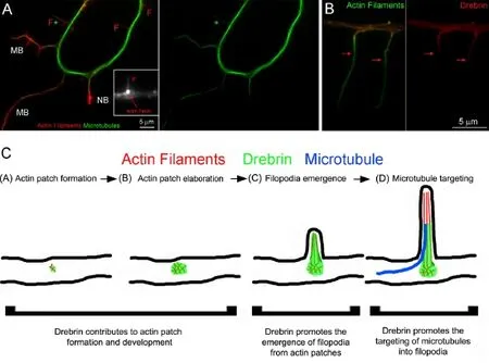
The formation of branches during development allows a single axon to make synaptic contacts with numerous target neurons, often in different parts of the nervous system, thereby allowing for the establishment of complex patterns of neuronal connectivity. Following injury, the formation of collateral branches contributes to the endogenous neuroplasticity which depending on the neuronal population can have beneficial (e.g., formation of compensatory relay circuitry) or adverse effects (e.g., development of autonomic dysreflexia). The formation of axon collateral branches from the axon shaft involves a complex series of signaling events and cytoskeletal reorganization. The initial step in the formation of a collateral branch is the emergence of axonal filopodia, supported by a bundle of actin filaments (Figure 1A). However, axonal filopodia are generated from precursor structures consisting of small meshworks of actin filaments, termed actin patches (Figure 1A inset). Actin patches are dynamic structures which form along the axon shaft, grow in size and subsequently are disassembled (for reviews on actin patches see Gallo, 2011, 2013). Although in live imaging studies using fluorescently labeled actin most axonal filopodia arise from a detectable actin patch, only a subset of patches gives rise to filopodia before dissipating. As the duration of filopodia is longer than that of patches, they are often not detected at the base of longer pre-existing filopodia, although they often re-emerge at the base of existing filopodia. In order for an axonal filopodium to mature into a collateral branch it must first become invaded by axonal microtubules (Figure 1A). Following the targeting and retention of axonal microtubules into the filopodium, the filopodium then begins to reorganize its actin filament cytoskeleton transitioning from a linear bundle of actin filaments to a distal accumulation of filaments giving rise to a nascent branch (Figure 1A), ultimately giving rise to a small growth cone structure at the tip of the branch. The transformation of a filopodium into a nascent branch is referred to as maturation of the branch which then grows in length (Figure 1A).
Although there are multiple neuronally expressed proteins that have the ability to bind and physically link microtubules and actin filaments, and the idea of microtubule-actin filament interactions in neuromorphogenesis has been proposed for a number of years, few studies have addressed if and how such molecules are involved in the mechanism of axon branching (see the recent review on actin-microtubule interactions in neurons by Cammarata et al., 2016). Specifically, there is a paucity of studies directly addressing both the actin filament and microtubule cytoskeleton in the context of the possible roles of actin-microtubule linking proteins during axon branching. The focus of this article is to review recent publications emphasizing a role of actin-microtubule interactions in the mechanism of axon branching.
In a recent paper, evidence has emerged for drebrin, a protein which can interact with actin filaments and microtubule plus tips, in mediating cytoskeletal interactions during the early phases of axon branching (Ketschek et al., 2016). Drebrin contains two actin filament binding domains and is under phosphoregulation by Cdk5 (Worth et al., 2013). Phosphorylation of drebrin by Cdk5 results in intramolecular changes which expose the second actin binding domain, which is otherwise cryptic, thereby allowing drebrin to bundle filaments. Microtubules are polarized polymers with plus and minus ends. The plus end of a microtubule undergoes cycles of dynamic instability characterized by rapid polymerization followed by depolymerization. Drebrin binds end binding protein-3 (EB3; Worth et al., 2013). EB3 is a protein that specifically accumulates on the plus tips of microtubules during periods of active polymerization. Thus, drebrin is well suited for serving multiple roles during the process of branch formation which involves the formation of filopodia supported by an actin filament bundle, and the targeting of microtubule plus tips into the filopodia.
In axonal filopodia, drebrin was found to target the proximal~5 microns of the filopodial shaft (Figure 1B). Drebrin also targets to the precursors of axonal filopodia, actin patches. As filopodia mature into branches drebrin extends further into the filopodium and the nascent branch, and in mature branches drebrin colocalizes with the actin filament distribution in the branch. Functional experiments depleting drebrin from embryonic sensory neurons using shRNA, or overexpression of drebrin, showed that drebrin promotes the formation of filopodia from actin patches. Depletion of drebrin also decreased the formation or actin patches, although overexpression did not affect patch formation, indicating that drebrin contributes to patch formation but it is not sufficient to initiate patches. The role of drebrin in regulating the emergence of filopodia from patches is likely in the formation of the actin filament bundle that supports filopodia. Drebrin was also found to promote the targeting of microtubule plus tips into axonal filopodia, the required event for the maturation of a filopodium into a branch. These data indicate that drebrin is a major regulator of the organization of the filopodial actin filament bundle and subsequently also serves to promote the targeting of microtubule tips into filopodia. The localization of drebrin in filopodia (Figure 1B) is consistent with drebrin serving this dual function during the early stages of branch formation.
Drebrin binding to filaments has been shown to alter the ability of other actin filament binding proteins to associate with filaments. Myosin II is a molecular motor protein that generates contractile forces on filaments and is generally considered to suppress axon extension and branching. The binding of drebrin to purified actin filaments has been reported to suppress the ability of myosin II to bind filaments and generate forces (Hayashi et al., 1996). Inhibition of the activity of myosin II, using the pharmacological inhibitor blebbistatin, further promoted axon branching under conditions of drebrin overexpression, which alone promotes branching. Unexpectedly, while under normal conditions drebrin is present only along the proximal portion of the filopodial shaft, inhibition of myosin II resulted in a redistribution of drebrin along the entire length of filopodia. The blebbistatin-induced redistribution of drebrin along the filopodial shaft correlated with an increase in the distance that microtubule plus tips penetrated into the filopodium. This observation suggests that the drebrin redistribution in filopodia, through its ability to bind EB3 at the plus tips of microtubules, may promote the continued extension of the plus tip into the distal-most portion of the filopodium.
Drebrin overexpression also increased the stability of axonal filopodia (i.e., filopodia exhibited longer durations). Microtubules can indirectly regulate the dynamics of the actin filament cytoskeleton (Cammarata et al., 2016). However, the effect of drebrin on filopodial stability was independent of the targeting of microtubules into filopodia, as indicated by the observation that pharmacologically preventing the entry of plus tips into filopodia did not affect the increase in filopodial stabilization induced by drebrin overexpression.

Figure 1 Sequence of cytoskeletal reorganization during branch formation and the intra-filopodial distribution of drebrin.
The induction of axon branches along sensory neurons by nerve growth factor (NGF) requires NGF-induced intra-axonal protein synthesis of actin regulatory proteins (Spillane et al., 2012, 2013), and drebrin mRNA has been detected in axons (Gumy et al., 2011). Acute treatment of cultures with NGF for 40 min, during which axons elaborate branches in response to NGF, increased the axonal levels of drebrin. However, inhibition of protein synthesis using two different inhibitors did not affect the NGF-induced increase in axonal drebrin levels, although the inhibitors were effective as shown by positive controls. Thus, the mechanism used by NGF to increase axonal levels of drebrin is not dependent of axonal protein synthesis. Drebrin levels in axons may be normally regulated by proteolytic/degradation mechanism which NGF may suppress. Indeed, the response of axons to extracellular signals often involve both axonal protein synthesis and degradation-based mechanism (Campbell and Holt, 2001). The recent demonstration of the regulated degradation of drebrin in neurons lends further credence to this notion (Chimura et al., 2015).
Finally, although not specifically addressing the manipulation of potential actin-microtubule crosslinkers on both the axonal actin and microtubule cytoskeleton, additional recent studies on MAP1B (microtubule associated protein 1B; Barnat et al., 2016) and MACF1 (microtubule actin crosslinking factor 1; Ka and Kim, 2015) have provided evidence for roles in axon branching. MAP1B has a suppressive role in branch formation, as evidenced by increased branching in MAP1B knock out neurons (Barnat et al., 2016). In the absence of branch inducting signals (e.g., NGF) MAP1B is found along microtubules that target into axonal filopodia, and also within filopodia but in the majority of filopodia it does not appear to be associated with actin filaments (Ketschek et al., 2015). However, its actin filament binding activity may be under phosphoregulation within individual filopodia thus regulating actin-microtubule interactions (Ketschek et al., 2015). The NGF induction of branches correlates with decreased levels of MAP1B along microtubules within filopodia, which however is then increased as the branch matures. The presence of the phosphorylated form of MAP1B, which is considered to promote actin filament binding, follows a similar trend. Thus, given the inhibitory role of MAP1B in branching, MAP1B may serve to regulate actin-microtubule interactions during later stages branch maturation and subsequent elongation, but perhaps not during the earliest stages of branch formation. A recent study addressed the functional consequences of knocking out MACF1 specifically in subsets of cortical and hippocampal neurons in vivo and in vitro on dendritic and axonal development (Ka and Kim, 2015). Although the study focused on dendritic branching, which was impaired by the absence of MACF1, effects on axonal development were also characterized. When MACF1 was knocked out axons grewshorter and formed less axon branches in vivo. MACF1 knock out also resulted in alterations in both the actin and microtubule cytoskeleton at the tips of neuronal processes, but the effects of MACF1 knock out on the axonal cytoskeleton was not specifically addressed.
In conclusion, evidence for the functional significance of actin-microtubule interactions in axon branch formation is mounting. Future studies will be required to further scrutinize how individual molecules which have the potential of crosslinking the two cytoskeletal systems impact the cytoskeleton and relevant morphogenetic processes. In particular, it will be important to analyze how deletion of microtubule and actin binding domains in the proteins affect their functions during branching. Furthermore, future studies ought to investigate the spatio-temporal dynamics of the localization of relevant proteins to filopodia, microtubules, nascent and mature branches. In the case of drebrin, this protein undergoes a developmentally regulated switch in isoform expression from its embryonic forms (E1/E2) to the adult form. It will be of interest to determine if the adult form is involved in branching in a manner similar to the embryonic form, and if so whether it may have a role in the sprouting of axon branches in the context of injury scenarios.
Ultimately, as our understanding of the mechanism of axon branching advances, the various molecular steps involved will become candidates for manipulation in therapeutic contexts. For example, depletion of drebrin in sensory axons has the potential to be used to prevent the sprouting of these axons in the context of autonomic dysreflexia. In this context, the development of pharmacological approaches, or perhaps rationally designed cell permeable peptides, that target drebrin function may provide an effective therapeutic venue. On the converse side, upregulation of drebrin levels or pharmacological/peptide mediated activation of drebrin activity could be used to promote axon branching in beneficial scenarios such as promoting the endogenous morphologic plasticity of spinal cord circuitry following injury. It should however also be considered that drebrin has important roles in synaptic structure and function and any attempt to target drebrin may have consequences on these additional aspects of neuronal function. Thus, considering alternative actin-microtubule interacting molecules underlying branching would also be cautious (e.g., MAP1B and MACF1).
This work has been supported by an NIH award (NS078030) to GG.
Gianluca Gallo*
Temple University, Lewis Kats School of Medicine, Department of Anatomy and cell Biology, Shriners Hospitals Pediatric Research Center, Philadelphia, PA, USA
*Correspondence to: Gianluca Gallo, Ph.D., tue86088@temple.edu.
Accepted: 2016-03-21
orcid: 0000-0002-0777-379X (Gianluca Gallo)
Barnat M, Benassy MN, Vincensini L, Soares S, Fassier C, Propst F, Andrieux A, von Boxberg Y, Nothias F (2016) The GSK3-MAP1B pathway controls neurite branching and microtubule dynamics. Mol Cell Neurosci 72:9-21.
Cammarata GM, Bearce EA, Lowery LA (2016) Cytoskeletal social networking in the growth cone: How +TIPs mediate microtubule-actin cross-linking to drive axon outgrowth and guidance. Cytoskeleton doi:10.1002/ cm.21272.
Campbell DS, Holt CE (2001) Chemotropic responses of retinal growth cones mediated by rapid local protein synthesis and degradation. Neuron 32:1013-1026.
Chimura T, Launey T, Yoshida N (2015) Calpain-mediated degradation of drebrin by excitotoxicity in vitro and in vivo. PLoS One 10:e0125119.
Gallo G (2011) The cytoskeletal and signaling mechanisms of axon collateral branching. Dev Neurobiol 71:201-220.
Gallo G (2013) Mechanisms underlying the initiation and dynamics of neuronal filopodia: from neurite formation to synaptogenesis. Int Rev Cell Mol Biol 301:95-156.
Gumy LF, Yeo GS, Tung YC, Zivraj KH, Willis D, Coppola G, Lam BY, Twiss JL, Holt CE, Fawcett JW (2011) Transcriptome analysis of embryonic and adult sensory axons reveals changes in mRNA repertoire localization. RNA 17:85-98.
Hayashi K, Ishikawa R, Ye LH, He XL, Takata K, Kohama K, Shirao T (1996) Modulatory role of drebrin on the cytoskeleton within dendritic spines in the rat cerebral cortex. J Neurosci 16:7161-7170.
Ka M, Kim WY (2015) Microtubule-actin crosslinking factor 1 is required for dendritic arborization and axon outgrowth in the developing brain. Mol Neurobiol doi:10.1007/s12035-015-9508-4.
Ketschek A, Jones S, Spillane M, Korobova F, Svitkina T, Gallo G (2015) Nerve growth factor promotes reorganization of the axonal microtubule array at sites of axon collateral branching. Dev Neurobiol 75:1441-1461.
Ketschek A, Spillane M, Dun XP, Hardy H, Chilton J, Gallo G (2016) Drebrin coordinates the actin and microtubule cytoskeleton during the initiation of axon collateral branches. Dev Neurobiol doi:10.1002/dneu.22377.
Spillane M, Ketschek A, Merianda TT, Twiss JL, Gallo G (2013) Mitochondria coordinate sites of axon branching through localized intra-axonal protein synthesis. Cell Rep 5:1564-1575.
Spillane M, Ketschek A, Donnelly CJ, Pacheco A, Twiss JL, Gallo G (2012) Nerve growth factor-induced formation of axonal filopodia and collateral branches involves the intra-axonal synthesis of regulators of the actin-nucleating Arp2/3 complex. J Neurosci 32:17671-17689.
Worth DC, Daly CN, Geraldo S, Oozeer F, Gordon-Weeks PR (2013) Drebrin contains a cryptic F-actin-bundling activity regulated by Cdk5 phosphorylation. J Cell Biol 202:793-806.
10.4103/1673-5374.182684 http∶//www.nrronline.org/
How to cite this article: Gallo G (2016) Coordination of the axonal cytoskeleton during the emergence of axon collateral branches. Neural Regen Res 11(5):709-711.
 中國(guó)神經(jīng)再生研究(英文版)2016年5期
- 中國(guó)神經(jīng)再生研究(英文版)的其它文章
- Self-assembling peptide nanofibrous hydrogel as a promising strategy in nerve repair after traumatic injury in the nervous system
- Recovery of injured fornical crura following neurosurgical operation of a brain tumor: a case report
- Possible application of apolipoprotein E-containing lipoproteins and polyunsaturated fatty acids in neural regeneration
- Antibody-based neuronal and axonal delivery vectors for targeted ligand delivery
- Alzheimer's disease: the silver tsunami of the 21stcentury
- Clinical trial perspective for adult and juvenile Huntington's disease using genetically-engineered mesenchymal stem cells
