
Impact of crosslinking/riboflavin-UVA-photodynamic inactivation on viability,apoptosis and activation of human keratocytesin vitro
2015-12-22 09:15:17TanjaStachonJiongWangXufeiSongAchimLangenbucherBertholdSeitzNoraSzentmary
Tanja Stachon,Jiong Wang,Xufei Song,Achim Langenbucher,Berthold Seitz, No′ra Szentma′ry
1Department of Ophthalmology,Saarland University Medical Center,Homburg,Saar 66424,Germany;
2Department of Ophthalmology,Renmin Hospital of Wuhan University,Wuhan,Hubei 43060,China;
3Department of Ophthalmology,The Second Hospital of Jilin University,Changchun,Jilin 130041,China;
4Experimental Ophthalmology,Saarland University,Homburg,Saar 66424,Germany.
Impact of crosslinking/riboflavin-UVA-photodynamic inactivation on viability,apoptosis and activation of human keratocytesin vitro
Tanja Stachon1,?,Jiong Wang1,2,Xufei Song1,3,Achim Langenbucher4,Berthold Seitz1, No′ra Szentma′ry1
1Department of Ophthalmology,Saarland University Medical Center,Homburg,Saar 66424,Germany;
2Department of Ophthalmology,Renmin Hospital of Wuhan University,Wuhan,Hubei 43060,China;
3Department of Ophthalmology,The Second Hospital of Jilin University,Changchun,Jilin 130041,China;
4Experimental Ophthalmology,Saarland University,Homburg,Saar 66424,Germany.
Riboflavin-UVA photodynamic inactivation is a potential treatment alternative in therapy resistant infectious keratitis. The purpose of our study was to determine the impact of riboflavin-UVA photodynamic inactivation on viability,apoptosis and activation of human keratocytes in vitro.Primary human keratocytes were isolated from human corneal buttons and cultured in DMEM/Ham′s F12 medium supplemented with 10%fetal calf serum.Keratocytes underwent UVA light illumination(375 nm)for 4.10 minutes(2 J/cm2)during exposure to different concentrations of riboflavin.Twenty-four hours after treatment,cell viability was evaluated photometrically,whereas apoptosis,CD34 and alpha-smooth muscle actin(α-SMA)expression were assessed using flow cytometry.We did not detect significant changes in cell viability, apoptosis,CD34 and α-SMA expression in groups only treated with riboflavin or UVA light.In the group treated with riboflavin-UVA-photodynamic inactivation,viability of keratocytes decreased significantly at 0.1%riboflavin(P<0.01) while the percentage of CD34(P<0.01 for both 0.05%and 0.1%riboflavin)and alpha-SMA positive keratocytes (P<0.01 and P<0.05 for 0.05%and 0.1%riboflavin,respectively)increased significantly compared to the controls. There was no significant change in the percentage of apoptotic keratocytes compared to controls at any of the used riboflavin concentrations(P=0.09 and P=0.13).We concluded that riboflavin-UVA-photodynamic-inactivation decreases viability of myofibroblastic transformation and multipotent haematopoietic stem cell transformation;however,it does not have an impact on apoptosis of human keratocytes in vitro.
crosslinking,human keratocytes,viability,apoptosis,α-SMA,CD34
Introduction
Photodynamic inactivation(PDI)is based on the use of a non-toxic photosensitizer,which is activated by light of appropriate wavelength.Activation of the photosensitizer through illumination leads to generation of singlet oxygen and free radicals,which are responsible for the cytotoxic effect on the target tissue[1].Corneal crosslinking uses riboflavin as photosensitizer,which is activated by 375 nm UVA-light illumination.A relative low concentration of free
oxygen radicals and singlet oxygen is produced following crosslinking as it has low photosensitizing efficacy, and crosslinking is called riboflavin-UVA-PDI[2].
As riboflavin-UVA-PDI has been reported to be capable of killing bacteria both in vitro[3-4]and in vivo[5-6],it may be a potential option in case of therapy resistant infectious keratitis.Several clinical studies have already reported the effectiveness of crosslinking such as PDI in curing bacterial[7-8]or even acanthamoeba keratitis[5].In contrast,some authors reported on occurrence of early bacterial keratitis[9-10]or acanthamoeba keratitis[11]after crosslinking therapy.The possible adverse effects of crosslinking on the corneal tissue may be the limits of usage of riboflavin-UVA-PDI in keratitis.Furthermore,crosslinking may also have an impact on activation of keratocytes or even on local immune response to pathogens.
Wollensak et al.[12]reported an abrupt cytotoxic effect on porcine keratocytes in vitro using collagen crosslinking,and a decrease in viability and an increase of apoptosis of porcine keratocytes[13].The impact of riboflavin-UVA-PDI was also tested in a human keratocyte cell line,which showed significantly decreased cell viability[13].However,properties of immortalized cell lines or porcine cells differ from primary human keratocytes,so we conducted this study to determine the impact of riboflavin-UVA PDI on viability,apoptosis and activation of human primary keratocytes in vitro.
Materials and methods
Reagents
Dulbecco’s Modified Eagle Medium(DMEM/F12), fetal calf serum(10%),P/S(1%of 10,000 U penicillin/10 mg streptomycin per mL),and 0.05%trypsin/ 0.02%ethylenediaminetetra-acetic acid(EDTA)were purchased from PPA Laboratories(Pasching, Austria).AlamarBlue?was obtained from Invitrogen (Karlsruhe,Germany).Collagenase A and Dispase II were obtained from Roche Diagnostics(Mannheim, Germany).The APO-DIRECTTMkit was from BD Biosciences(Heidelberg,Germany).Mouse antihuman CD34-FITC anti-alpha smooth muscle actin antibody(FITC)was obtained from Biozol(Eching, Germany)and Abcam(Cambridge,USA),respectively.Riboflavin-5-phosphate and Dextran were both purchased from Sigma-Aldrich(Heidelberg,Germany).
Isolation of primary human corneal keratocytes
Human corneas were obtained from the Saarland University Hospital Eye Bank.Keratocytes were isolated as described previously[14].The human corneoscleral buttons were aseptically rinsed in phosphate-buffered saline (PBS)before removal of the endothelium including Descemet’s membrane by sterile surgical disposable scalpel.A central corneal button with epithelium was cut using a 8.0 mm Barron’s trephine and thereafter incubated in culture medium containing 2.4 U/mL Dispase II for 4 hours at 37°C.Thereafter,the corneal button was washed with PBS for several times and the loose corneal epithelium was removed with surgical disposable scalpel.The remaining corneal stroma was incubated in culture medium with 1.0 mg/mL collagenase A for 8-10 hours at 37°C.The digested tissues and cells were centrifuged at 800 g for 7 minutes and finally resuspended in 1.0 mL basic medium(DMEM/F12)supplemented with 10%FCS and 1%P/S.The cell suspension was seeded in 6-well plates and the medium was changed 24 hours after seeding.Medium was changed every 2 to 3 days until keratocytes reached confluence.The cells were sub-cultured in 25 cm2culture flasks after 5 to 10 days following dispersal with 0.05%trypsin-EDTA for 3 to 5 minutes and passage 4 to 8 of cells was used for experiments.
Riboflavin-UVA PDI
Human keratocytes were seeded in tissue culture plates and were allowed to grow for 48 hours before photodynamic treatment.For viability,apoptosis, CD34 and α-SMA expression measurements,final 0.05%and 0.1%concentration of riboflavin-5-phospate was diluted using 20%Dextran-PBS,as the 0 and 1% concentration of riboflavin were used in clinical settings.Cells were exposed directly to UVA light(375 nm)for 4.10 minutes(2 J/cm2)following wash twice with PBS,and fed with culture medium and cultured at 37°C for 24 hours before measurements.
AlamarBue?assays
Cell viability was investigated using the AlamarBue? assay as follows:Human keratocytes were seeded in 24-well cell culture plates at a concentration of 7.5×10³cells/cm2.At 24 hours after illumination, AlamarBlue?solution was diluted with culture medium to a final concentration of 10%and 500 μL of the solution was added to each well.AlamarBlue?solution was added to a well without cells as a negative control. Thereafter,all plates were exposed to an excitation wavelength of 560 nm,and the emission at 616 nm was recorded using a 96-well microplate reader(WALLAC 1420 Multilabel Counter,Perkin Elmer,Life Sciences, Wellesley,MA,USA).
Determination of apoptosis
APO-DIRECTTMkit was used to determine the relative number of apoptotic cells.Cells were seeded in 6-well cell culture plates with a concentration of 7.5×10³cells/cm2and exposed to PDI as described above.After 24-hour exposure to PDI,the culture medium was discarded and the cells were trypsinized before centrifugation,and then the cells were suspended in 1%paraformaldehyd at a concentration of 1.0×10⁶cells/mL and placed on ice for 30-60 minutes.Thereafter,cells were washed twice with PBS and stored for 30 minutes at-20°C following adding 1.0 mL ice cold 70%ethanol.After removing ethanol carefully by aspiration,fixed cells were resuspended twice in 1.0 mL Wash-Buffer.The control cells and the probes were resuspended in 50 μL DNALabeling-Solution(FITC marked dUTP)and the cells were washed twice before resuspending the pellet in 500 μL PI/RNase Staining Buffer.Cells were incubated in the dark for at least 30 minutes at room temperature prior to analysis using a FACSCanto flow cytometer(BD Biosciences,Heidelberg,Germany).
Determination of CD34 and α-SMA expression
Cells were seeded in 6-well cell culture plates with a final concentration of 4.0×10³cells/cm2and underwent PDI as described above.After 24 hours exposed to PDI,the cells were trypsinized and washed with PBS.To demonstrate α-SMA expression,the cells were incubated with 0.5 mL PERM solution for 10 minutes, and then washed once with PBS followed by incubation with FITC-conjugated mouse monoclonal antibodies (IgG2a)against human α-SMA(100 μg/104cells)for 30 minutes in the dark at room temperature.For CD34 detection,aFITC-conjugatedmonoclonalantibody (IgG1)was used directly at a concentration of 200 μg/mL followed by incubation for 30 minutes in the dark at room temperature.To prove the specificity of the staining,isotype control experiment for each primary IgG-subtype antibody was performed.Cells were washed twice with PBS and analyzed using a FACSCanto flow cytometer(BDBiosciences,Heidelberg,Germany),and the evaluation was performed with WinMDI software (Version 2.9).
Statistical analysis
The GraphPad Prism 2.01(GraphPad Software Inc.,La Jolla,USA)was used for statistical analysis.Data are presentedasmean±standarddeviation(SD).Kruskal-Wallis one-way analysis of variance followed by Wilcoxon-Mann-Whitney Test was performed.P values below 0.05 were considered statistically significant.
Results
Keratocyte viability
The results of the AlamarBlue?assays are shown in Fig.1A(n=5).The Kruskal-Wallis one-way analysis of variance indicated a significant P-value of<0.001 between groups with and without UVA-light illumination.With separate use of riboflavin or UVA-light, there was no significant change in the viability of keratocytes compared to controls(P>0.25).Following riboflavin-UVA-PDI,keratocyte viability was significantly decreased in groups treated with 0.05%and 0.1%riboflavin(both P<0.01)compared to controls.
Furthermore,there was no significant difference in the percentage of apoptotic keratocytes using riboflavin,UVA-light illumination or riboflavin-UVA-PDI compared to controls(Fig.1B,n=6)(P=0.581)
CD34 and α-SMA expression in keratocytes
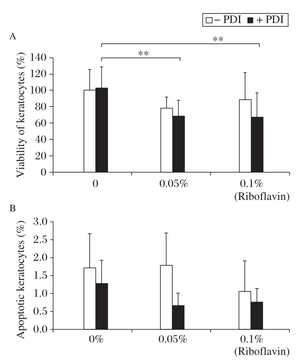
Fig.1(A)Viability of keratocytes 24 hours after Photodynamic inactivation(PDI)(Riboflavin).Viability of keratocytes were examined by AlamarBlue assays as described in Methods.**P<0.01 (B)Apoptosis of keratocytes 24 hours after PDI(Riboflavin).

Fig.2CD34(A)and a-SMA(B)expression of keratocytes 24 hours after PDI(Riboflavin).**P<0.01
CD34 and α-SMA expression of keratocytes 24 hours following PDI is summarized inFig.2Aand 2B(n=6).CD34 and α-SMA expression between groups treated with and without UVA-light showed a significant difference with P-value of 0.004 and 0.012,respectively.CD34 and α-SMA expression in cells treated with riboflavin or UVA did not change significantly compared to controls.Twenty-four hours after riboflavin-UVA-PDI,the expression of CD34 increased significantly in groups treated with 0.05% (8.7%positive)and 0.1%(6.6%positive)riboflavin compared to controls(2.6%positive;P<0.01 for both).The percentage of α-SMA positive keratocytes increased significantly in groups treated with riboflavin-UVA-PDI at 0.05%(35.8%positive,P<0.01) and 0.1%(29.1%α-SMA positive,P<0.05)compared to untreated cells(14.5%positive).
DISCUSSION
Corneal collagen crosslinking is currently used to reduce or stop the progression of keratoconus[8],or further applied as riboflavin-UVA-PDI,which has a potential to be an alternative therapeutic option in infectious keratitis[2].Even though clinical use of crosslinking started several years ago,this is the first study of illustration of viability,apoptosis and activation using primary human keratocytes following crosslinking/riboflavin-UVA-PDI.
In the present in vitro study,we demonstrated that PDI using riboflavin-5-phosphate reduced the viability of primary human keratocytes 24 hours following UVA-illumination,but did not induce cell apoptosis, In our previous experiments,we found significantly decreased viability and increased apoptosis of primary human keratocytes in vitro 24 hours after PDI using chlorine e6 as photosensitizer and 675 nm wavelength illumination[15].The decrease of viability and the increase of apoptosis were photosensitizer dose dependent.Chlorin e6 is a photosensitizer with high photosensitizing efficacy,therefore,with more reactive oxygen species production during photodynamic inactivation,compared to riboflavin-UVA-PDI.This may result in a decreased viability and triggered apoptosis of keratocytes at even lower concentrations of the photosensitizer,by the use of chlorine e6-PDI[15].
A study of Dua et al.[16]confirmed that CD34 positive keratocytes were multi-potent hematopoietic stem cells.The role of CD34 positive hematopoietic stem cells may be to replenish tissue resident macrophages and plasmacytoid dendritic cells[17-18].A study on CD34 positive corneal cells suggested that they may also play roles in cytoadhesion and signalling related to differentiation and proliferation[19].
Increased α-smooth actin expression was detected in myofibroblasts,but not in keratocytes[20].Corneal myofibroblasts play an important role in corneal wound healing and contraction.Recent studies have shown that corneal myofibroblasts are also involved in bacterial and viral clearance by the expression of toll-like receptors(TLRs),which are receptors for lipopolysaccharides produced by bacteria[21].CD34 expression in human keratocytes was reported to be down-regulated during myofibroblast differentiation[22].
The most conspicuous finding of our study is that not only α-smooth actin expression,but also CD34 expression were triggered 24 hours following riboflavin-UVA-PDI.In other words,crosslinking promoted myofibroblastic transformation and transformation of keratocytes into multi-potent hematopoietic stem cells in short term.This means that the reported early bacterial or even acanthamoeba infections after crosslinking did not occur through suppression of myofibroblastic or hematopoietic stem cell transformation.The use of crosslinking as antimicrobial therapy may be appropriate,because that it may promote instead of suppressing the cellular defense mechanisms against pathogens.
Interestingly,chlorine e6-PDI inhibits myofibroblastic transformation of keratocytes,but does not have an impact on activation of CD34-positive keratocytes[23]. The triggered molecular pathways in human cells or even in microorganisms following PDI using distinct photosensitizers needs further analyses.
In conclusion,we confirmed that crosslinking/riboflavin-UVA-PDI decreases viability but does not have an impact on apoptosis of human keratocytes in vitro.
Acknowledgments
We thank the supports of the China Scholarship Council(CSC)for the author’s study(J Wang and X Song)and the Alexander von Humboldt Foundation for supporting the work of Dr.N.Szentma′ry at the Department of Ophthalmology of Saarland University,Homburg/Saar,Germany.This project was also supported by‘‘ZentralesInnovationsprogramMittelstand(ZIM)’’ofthe German Federal Ministry of Economics and Technology (Projectnumber:KF2152004MD0).WealsothankCarole Simon andHans-JochenFoth(ZIMproject partners at the Department of Physics,University of Kaiserslautern, Germany)for development of the illumination chamber.
[1] Dougherty TJ,Gomer CJ,Henderson BW,et al. Photodynamic therapy[J].J Natl Cancer Inst,1998,90: 889-905.
[2] Szentma′ryN,GoebelsS,BischoffM,SeitzB.Photodynamic therapy for infectious keratitis[J].Ophthalmologe,2012, 109(2):165-170.
[3] Fimple JL,Fontana CR,Foschi F,et al.Photodynamic treatment of endodontic polymicrobial infection in vitro[J]. J Endod,2008,34(6):728-734.
[4] Martins SA,Combs JC,Noguera G,et al.Antimicrobial efficacy of riboflavin/UVA combination(365 nm)in vitro for bacterial and fungal isolates:a potential new treatment for infectious keratitis[J].Invest Ophthalmol Vis Sci,2008, 49(8):3402-3408.
[5] Khan YA,Kashiwabuchi RT,Martins SA,et al. Riboflavin and ultraviolet light a therapy as an adjuvant treatment for medically refractive Acanthamoeba keratitis: report of 3 cases[J].Ophthalmology,2011,118(2):324-331.
[6] Goto B,Iriuchishima T,Horaguchi T,et al.Therapeutic effect of photodynamic therapy using Na-pheophorbide a on osteomyelitis models in rats[J].Photomed Laser Surg,2011,29(3):183-189.
[7] Lang GE.Photodynamische Therapie in der Augenheilkunde[M].Springer Verlag,1st Edition,Heidelberg 2008,1-5.
[8] Cursiefen C.Corneal crosslinking:‘‘Safe and effective’’[J]? Ophthalmologe,2009,106(2):164-5;author reply 65-66.
[9] Kymionis GD,Portaliou DM,Bouzoukis DI,et al. Herpetic keratitis with iritis after corneal crosslinking with riboflavin and ultraviolet A for keratoconus[J].J Cataract Refract Surg,2007,33(11):1982-1984.
[10]Kymionis GD,Bouzoukis DI,Diakonis VF,et al.Diffuse lamellar keratitis after corneal crosslinking in a patient with post-laser in situ keratomileusis corneal ectasia[J].J Cataract Refract Surg,2007,33(12):2135-2137.
[11]Rama P,Di Matteo F,Matuska S,et al.Acanthamoeba keratitis with perforation after corneal crosslinking and bandage contact lens use[J].J Cataract Refract Surg,2009, 35(4):788-791.
[12]Wollensak G,Spoerl E,Reber F,Seiler T.Keratocyte cytotoxicity of riboflavin/UVA-treatment in vitro[J].Eye (Lond),2004,18(7):718-722.
[13]Grobe GM,Reichl S.Examining the suitability of Riboflavin/UVA treatment for strengthening the stromal bioequivalent of a human cornea construct[J].Curr Eye Res,2011,36(3):217-231.
[14]Seitz B,Hayashi S,Wee WR,et al.In vitro effects of aminoglycosides and fluoroquinolones on keratocytes[J].Invest Ophthalmol Vis Sci,1996,37(4):656-665.
[15]Wang J,Stachon T,Eppig T,et al.Impact of photodynamic inactivation(PDI)on viability,apoptosis and proliferation of human keratocytes in vitro[J].Invest Ophthalmol Vis Sci,2012,53:E-Abstract 1092.
[16]Dua HS.Epithelial mesenchymal transition in the cornea III[J].EuCornea Congress,2012,Milan,Italy.
[17]Brissette-Storkus CS,Reynolds SM,Lepisto AJ,Hendricks RL.Identification of a novel macrophage population in the normal mouse corneal stroma[J].Invest Ophthalmol Vis Sci, 2002,43(7):2264-2271.
[18]Hamrah P,Huq SO,Liu Y,et al.Corneal immunity is mediated by heterogeneous population of antigen-presenting cells[J].J Leukoc Biol,2003,74(2):172-178.
[19]Hu MC,Chien SL.The cytoplasmic domain of stem cell antigen CD34 is essential for cytoadhesion signaling but not sufficient for proliferation signaling[J].Blood,1998, 91(4):1152-1162.
[20]Jester JV,Ho-Chang J.Modulation of cultured corneal keratocyte phenotype by growth factors/cytokines control in vitro contractility and extracellular matrix contraction[J]. Exp Eye Res,2003,77(11):581-592.
[21]Ebihara N,Yamagami S,Chen L,et al.Expression and function of toll-like receptor-3 and-9 in human corneal myofibroblasts[J].Invest Ophthalmol Vis Sci,2007,48(7): 3069-3076.
[22]Espana EM,Kawakita T,Liu CY,Tseng SC.CD-34 expression by cultured human keratocytes is downregulated during myofibroblastdifferentiationinducedbyTGF-beta1[J].Invest Ophthalmol Vis Sci,2004,45(9):2985-2991.
[23]Szentma′ry N,Wang J,Stachon T,et al.CD34 andAlphasmooth-Muscle-Actin expression of keratocytes following photodynamic inactivation(PDI)[J].Klin Monbl Augenheilkd,2013,230(6):570-574.
?Corresponding author:Tanja Stachon,Department of Ophthalmology,Saarland University Medical Center,Kirrberger Str 100,D-66424 Homburg,Saar,Germany.Tel/Fax:0049-6841-1622387/0049-6841-1622400.E-mail:tanja.stachon@uks.eu
Received 08 November 2013,Revised 01 January 2014,Accepted 15 January 2015,Epub 01 May 2015
R772.2,Document code:A
The authors reported no conflicts of interests.
 THE JOURNAL OF BIOMEDICAL RESEARCH2015年4期
- THE JOURNAL OF BIOMEDICAL RESEARCH的其它文章
- Class II transactivator(CIITA)mediates transcriptional repression of pdk4gene by interacting with hypermethylated in cancer 1(HIC1)
- TYMSgene 5′-and 3′-untranslated region polymorphisms and risk of non-syndromic cleft lip and palate in an Indian population
- Distraction osteogenesis for correction of post ankylosis mandibular deformities
- Delorme's operation plus sphincteroplasty for complete rectal prolapse associated with traumatic fecal incontinence
- Co-firing of levator palpebrae and masseter muscles links the masticatory and oculomotor system in humans
- Comparison of dimension reduction-based logistic regression models for case-control genome-wide association study:principal components analysisvs.partial least squares