Evaluation of kidney allograft status using novel ultrasonic technologies
Cheng Yng,Mushung Hu,Tongyu Zhu,Wnyun He*
aDepartment of Urology,Zhongshan Hospital,Fudan University,Shanghai Key Laboratory of Organ Transplantation,Shanghai,China
bDepartment of Ultrasound,Zhongshan Hospital,Fudan University,Shanghai Institute of Imaging Medicine,Shanghai,China
Evaluation of kidney allograft status using novel ultrasonic technologies
Cheng Yanga,1,Mushuang Hua,1,Tongyu Zhua,Wanyuan Heb,*
aDepartment of Urology,Zhongshan Hospital,Fudan University,Shanghai Key Laboratory of Organ Transplantation,Shanghai,China
bDepartment of Ultrasound,Zhongshan Hospital,Fudan University,Shanghai Institute of Imaging Medicine,Shanghai,China
Early diagnosis of kidney allograft injury contributes to proper decisions regarding treatment strategy and promotes the long-term survival of both the recipients and the allografts.Although biopsy remains the gold standard,non-invasive methods of kidney allograft evaluation are required for clinical practice.Recently,novel ultrasonic technologies have been applied in the evaluation and diagnosis of kidney allograft status,including tissue elasticity quantif i cation using acoustic radiation force impulse(ARFI)and contrast-enhanced ultrasonography(CEUS).In this review,we discuss current opinions on the application of ARFI and CEUS for evaluating kidney allograft function and their possible inf l uencing factors,advantages and limitations.We also compare these two technologies with other non-invasive diagnostic methods,including nuclear medicine and radiology.While the role of novel non-invasive ultrasonic technologies in the assessment of kidney allografts requires further investigation,the use of such technologies remains highly promising.
Ultrasound;
Acoustic radiation force impulse;
Contrast-enhanced ultrasonography; Kidney
transplantation;
Non-invasive
1.Introduction
Renal transplantation is considered the best treatment for patients with end-stage renal dysfunction.Kidney allograft dysfunction and malfunction none the less remain major threats to the long-term survival of the graft and the recipient.Early diagnosis of allograft injury enables proper treatment to prevent further damage to the transplantedkidney.However,it is often diff i cult to differentiate the cause of kidney allograft injury.Although biopsy remains the gold standard for the diagnosis of kidney allograft dysfunction,it carries the risks and complications of any invasive examination,including hemorrhage,hematuria, perirenal hematoma and arteriovenous f i stula[1].In addition,renal allograft biopsies require adequate routine laboratory test results(routine blood and coagulation tests) prior to the operation,as well as a signif i cant period of strict bed rest and monitoring.The patient is also required to be hospitalized and treated with additional care.
Given the inconvenience and potential risks inherent in allograft biopsies,non-invasive methods are important for clinical decision-making,particularly during outpatient follow-up of recipients[2].Ultrasound(US),an economical and non-invasive technique,plays an important role in the assessment of renal allograft function.Recently,in addition to routine B-mode ultrasound,attempts to evaluate kidney allograft function through novel ultrasonic technologies have shown promise.Acoustic radiation force impulse (ARFI)has been integrated into a conventional ultrasound instrument.ARFI quantif i cation estimates tissue stiffness by measuring shear wave velocity(SWV)in a region of interest(ROI).This technology has been used for the detection of inf l ammation[3],tumors[4]and f i brosis[5]due to its advantages of safety,accuracy and reproducibility. Another novel ultrasonic technology,contrast-enhanced ultrasonography(CEUS),uses microbubble contrast agents and complementary harmonic pulse sequences to demonstrate blood perfusion.The f i rst attempts using these novel ultrasonic technologies to diagnose kidney allograft function have shown promise.
2.ARFI
2.1.The mechanisms of ARFI technology
ARFI technology quantif i es tissue elasticity through the SWV (m/s)within an ROI[6].Shear waves are created by a shortduration,high-intensity acoustic pulse.SWV has been documented to be correlated strongly with grade of f i brosis [7];the stiffer the tissue is,the higher the shear wave velocity is.
2.2.Evaluation of kidney allograft status
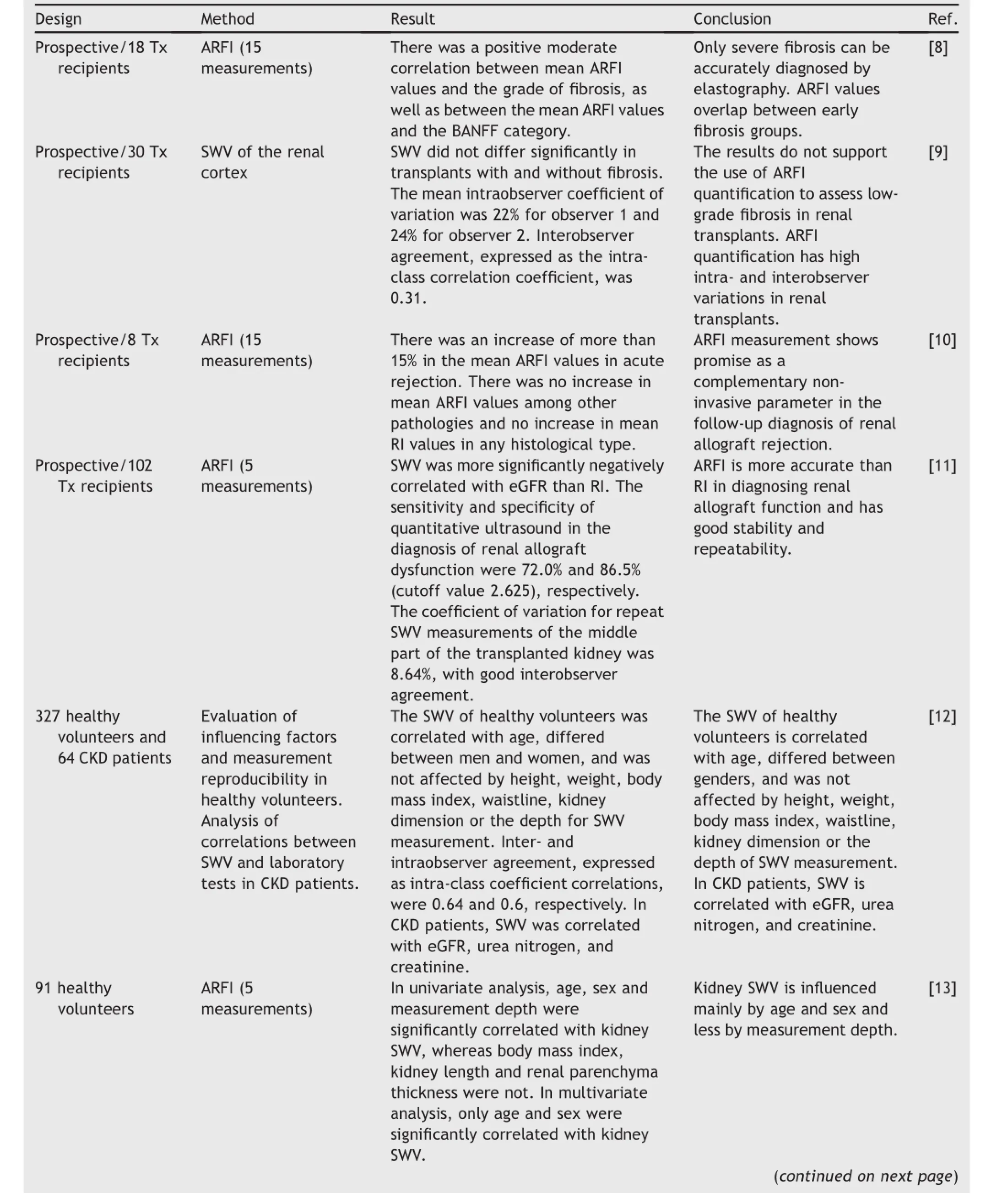
In 2010,the fi rst study by Stock et al.[8]of renal allograft fi brosis using ARFI reported a signi fi cant,positive,moderate correlation between mean SWV values and the grade of fi brosis in renal allografts,as well as the BANFF category. However,the next pilot study by Syversveen et al.[9] showed interfering factors and opposite results,and this study did not support the use of ARFI quanti fi cation to assess low-grade fi brosis in renal transplants.In 2011,the fi rst clinical experience with ARFI-based tissue elasticity quanti fi cation for the examination of kidney allograft dysfunction was reported by Stock’s group.The mean ARFI values showed an average increase of more than 15%in fi ve acute rejected kidneys,whereas no increase was observed in the other three dysfunctional kidneys,including two cases of acute tubular necrosis(ATN)and one case of drugrelated toxicity[10].
Our recent study compared the diagnostic eff i cacy of SWV and resistive index(RI)in an expanded sample.Fifty-two patientswithstablerenalfunctionand50patientswithacute rejection(AR)were enrolled.Our results indicated that the mean SWV was more signif i cantly negatively correlated with estimated glomerular f i ltration rate(eGFR).The sensitivity and specif i city of SWV in the diagnosis of renal allograft dysfunction were 72.0%and 86.5%(cutoff value Z 2.625), respectively,and were better than those of RI,which were 62.0%and 69.2%(cutoff value Z 0.625)[11],respectively.
The results from our and other groups revealed good inter-and intraobserver agreement in both kidney allografts[11]and native kidneys[12].However,Syversveen et al.[9]raised concerns regarding the intra-and interobserver agreement in renal allograft ARFI evaluation.Their group found no signif i cant difference in median SWV between patients without and with renal allograft f i brosis,as well as low intra-and interobserver agreement rates.It is diff i cult to ascertain the reason for these results.However, because of the limited number of enrolled subjects and less detailed descriptions of observer training,one must question the different conclusions drawn.Given the importance of inter-and intraobserver agreement in any type of ultrasonic examination,attention is certainly warranted. However,studies with larger samples are needed to conf i rm any conclusion,and these studies preferably should use experienced doctors who have participated in standard training and have proved to be qualif i ed in ARFI performance(Table 1).
2.3.Possible factors inf l uencing ARFI examinations
Recent studies have reported various factors that could interfere with measurement using ARFI.Such factors have included targetdepth[13],appliedtransducer force [14,15],medium between target and probe[16],probe machines and examiner differences[17,18],and diminution of organ blood f l ow[19].Syversveen et al.[9]found that SWV measurements were dependent on the applied transducer force and that SWV measurements were not different in kidney allografts with different grades of f i brosis.The experiment was scientif i cally credible,yet part of the conclusion contrasted with the well-known relationship between SWV measurements and organ f i brosis.Through a phantom study,Yamanaka et al.[17]discovered that targets with deep ROI shad slightly lower SWV values than superf i cial targets.This conclusion is consistent with the results reported by Kaminuma et al.[16].Because patients possess different tissue thickness and therefore organ depth,further research in larger samples is needed to investigate and prove the effects of each suspected factor on ARFI values.
Regarding factors known to not affect ARFI values,the study from our group proved that kidney volume did not affect SWV and RI measurements or eGFR[11].Goertz et al. [20]reported that age,sex,height,weight,BMI and kidney volume did not affect SWV measurements.Lee et al.[21], however,discovered an age-related increase in SWV in the kidneys of children younger than 5 years old,suggesting anage-related effect on SWV results within a limited pediatric population.The conclusion drawn by Goertz et al.[20]did not entirely agree with the experimental results of Nightingale et al.[22],who estimated mean Young’s modulus values of between 3.8 and 5.6 kPa,11.7 kPa and 14.0 kPa for fat,f i broadenoma and skin,respectively.Given that the formula for body mass index(BMI)calculation contains only height and body weight and that no index of body type or tissue proportion was considered,these f i ndings indicated somewhat that two patients with the same BMI yet with very different fat and muscle ratios should have the same SVW.An explanation for such a conclusion could be the limited subject distribution.More research on the matter with broad subject distribution might provide a satisfactory answer.While debate continues over the inf l uences of surface tissue and target depth,most researchers agree that transplanted kidneys are usually located in the iliac fossa.This knowledge ensures a relatively consistent anatomic position and small target depth.Because of the superf i cial and consistent locations of transplanted kidneys,the possible inf l uences of tissue depth and type are relatively lower among kidney allografts,compared to normal kidneys.

Table 1 AFRI studies.

Table 1 (continued)
3.CEUS
3.1.The mechanisms of CEUS technology
CEUS employs microbubble contrast agents and complementary harmonic pulse sequences to demonstrate parenchymal perfusion,detecting microvascular blood f l ow in real time without affecting renal function[23].CEUS enablesthedynamicassessmentandquantif i cationof microvascularization up to capillary perfusion.
3.2.Evaluation of kidney allograft status
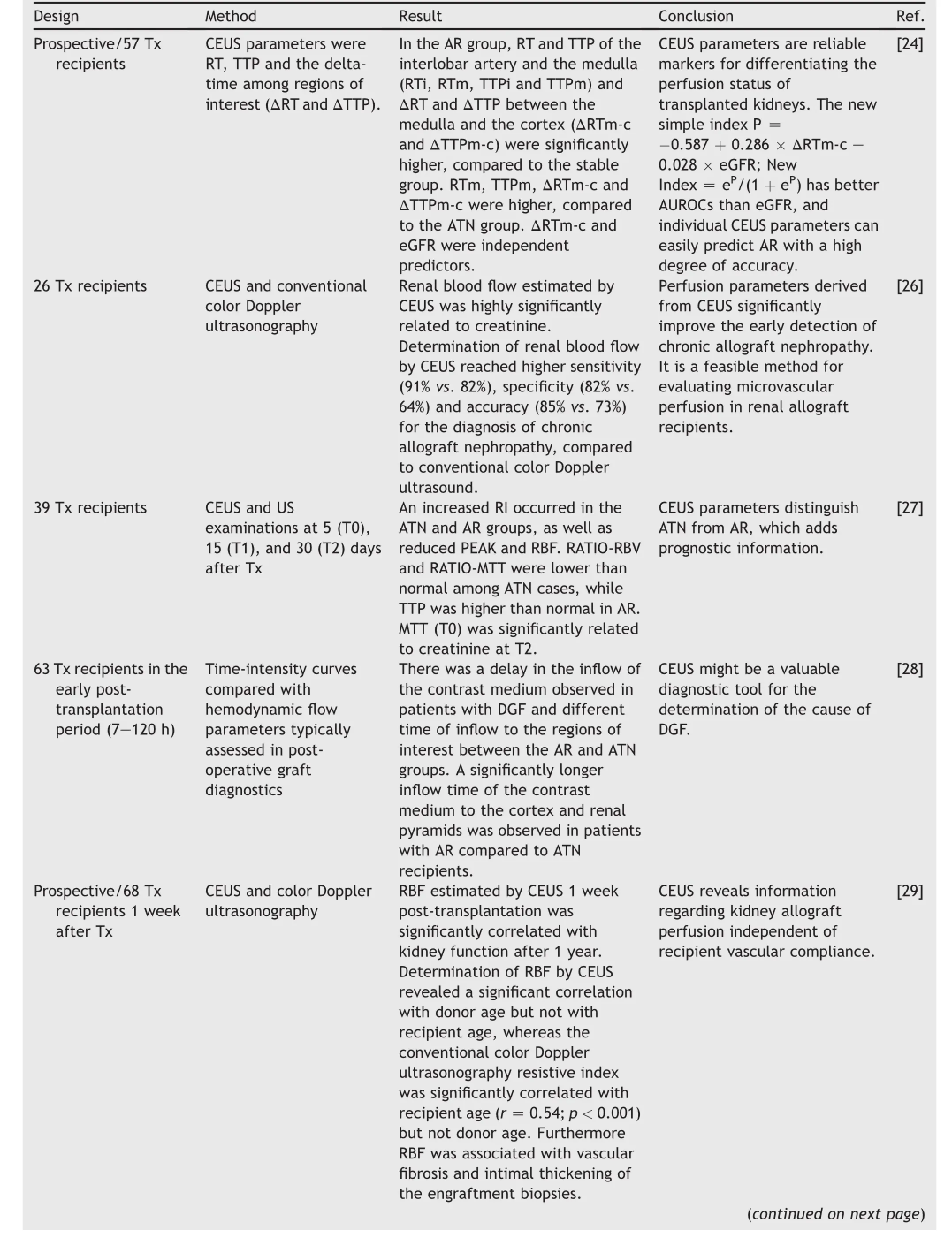
As a novel ultrasonography diagnostic method,CEUS has been reported to be promising for the assessment of renal allograft dysfunction[24,25].Its parameters include rising time(RT),time to peak(TTP)and the delta-time among ROIs(DRT and DTTP).In a pilot study in renal allograft patients in 2006,the Schwenger group demonstrated the diagnostic value of CEUS in diagnosing chronic allograft nephropathy(CAN).The determination of renal blood f l ow byCEUSdemonstratedsignif i cantlyhighersensitivity, specif i city and overall accuracy compared with the RI for the detection of chronic allograft nephropathy,and CEUS was the most signif i cant test for the detection of CAN, compared with all of the other conventional parameters [26].Benozzi et al.[27]later found that,in addition to CAN,some CEUS-derived parameters were able to distinguishATNandAR episodes,includingreducedpeak enhancement,TTP,cortical to medullary ratio of regional blood f l ow and cortical to medullary ratio of mean transit time.
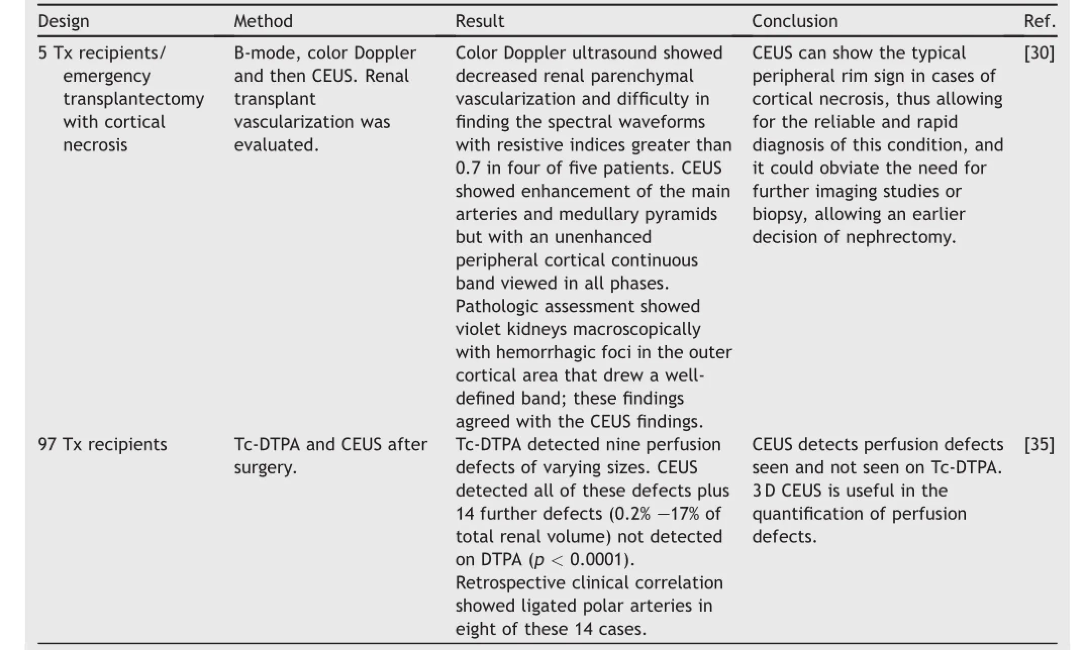
Delayed graft function(DGF)could also be distinguished by CEUS.Grzelak et al.[28]assessed graft perfusion in the early period after kidney transplantation(72e120 h)in 63 kidney allograft recipients;35 had good early graft function (EGF),and 28 had DGF.In patients with DGF,a delay in the inf l ow of the contrast medium was observed,as well as signif i cant differences in the time of inf l ow to the ROIs between the two groups.Schwenger et al.[29]recently discovered in a study that consists of 69 kidney transplant recipients that renal blood f l ow,estimated by CEUS 1 week post-transplantation,is signif i cantly correlated with kidney function after 1 year,suggesting a prognostic value of CEUS soon after kidney transplantation.The renal blood f l ow assessed through CEUS resembled a signif i cant correlation with donor age but not recipient age,and was associated withvascularf i brosisandintimalthickeningofthe engraftment biopsies from their study,implying the ability of CEUS to reveal information on kidney allograft perfusion independent of recipient vascular compliance.Fernandez et al.[30]concluded,after a retrospective analysis of f i ve patients who underwent emergency transplantectomy, with the cases later proving to be acute cortical necrosis, that CEUS could show the typical peripheral rim sign in cases of cortical necrosis,allowing for the reliable and rapid diagnosis of this condition and earlier decisions for nephrectomy,obviating the need for further imaging studies or biopsy.
Recently,our research group enrolled 57 renal transplant recipients in a prospective study.CEUS examinations were performed before renal allograft biopsy.The biopsy results proved 23 cases of AR,10 cases of ATN,and 24 cases with normal evolution(stable).In addition to the f i ndings of the CEUS parameters among these groups,we furtherestablished the following novel simple index to distinguish AR:P Z?0.587 t 0.286?DRTm-c(medulla to cortex)e 0.028?eGFR;New Index Z eP/(1 t eP).This index could easily predict AR with a high degree of accuracy[24](Table 2)(Fig.1).

Table 2 CEUS studies.

Table 2 (continued)
3.3.Advantages and limitations of CEUS
Like all ultrasonic examinations,CEUS is believed to be safe and simple,with wide tolerance and no proven nephrotoxicity.The contrast medium can be safely injected in patients with renal failure because there is no renal excretion of the contrast agent[31],making CEUS a top choiceforpatientswithsuspectedrenalallograft dysfunction.US contrast agents are known to be very safe, with very low anaphylactic reaction rates(1:7000 patients, 0.014%)thataremuchlowerthanthecomparable anaphylactic reaction rate with CT agents(0.035%e0.095%) [23].
However,CEUS,like other ultrasonic techniques for diagnosis,is restricted by lesion location.Obesity and bowel gas interposition are interfering factors or even contraindications.While the use of CEUS is highly promising in kidney allograft recipients,experience remains limited in a clinical setting.
4.Comparison between ARFI and CEUS with other non-invasive diagnostic methods
4.1.Comparison between ARFI and CEUS with other ultrasonic indicators
Real-time color Doppler ultrasound provides parameters including the RI and pulsatility index(PI).The RI ref l ects the vascular status of a transplanted kidney,and it has been used in the early diagnosis of acute renal allograft rejection in initial Doppler studies.It has been reported that during the early post-transplantation period,RI and PI were correlated with long-term allograft function and could potentially be used as prognostic factors to aid in risk stratif i cation for future transplant dysfunction[2].An RI value greater than 0.8 was shown to be predictive of death and poor long-term prognosis for the renal allograft[32]. However,it was also reported that an increased RI was observed in patients with stable renal allograft function [33].It has been accepted that RI has a lack of sensitivity and specif i city for the diagnosis of renal allograft function, except in acute rejection,because it varies with different factors,such as age and blood f l ow velocity[34].A higher RI level indicates a dysfunctional allograft kidney,but it does not differentiate AR from ATN,which are two importantand common causes of allograft kidney dysfunction during the early post-transplantation stage.This fact reduces the clinical value of RI because the therapeutic strategies for the two diseases are different.As mentioned above,ARFI and CEUS parameters have shown higher sensitivity and specif i city for the diagnosis of kidney allograft dysfunction, compared to conventional ultrasound parameters.
4.2.Comparison between ARFI and CEUS and other non-ultrasonic diagnostic methods
Nuclear medicine:Tc-DTPA is a common post-transplant renogram examination performed to assess perfusion.It provides some functional information,but overall,it is a lengthy,immobile and costly examination method with low spatial resolution that uses ionizing radiation[35].A recent study by Stenberg et al.[35]compared the detection of post-surgical perfusion defects in kidney transplants between 3-dimensional CEUS and Tc99m-DTPA.They found that Tc-DTPA detected nine perfusion defects of varying sizes.CEUS,however,detected all of these defects plus 14 further defects not detected by DTPA.This f i nding likely occurred because CEUS detected perfusion defects seen with Tc-DTPA,as well as further perfusion defects not seen on Tc-DTPA,possibly due to increased spatial and temporal resolution and multiple scanning angles.
Computerized tomography(CT):With regard to the kidney,CEUS can obviate the need for CT.The greatest advantage of ARFI and CEUS is their lack of nephrotoxicity caused by CT contrast agents,affording kidney recipients with impaired renal function a simple method of evaluation [36].In addition,it is estimated that 1.5%e2%of all cancers might be attributable to radiation from CT examinations. Neither ARFI nor CEUS exposes a patient to radiation as CT does,making it signif i cantly safer even among special patient populations.
Functional magnetic resonance imaging(MRI):Functional MRI contributes to multilateral,noninvasive,in vivo assessment of kidney function.Two promising functional MRI techniques for assessing kidney function are diffusionweighted(DW)MRI and blood oxygen level-dependent (BOLD)MRI[37].DW MRI is based on the thermally induced Brownian motion of water molecules in tissue,and it does not require a contrast medium.Kaul et al.[38] found that apparent diffusion coeff i cient(ADC)values decreased signif i cantly when rejection occurred,and this decrease was correlated with the degree of rejection on kidney biopsies.However,the reduction in ADC was also observed in kidney allografts with ATN in animal models [39].Park et al.[40]revealed that DW MRI at 3T could demonstrate the early functional state of renal allografts, but it might also be limited in characterizing the cause of early renal allograft dysfunction.In contrast,CEUS is able to distinguish more of the possible causes of kidney allograft function impairment due to its display of blood f l ow (such as thrombosis),making it a more ideal choice as a non-invasive method for allograft function assessment,in the hope of replacing renal biopsy.
BOLD MRI utilizes deoxygenated hemoglobin as an endogenous marker of tissue oxygenation.Xiao et al.[41] reported that decreased R2 values of the cortex and medulla and the R2 ratio of M/C suggested AR in renal allografts.Although this tool represents a major addition to our armamentarium of methodologies to investigate the role of hypoxia in the pathogenesis of acute kidney injury and progressive chronic kidney disease,numerous technical limitations have confounded the interpretation of data derived from this approach.Attempts using BOLD MRI have been undertaken to assess acute and chronic rejection,as well as ATN,but the experimental results and conclusions have lacked agreement[42].
SeveralotherMR-basednon-invasivetechnologies, including arterial spin labeling MRI[43],diffusion-tensor MRI[44]and ferumoxytol-enhanced MRA[45]have also been reported to have potential in kidney allograft function evaluation.However,until now,there have been nocomparison studies between ARFI or CEUS and these new MRI technologies.
5.Perspectives and conclusions
ARFI and CEUS are both considered promising because they share features,including safety,convenience and eff iciency,and they are non-invasive,have low costs and do not require hospitalization.It is also possible that better evaluation of kidney allograft function lies in a formula containing multiple ultrasonic and clinical indicators.
Ultrasonicexaminationscanbeperformedinthe outpatient department,making them a top choice for economical kidney allograft assessment.They are highly convenient and eff i cient,requiring an average of 2e5 min for a trained physician to evaluate a single patient through ARFI.Clinical application of ultrasonic renal allograft function evaluation also avoids the potential risks and complications of renal biopsies,lessening the concern among patients during both routine post-operative evaluations and the conf i rmation of suspected allograft injury.To achieve an agreeable conclusion despite possible interfering factors,more research with a much larger sample size is required,as well as a standard for ensuring proper interobserver agreement.With a unif i ed diagnostic standard and controlled environment,minimal interference during evaluation can be achieved.The current value of ARFI and CEUS in kidney allograft function assessment remains experimental,yet these technologies have a hopeful future.
Conf l icts of interest
The authors declare no conf l ict of interest.
Acknowledgments
This study was supported by the Science and Technology CommissionofShanghaiMunicipality(13ZR1406400, 15411964900 to Wanyuan He)and the National Natural Science Foundation of China(81270833 to Tongyu Zhu; 81400752 to Cheng Yang).
[1]Torres Munoz A,Valdez-Ortiz R,Gonzalez-Parra C,Espinoza-Davila E,Morales-Buenrostro LE,Correa-Rotter R.Percutaneous renal biopsy of native kidneys:eff i ciency,safety and risk factors associated with major complications.Arch Med Sci 2011;7:823e31.
[2]McArthur C,Geddes CC,Baxter GM.Early measurement of pulsatility and resistive indexes:correlation with long-term renal transplant function.Radiology 2011;259:278e85.
[3]Goya C,Hamidi C,Hattapoglu S,Cetincakmak MG,Teke M, Degirmenci MS,et al.Use of acoustic radiation force impulse elastography to diagnose acute pancreatitis athospital admission:comparison with sonography and computed tomography.J Ultrasound Med 2014;33:1453e60.
[4]Park MK,Jo J,Kwon H,Cho JH,Oh JY,Noh MH,et al.Usefulness of acoustic radiation force impulse elastography in the differential diagnosis of benign and malignant solid pancreatic lesions.Ultrasonography 2014;33:26e33.
[5]Wu YM,Xu N,Hu JY,Xu XF,Wu WX,Gao SX,et al.A simple noninvasive index to predict signif i cant liver f i brosis in patients with advanced schistosomiasis japonica.Parasitol Int 2013;62:283e8.
[6]Ozkan F,Menzilcioglu MS,Duymus M,Yildiz S,Avcu S.Acoustic radiation force impulse elastography for evaluating renal parenchymal stiffness in children.Pediatr Radiol 2015;45:461.
[7]TakahashiH,OnoN,EguchiY,EguchiT,KitajimaY, Kawaguchi Y,et al.Evaluation of acoustic radiation force impulse elastography for f i brosis staging of chronic liver disease:a pilot study.Liver Int 2010;30:538e45.
[8]Stock KF,Klein BS,Vo Cong MT,Sarkar O,Romisch M, Regenbogen C,et al.ARFI-based tissue elasticity quantif i cation in comparison to histology for the diagnosis of renal transplant f i brosis.Clin Hemorheol Microcirc 2010;46:139e48.
[9]Syversveen T,Brabrand K,Midtvedt K,Strom EH,Hartmann A, Jakobsen JA,et al.Assessment of renal allograft f i brosis by acoustic radiation force impulse quantif i cationea pilot study. Transpl Int 2011;24:100e5.
[10]Stock KF,Klein BS,Cong MT,Regenbogen C,Kemmner S, Buttner M,et al.ARFI-based tissue elasticity quantif i cation and kidney graft dysfunction:f i rst clinical experiences.Clin Hemorheol Microcirc 2011;49:527e35.
[11]He WY,Jin YJ,Wang WP,Li CL,Ji ZB,Yang C.Tissue elasticity quantif i cation by acoustic radiation force impulse for the assessment of renal allograft function.Ultrasound Med Biol 2014;40:322e9.
[12]Guo LH,Xu HX,Fu HJ,Peng A,Zhang YF,Liu LN.Acoustic radiation force impulse imaging for noninvasive evaluation of renal parenchyma elasticity:preliminary f i ndings.PLoS One 2013;8:e68925.
[13]Bota S,Bob F,Sporea I,Sirli R,Popescu A.Factors that infl uence kidney shear wave speed assessed by acoustic radiation force impulse elastography in patients without kidney pathology.Ultrasound Med Biol 2015;41:1e6.
[14]Syversveen T,Midtvedt K,Berstad AE,Brabrand K,Strom EH, Abildgaard A.Tissue elasticity estimated by acoustic radiation force impulse quantif i cation depends on the applied transducer force:an experimental study in kidney transplant patients.Eur Radiol 2012;22:2130e7.
[15]Tozaki M,Isobe S,Fukuma E.Preliminary study of ultrasonographic tissue quantif i cation of the breast using the acoustic radiation force impulse(ARFI)technology.Eur J Radiol 2011; 80:e182e7.
[16]Kaminuma C,Tsushima Y,Matsumoto N,Kurabayashi T,Taketomi-Takahashi A,Endo K.Reliable measurement procedure of virtual touch tissue quantif i cation with acoustic radiation force impulse imaging.J Ultrasound Med 2011;30:745e51.
[17]Yamanaka N,Kaminuma C,Taketomi-Takahashi A,Tsushima Y. Reliable measurement by virtual touch tissue quantif i cation with acoustic radiation force impulse imaging:phantom study. J Ultrasound Med 2012;31:1239e44.
[18]Potthoff A,Attia D,Pischke S,Kirschner J,Mederacke I, Wedemeyer H,et al.Inf l uence of different frequencies and insertion depths on the diagnostic accuracy of liver elastography by acoustic radiation force impulse imaging(ARFI).Eur J Radiol 2013;82:1207e12.
[19]Asano K,Ogata A,Tanaka K,Ide Y,Sankoda A,Kawakita C, et al.Acoustic radiation force impulse elastography of the kidneys:is shear wave velocity affected by tissue f i brosis or renal blood f l ow?J Ultrasound Med 2014;33:793e801.
[20]Goertz RS,Amann K,Heide R,Bernatik T,Neurath MF, Strobel D.An abdominal and thyroid status with acoustic radiation force impulse elastometryea feasibility study:acoustic radiation force impulse elastometry of human organs.Eur J Radiol 2011;80:e226e30.
[21]Lee MJ,Kim MJ,Han KH,Yoon CS.Age-related changes in liver,kidney,andspleenstiffnessinhealthychildren measured with acoustic radiation force impulse imaging.Eur J Radiol 2013;82:e290e4.
[22]Nightingale K,McAleavey S,Trahey G.Shear-wave generation using acoustic radiation force:in vivo and ex vivo results. Ultrasound Med Biol 2003;29:1715e23.
[23]CokkinosDD,AntypaEG,SkilakakiM,KriketouD, Tavernaraki E,Piperopoulos PN.Contrast enhanced ultrasound of the kidneys:what is it capable of?BioMed Res Int 2013;2013:595873.
[24]Jin Y,Yang C,Wu S,Zhou S,Ji Z,Zhu T,et al.A novel simple noninvasive index to predict renal transplant acute rejection by contrast-enhanced ultrasonography.Transplantation 2015; 99:636e49.
[25]Harvey CJ,Sidhu PS,Bachmann Nielsen M.Contrast-enhanced ultrasound in renal transplants:applications and future directions.Ultraschall Med 2013;34:319e21.
[26]Schwenger V,Korosoglou G,Hinkel UP,Morath C,Hansen A, Sommerer C,et al.Real-time contrast-enhanced sonography of renal transplant recipients predicts chronic allograft nephropathy.Am J Transplant 2006;6:609e15.
[27]Benozzi L,Cappelli G,Granito M,Davoli D,Favali D, Montecchi MG.Contrast-enhanced sonography in early kidney graft dysfunction.Transplant Proc 2009;41:1214e5.
[28]GrzelakP,SzymczykK,StrzelczykJ,KurnatowskaI, Sapieha M,Nowicki M,et al.Perfusion of kidney graft pyramids and cortex in contrast-enhanced ultrasonography in the determination of the cause of delayed graft function.Ann Transplant Q Pol Transplant Soc 2011;16:48e53.
[29]Schwenger V,Hankel V,Seckinger J,Macher-Goppinger S, Morath C,Zeisbrich M,et al.Contrast-enhanced ultrasonography in the early period after kidney transplantation predicts long-term allograft function.Transplant Proc 2014;46:3352e7.
[30]Fernandez CP,Ripolles T,Martinez MJ,Blay J,Pallardo L, Gavela E.Diagnosis of acute cortical necrosis in renal transplantation by contrast-enhanced ultrasound:a preliminary experience.Ultraschall Med 2013;34:340e4.
[31]Wilson SR,Burns PN.Microbubble-enhanced US in body imaging:what role?Radiology 2010;257:24e39.
[32]PerrellaRR,DuerinckxAJ,TesslerFN,DanovitchGM, Wilkinson A,Gonzalez S,et al.Evaluation of renal transplant dysfunction by duplex Doppler sonography:a prospective study and review of the literature.Am J Kidney Dis 1990;15: 544e50.
[33]Kocabas B,Aktas A,Aras M,Isiklar I,Gencoglu A.Renal scintigraphyf i ndingsinallograftrecipientswithincreased resistance index on Doppler sonography.Transplant Proc 2008; 40:100e3.
[34]Schwenger V,Keller T,Hofmann N,Hoffmann O,Sommerer C, Nahm AM,et al.Color Doppler indices of renal allografts depend on vascular stiffness of the transplant recipients.Am J Transplant 2006;6:2721e4.
[35]Stenberg B,Chandler C,Wyrley-Birch H,Elliott ST.Postoperative 3-dimensional contrast-enhanced ultrasound(CEUS) versus Tc99m-DTPA in the detection of post-surgical perfusion defects in kidney transplants e preliminary f i ndings.2014;35: 273e8.
[36]McArthur C,Baxter GM.Current and potential renal applications of contrast-enhanced ultrasound.Clin Radiol 2012;67: 909e22.
[37]Inoue T,Kozawa E,Okada H,Inukai K,Watanabe S,Kikuta T, et al.Noninvasive evaluation of kidney hypoxia and f i brosis using magnetic resonance imaging.J Am Soc Nephrol 2011;22: 1429e34.
[38]Kaul A,Sharma RK,Gupta RK,Lal H,Jaisuresh,Yadav A,et al. Assessment of allograft function using diffusion-weighted magnetic resonance imaging in kidney transplant patients. Saudi J Kidney Dis Transplant 2014;25:1143e7.
[39]Yang D,Ye Q,Williams DS,Hitchens TK,Ho C.Normal and transplanted rat kidneys:diffusion MR imaging at 7 T.Radiology 2004;231:702e9.
[40]Park SY,Kim CK,Park BK,Kim SJ,Lee S,Huh W.Assessment of early renal allograft dysfunction with blood oxygenation leveldependent MRI and diffusion-weighted imaging.Eur J Radiol 2014;83:2114e21.
[41]Xiao W,Xu J,Wang Q,Xu Y,Zhang M.Functional evaluation of transplanted kidneys in normal function and acute rejection using BOLD MR imaging.Eur J Radiol 2012;81:838e45.
[42]NeugartenJ,GolestanehL.Bloodoxygenationleveldependent MRI for assessment of renal oxygenation.Int J Nephrol Renovasc Dis 2014;7:421e35.
[43]Heusch P,Wittsack HJ,Blondin D,Ljimani A,Nguyen-Quang M, Martirosian P,et al.Functional evaluation of transplanted kidneys using arterial spin labeling MRI.J Magn Reson Imaging 2014;40:84e9.
[44]Lanzman RS,Ljimani A,Pentang G,Zgoura P,Zenginli H, Kropil P,et al.Kidney transplant:functional assessment with diffusion-tensorMRimagingat3T.Radiology2013;266: 218e25.
[45]Bashir MR,Jaffe TA,Brennan TV,Patel UD,Ellis MJ.Renal transplant imaging using magnetic resonance angiography with a nonnephrotoxic contrast agent.Transplantation 2013; 96:91e6.
Received 26 June 2015;accepted 29 June 2015 Available online 6 July 2015
*Corresponding author.
E-mail addresses:he.wanyuan@zs-hospital.sh.cn,wanyuanhe@126.com(W.He).
Peer review under responsibility of Shanghai Medical Association and SMMU.
1These authors contributed equally to this study.
http://dx.doi.org/10.1016/j.ajur.2015.06.008
2214-3882/a2015 Editorial Off i ce of Asian Journal of Urology.Production and hosting by Elsevier(Singapore)Pte Ltd.This is an open access article under the CC BY-NC-ND license(http://creativecommons.org/licenses/by-nc-nd/4.0/).
 Asian Journal of Urology2015年3期
- Asian Journal of Urology的其它文章
- The men’s health center:Disparities in gender specif i c health services among the top 50“best hospitals”in America
- Flexible ureteroscopy:Technological advancements,current indications and outcomes in the treatment of urolithiasis
- The older the better:The characteristic of localized prostate cancer in Chinese men
- Clear cell renal cell carcinoma located in sinus renalis confused with renal pelvis mass in image
- Giant adrenal tumor presenting as Cushing’s syndrome and pheochromocytoma:A case report
- Extensive prostatic calculi in alkaptonuria: An unusual manifestation of rare disease
