Effect of Phenylephrine on Alveolar Fluid Clearance in Ventilator-induced Lung Injury△
Nai-jing Li *,Xiu Gu ,Wei Li ,Yan Li ,Sheng-qi Li ,and Ping He
1Department of Gerontology,2Department of Respiratory,Shengjing Hospital,China Medical University,Shenyang 110004,China
3College of Pharmacy,Shenyang Pharmaceutical University,Shenyang 110004,China
THE development of pulmonary edema can be considered as a combination of alveolar floodingviaincreased fluid filtration,impaired alveolar-capillary barrier integrity,and disturbed resolution due to decreased alveolar fluid clearance (AFC).An important mechanism regulating AFC is sodium transport across the alveolar epithelium.1-3Na+enters the apical membranes of alveolar type II cells through amiloride-sensitive ion channels and is actively transported across the basolateral membranes of these cells by the ouabain-sensitive Na+/K+-ATPase.4,5The stimulation of AFC could accelerate the resolution of pulmonary edema and facilitate gas exchange across the alveolar epithelium.
It has been known that β-adrenergic agonists increase AFC under both physiological and pathological conditions.6-8However,it is unclear whether α-adrenergic agonists have similar effects under pathological conditions.For patients with respiratory failure,mechanical ventilation is an indispensable support.However,the use of this technique has adverse effects,including increased risk of lung injury.9,10The purpose of this study was to investigate the possible effect of phenylephrine,an α-adrenergic agonist,on AFC in rats with ventilator-associated lung injury.
MATERIALS AND METHODS
Materials
The (14±2)-week specific-pathogen-free male Wistar rats(n=170) weighing 330±30 g were provided by animal center of Shengjing Hospital affiliated to China Medical University.The study protocol was approved by the institutional ethics committee.
Phenylephrine,prazosin (an α1-adrenergic antagonist),yohimbine (an α2-adrenergic antagonist),atenolol (a β1-adrenergic antagonist),ICI-118551 (a β2-adrenergic antagonist),amiloride (a Na+channel blocker),ouabain (a Na+/K+-ATPase blocker),colchicine (a microtubular disrupting agent),β-lumicolchicine,and Evans blue were purchased from Sigma (St.Louis,MO,USA).Albumin bovine serum,chloral hydrate,phosphoric acid,osmium teroxide,propylene oxide,uranyl acetate were purchased from Beijing Superior Chemicals &Instruments.
Grouping of the rats
The rats were randomly allocated into 17 groups (n=10)using random number tables.Specific treatments for each group were as follows.
Effects of high tidal volume (HVT) ventilation on lung morphological change and total lung water (TLW)∶Tissue samples were taken from the left lung in rats ventilated with HVT for 40 minutes (n=10) and unventilated rats as controls (n=10) for morphological observation and TLW measure.
Effects of HVT ventilation on AFC∶Isomolar albumin solutions were injected into the alveolar spaces in rats ventilated with HVT for 40 minutes (n=10) and unventilated rats as controls (n=10).
Effects of phenylephrine on AFC∶Isomolar albumin solutions with 10-5,10-6,10-7,10-8,and 10-9mol/L phenylephrine were injected into the alveolar spaces in rats ventilated with HVT for 40 minutes (n=10 for each concentration of phenylphrine).
Effects of adrenergic antagonists on AFC modulation by phenylephrine∶Prazosin (1×10⁻⁴mol/L,n=10),yohimbine(1×10-4mol/L,n=10),atenolol (1×10⁻⁵mol/L,n=10),or ICI-118551 (1×10⁻⁵mol/L,n=10) was added to the albumin solution containing phenylephrine (1×10⁻⁵mol/L)and injected into the alveolar spaces in rats ventilated with HVT for 40 minutes.
Effects of phenylephrine on amiloride-sensitive and amiloride-insensitive AFC∶Isomolar albumin solution with 1×10⁻⁵mol/L phenylephrine and 5×10⁻⁴mol/L amiloride(n=10) or 5×10⁻⁴mol/L ouabain (n=10) was injected into the alveolar spaces in rats ventilated with HVT for 40 minutes.
Effects of the cellular microtubular system on AFC modulation by phenylephrine∶Isomolar albumin solution with 1×10⁻⁵mol/L phenylephrine was injected into the alveolar spaces in rats ventilated with HVT for 40 minutes and treated with colchicine (0.25 mg/100 g body weight injected intraperitoneally 15 hours before the isolation and perfusion of rat lung,n=10).As controls,β-lumicolchicine of the same dose was injected intraperitoneally at the same time-point (n=10).β-lumicolchicine is an isomer of colchicine that does not bind tubulin or depolymerize microtubules.11However,it shares other properties of colchicine,such as inhibiting protein synthesis,therefore an appropriate control to demonstrate the effect of colchicine which is caused by microtubular disruption.
Mechanical ventilation
Mechanical ventilation in the rats was performed following the method in a previous report.12The rats were anesthetized by intraperitoneal administration of chloral hydrate(0.03 mL/10 g),tracheotomized to insert an endotracheal tube,and mechanically ventilated with a rodent ventilator(TRR-200C,Jiangxi Teli Anaesthesia &Respiration Equipment Co.Ltd.,China).The rats were ventilated with HVT of 40 mL/kg for 40 minutes,with peak airway pressure at 35 cm H2O and respiratory rate at 40 breathes/minute without positive end-expiratory pressure.
Measurement of AFC
AFC was measured as previously described.13The rats were exsanguinated through the abdominal aorta.The trachea,lungs,and heart were excised and placed in a humidified incubator at 37°C.The lungs were ventilated with 100% nitrogen.Isomolar albumin solution,that is,physiological saline solution (5 mL/kg) containing 5% albumin and Evans blue dye (0.15 mg/mL) was injected into the alveolar spaces through the endotracheal tube.After injection,the lungs were inflated with 100% nitrogen at an airway pressure of 7 cm H2O.Alveolar fluid was aspirated 1 hour after injection.The concentrations of Evans bluelabeled albumin in the injected and aspirated solutions were measured using a spectrophotometer (Shimadzu Corporation,UV-160A,Tokyo,Japan) at 621 nm.AFC was estimated based on the progressive increase in the concentration of alveolar Evans blue-labeled albumin and calculated as follows∶13AFC=[(Vi-Vf)/ Vi]×100,Vf=Vi×Pi/Pf.Vistands for the volume of injected solution,Vffor the volume of final alveolar fluid,Pifor the concentration of Evans blue in the injected solution,and Pffor the concentration of Evans blue in the final alveolar fluid.
Morphological changes
Tissue samples for electron microscopy and light microscopy were taken from left lung of the rats.Samples for electron microscopy were washed in 1% phosphoric acid buffer,fixed with 4% osmium tetroxide,and dehydrated in a graded series of alcohol,transferred to propylene oxide,and embedded in Epon 618.The samples were cut into 1-mm thin sections using a diamond knife,and stained with uranyl acetate and lead citrate for electron microscopy.
The methods for evaluating the number of endothelial cells and alveolar epitheliai cells were described previously in detail.14The numerical density of lung cells (Nv) was determined following the equation,where NA stands for the frequency of occurrence of nuclear profiles per unit area of a random sectioning plane,and D for the mean caliper diameter of pulmonary cell nuclei.NA was determined using the electron microscope,and D was determined using previously described techniques.14
Tissue samples for light microscopy were embedded in paraffin,cut to 10-μm sections,and stained with hematoxylin and eosin following standard procedures.
Measurement of TLW
The TLW of the rats was measured by drying the lungs to a constant weight at 60°C for 72 hours.The TLW was calculated using the previously described method∶15TLW=(wet lung weight-dry lung weight)/ dry lung weight.
Statistical analysis
Prism 4 (GraphPad Software,San Diego,CA,USA) was applied for statistical analysis of this study.The data are presented as means±SD.Kolmogorov-Smirnov test was used for normality of variables.Bartlett’s method andFtest were used for homogeneity of variances.All data had confirmed normality of the population and equal variances among different experiment groups.Statistical significance was evaluated byttest between two groups and analysis of variance post-hoc Student-Newman-Keuls method among multiple groups.The level of statistical significance was set atP<0.05.
RESULTS
Morphological findings
In the control group,the alveolar space was basically dry,the lung interstitium had no edema,and the peri-alveolar blood vessels showed no dilations or hyperemia (Fig.1A);the lung epithelial cells remained normal (Fig.1B).In HVT ventilated rats,the peri-alveolar blood vessels were dilated,intra-alveolar and interstitial hemorrhage and edema were observed (Fig.2A).Other findings included structural damage of lamellar bodies,vacuole-like changes of mitochondria,edematous cytoplasm,condensed nuclei,and large and blunt protrusions in replace of microvilli in the alveolar type II epithelial cells (Fig.2B).
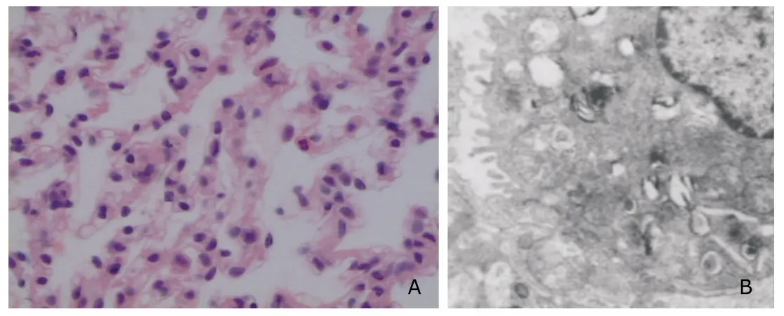
Figure 1.Light microscopic (A) and electron microscopic findings (B) in rats not ventilated (n=10).
The total number of alveolar type II epithelial cells in HVT ventilated rats decreased significantly compared with control rats (P<0.05,Fig.3).The TLW significantly increased from 2.57±0.26 g/g in the control rats to 5.26±0.87 g/g in HVT ventilated rats (P=0.019).
Basal AFC in the control rats was (17.47±2.56)%/hour,which decreased to (9.64±1.32)%/hour in HVT ventilated rats (P=0.003).The presence of phenylephrine at 1×10-8,1×10-7,1×10⁻⁶,and 1×10⁻⁵mol/L increased AFC significantly in HVT ventilated rats (allP<0.05,Fig.4).
Prazosin,atenolol,and ICI-118551 significantly inhibited the increase in AFC by 1×10⁻⁵mol/L phenylephrine in HVT ventilated rats by 53%,31%,and 37%,respectively(allP<0.05,Fig.5).No such effect was observed in yohimbine treated rats.Amiloride and ouabain significantly inhibited AFC stimulation by phenylephrine in HVT ventilated rats by 33% and 42%,respectively (bothP<0.05,Fig.5).Colchicine inhibited the stimulatory effect of phenylephrine on AFC in HVT ventilated rats (P=0.031),whereas the isomer β-lumicolchicine did not influence the effect of phenylephrine on AFC (Fig.5).
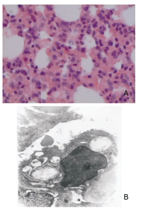
Figure 2.Light microscopic (A) and electron microscopic findings (B) in rats ventilated with high tidal volume(HVT) for 40 minutes (n=10).
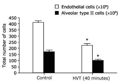
Figure 3.Total number of endothelial and alveolar type Ⅱepithelial cells in the lungs of unventilated rats (n=10) or HVT ventilated rats (n=10).
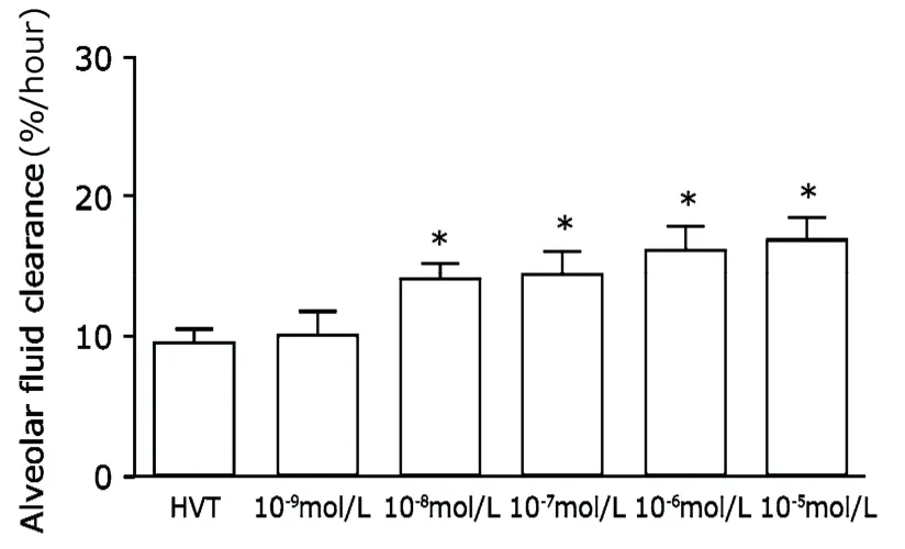
Figure 4.Effects of phenylephrine on alveolar fluid clearance(AFC) in rats ventilated with HVT for 40 minutes(n=10 in each group).
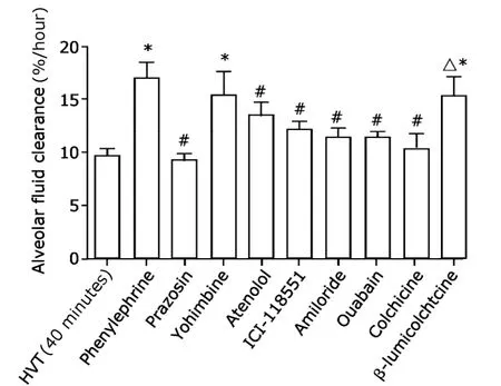
Figure 5.Effects of adrenergic antagonists,amiloride,and microtubular system disrupting agent on AFC modulation by phenylephrine (n=10 in each group).
DISCUSSION
Mechanical ventilators are increasingly used in intensive care units.However,they may cause lung injury and pulmonary edema due to increased permeability.16,17Impaired AFC is associated with mortality in patients with pulmonary edema.It is well accepted that β-adrenergic agonists can stimulate AFC.6-8The results of these studies in rats with lung injury demonstrated a marked upregulation in alveolar epithelial fluid transport capacity by α-adrenergic agonist.
After HVT ventilation to 35 cm H2O for 40 minutes,TLW increased and AFC decreased markedly in ventilated rats compared to the unventilated rats.The alveolar epithelial cells showed structural changes.The number of endothelial cells and alveolar type II epithelial cells decreased significantly.These findings illustrated that the disruption in alveolar fluid transport capacity might depend on the structural damage and functional disorder of epithelial sodium channels in type II epithelial cells and on the decreased number of type II cells.The destruction of pulmonary epithelial structures must accompany with the decreases of sodium channel and Na+/K+-ATPase activity.18Normal structural morphology of alveolar epithelium is indispensable for maintaining the normal AFC.
Alveolar epithelial fluid transport is mainly regulated by a catecholamine-dependent regulatory mechanism.19-21Both endogenous and exogenous catecholamine can upregulate the alveolar epithelial fluid clearance.But it is unknown whether α-adrenergic agonists can stimulate AFC in lung injury.As demonstrated by the present study,although 1×10⁻⁹mol/L phenylephrine did not increase AFC,phenylephrine at 1×10⁻⁸mol/L to 1×10⁻⁵mol/L increased AFC significantly in HVT ventilated rats.Therefore,it can be speculated that the effect of phenylephrine on AFC is dosedependent.Furthermore,the present study showed that α1-adrenergic antagonist prazosin,β1-adrenergic antagonist atenolol,and β2-adrenergic antagonist ICI-118-551 significantly inhibited the increase of AFC by phen-ylephrine in HVT ventilated rats (allP<0.05).In contrast,α2receptor antagonist yohimbine had no effect on AFC.It suggests that the stimulatory effect of phenylephrine on AFC is mediated by stimulating α1,β1,and β2adrenergic receptors.
As demonstrated by the present study,the Na+channel blocker amiloride and the Na+/K+-ATPase inhibitor ouabain reduced the stimulatory effects of phenylephrine on AFC by 33% and 42% respectively (bothP<0.05),suggesting that phenylephrine acts mainly through stimulating Na+channel and Na+/K+-ATPase.The ratio of amiloride-sensitive sodium channel to Na+/K+-ATPase are different among different animals.The amiloride-sensitive sodium channel is 40%-50% in human and rat,but over 80% in rabbit and mouse.22The biological function of Na+/K+-ATPase in human is not negligible.
Saldías et al11reported that Na+/K+-ATPase exists in intracellular pools and can be rapidly recruited in response to specific signalsviamicrotubular transport into the plasma membrane.Therefore we studied whether colchicine,an inhibitor of cell microtubular transport of Na+/K+-ATPase from intracellular pools to the plasma membrane,could hamper the stimulatory effects of phenylephrine on active Na+transport.The results suggest that phenylephrine stimulation of AFC is probably mediated by recruitment of ion-transporting proteins from inner pools to the plasma membrane of the alveolar epithelium.
In conclusion,phenylephrine regulates the AFC in rats with ventilator-associated lung injury,possibly through promoting the intracellular transport of Na+/K+-ATPase.This substance can accelerate the resolution of pulmonary edema and thus may be beneficial in the management of patients with acute ventilator-associated lung injury.
1.Althaus M,Clauss WG,Fronius M.Amiloride-sensitive sodium channels and pulmonary edema.Pulm Med2011;2011∶830320.
2.Mac Sweeney R,Fischer H,McAuley DF.Nasal potential difference to detect Na+channel dysfunction in acute lung injury.Am J Physiol Lung Cell Mol Physiol2011;300∶L305-18.
3.Li NJ,Li W,He P,et al.Effect of beta3-adrenergic agonists on alveolar fluid clearance in hypoxic rat lungs.Chin Med J(Engl)2010;123∶1028-33.
4.Rahman MS,Gandhi S,Otulakowski G,et al.Long-term terbutaline exposure stimulates alpha1-Na+-K+-ATPase expression at postteanscriptional level in rat fetal distal lung epithelial cells.Am J Physiol Lung Cell Mol Physiol 2010;298∶L96-104.
5.Mehta D,Bhattacharya J,Matthay MA,et al.Integrated control of lung fluid balance.Am J Physiol Lung Cell Mol Physiol 2004;287∶L1081-90.
6.Li NJ,Li W,Gu X,et al.Structural changes and effect of denopamine on alveolar fluid clearance in hypoxic rat lungs.Afr J Biotechnol2010;9∶6213-20.
7.Maron MB,Luther NJ,Pilati CF,et al.Beta-adrenoceptor stimulation of alveolar fluid clearance is increased in rats with heart failure.Am J Physiol Lung Cell Mol Physiol 2009;297∶L487-95.
8.Sakuma T,Gu X,Sugita M,et al.Catecholamine clearance from alveolar spaces of rat and human lungs.Respiration 2005;72∶189-96.
9.Pinhu L,Whitehead T,Evans T,et al.Ventilator-associated lung injury.Lancet 2003;361∶332-40.
10.Ngiam N,Kavanagh BP.Ventilator-induced lung injury∶the role of gene activation.Curr Opin Crit Care2012;18∶16-22.
11.Saldías FJ,Comellas AP,Pesce L,et al.Dopamine increases lung luquid clearance during mechanical ventilation.Am J Physiol Lung Cell Mol Physiol 2002;283∶L136-43.
12.Wang Z,Xu J,Ma G,et al.Chronic pulmonary artery occlusion increases alveolar fluid clearance in rats.J Thorac Cardiovasc Surg 2007;134∶1213-9.
13.Sakuma T,Zhao Y,Sugita M,et al.Malnutration impairs alveolar fluid clearance in rat lungs.Am J Physiol Lung Cell Mol Physiol 2004;286∶L1268-74.
14.Crapo JD,Barry BE,Foscue HA,et al.Structural and biochemical changes in rat lungs occurring during exposures to lethal and adaptive doses of oxygen.Am Rev Respir Dis 1980;122∶123-43.
15.Sakuma T,Hida M,Nambu Y,et al.Effects of hypoxia on alveolar fluid transport capacity in rat lungs.J Appl Physiol 2001;91∶1766-74.
16.Simpson PM,Bendal JC.Prehospital non-invasive ventilation for acute cardiogenic pulmonary oedema∶an evidence-based review.Emerg Med J 2011;28∶609-12.
17.Joshi A.Postobstructive pulmonary edema after chronic airway obstruction relief∶a positive end-expiratory pressure effect.Chest 2010;137∶495.
18.Dada LA,Sznajder JI.Mechanisms of pulmonary edema clearance during acute hypoxemic respiratory failure∶role of the Na,K-ATPase.Cri Care Med 2003;31∶S248-52.
19.Groshaus HE,Manocha S,Walley KR,et al.Mechanisms of beta-receptor stimulation-induced improvement of acute lung injury and pulmonary edema.Cri Care 2004;8∶234-42.
20.Perkins GD,McAsuley DF,Richter A,et al.Bench-tobedside review∶beta2-agonists and the acute respiratory distress syndrome.Crit Care 2004;8∶25-32.
21.Matthay MA,Robriquet L,Fang X.Alveolar epithelium∶role in lung fluid balance and acute lung injury.Proc Am Thorac Soc 2005;2∶206-13.
22.Vivona ML,Matthay M,Chabaud MB,et al.Hypoxia reduces alveolar epithelial sodium and fluid transport in rats.Am J Respir Cell Mol Biol 2001;25∶554-61.
 Chinese Medical Sciences Journal2013年1期
- Chinese Medical Sciences Journal的其它文章
- Effect of Nitric Oxide on Esophageal Cancer Cell Line TE-1
- Clinical Study on Suspension Pancreatic-Duct-Jejunum End-to-Side Continuous Suture Anastomosis in Pancreaticoduodenectomy
- Awareness of Cornea Donation of Registered Tissue Donors in Nanjing△
- Breast Milk Lead and Cadmium Levels in Suburban Areas of Nanjing,China
- Possible Role of Mast Cells and Neuropeptides in the Recovery Process of Dextran Sulfate Sodium-induced Colitis in Rats△
- Arthroscopic Debridement and Synovium Resection for Inflammatory Hip Arthritis
